MSK, Skin, & Joints
Heart & Major Vessels
Microcirculation & Liver
Renal & Filtration
Respiratory
100
Which structure connects muscle to bone?
A. Ligament
B. Tendon
C. Deep fascia
D. Superficial fascia
E. Joint capsule
Answer: B — Tendon
Explanation: Tendons connect muscle to bone and transfer muscle-generated force to bone to facilitate movement around a joint. Ligaments connect bone to bone and stabilize joints.
100
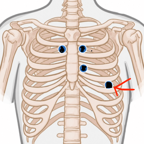
Which valve is best auscultated at the indicated location?
A. Aortic
B. Pulmonary
C. Tricuspid
D. Mitral
E. Pulmonary semilunar
Answer: D — Mitral
Explanation:
To | Please | Be |Aware
Tricuspid | Pulmonary | Bicuspid/Mitral | Aorta
3/4th | 2 | 5 | 2
100
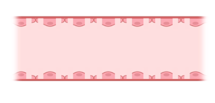
Identify the indicated capillary type.
A. Continuous
B. Fenestrated
C. Discontinuous
D. Sinusoid
E. Portal
Answer: B — Fenestrated
Explanation: Fenestrated capillaries contain pores in their endothelial cytoplasm but retain a complete basement membrane
100
Identify the indicated vessel.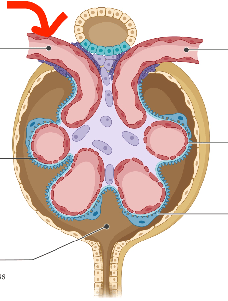
A. Efferent arteriole
B. Afferent arteriole
C. Portal venule
D. Hepatic arteriole
E. Fenestrated vein
Answer: B — Afferent arteriole
Explanation: Abbott's mnemonic is essentially afferent arrives: the afferent arteriole carries blood into the glomerulus, while the efferent arteriole exits it.
100
What structure is ONLY found in the right lung?
A. Oblique fissure
B. Horizontal fissure
C. Cardiac notch
D. Lingula
E. Costodiaphragmatic recess
Answer: B — Horizontal fissure
Explanation: The horizontal fissure is found only in the right lung and separates the superior and middle lobes.
200
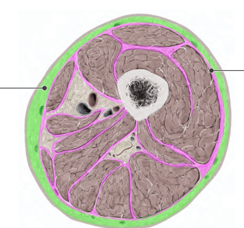
The indicated tissue lies immediately deep to the skin. Which additional feature is characteristic of this layer?
 A. Thin fibrous covering of deeper structuresB. Limits spread of malignant diseaseC. Contains fat, blood vessels, and nerves
D. Forms the wall of a synovial cavity
E. Connects muscle directly to bone
Answer: C — Contains fat, blood vessels, and nerves
Explanation: This is superficial fascia, also called subcutaneous tissue/hypodermis. Abbott describes it as immediately deep to skin, typically containing abundant fat, blood vessels, and nerves, and providing insulation and cushioning.
200
Which sequence correctly follows blood leaving the right ventricle?
A. Aortic valve → aorta → lungs
B. Pulmonary valve → pulmonary trunk → pulmonary arteries
C. Tricuspid valve → pulmonary trunk → pulmonary veins
D. Mitral valve → pulmonary arteries → lungs
E. Pulmonary valve → pulmonary veins → lungs
Answer: B — Pulmonary valve → pulmonary trunk → pulmonary arteries
Explanation: Abbott's right pump is right ventricle → pulmonary semilunar valve → pulmonary trunk → right/left pulmonary arteries → lungs.
200
Which capillary type is the least selective?
A. Continuous
B. Fenestrated
C. Discontinuous/sinusoid
D. Pulmonary
E. Glomerular
Answer: C — Discontinuous/sinusoid
Explanation: Abbott's discontinuous capillary has endothelial pores and an incomplete basement membrane and can allow plasma proteins, RBCs, WBCs, and other large molecules to pass
200
What type of capillary forms the glomerulus?
A. Continuous
B. Fenestrated
C. Discontinuous
D. Sinusoidal
E. Portal
Answer: B — Fenestrated
Explanation: The glomerular endothelium contains small circular pores and sits on a complete basement membrane, making it a fenestrated capillary.
200
The vomer forms which component of the nasal septum?
A. Cartilaginous component
B. Bony component
C. Muscular component
D. Pleural component
E. Fascial component
Answer: B — Bony component
Explanation: Abbott divides the septum into septal cartilage anteriorly and the bony components vomer + perpendicular plate of the ethmoid.
300
A patient receives a permanent tattoo. Several weeks later, the superficial cells of the skin continue to be shed normally, but the tattoo remains visible. According to Abbott's review, into which layer was the ink deposited?
A. Epidermis
B. Dermis
C. Superficial fascia
D. Deep fascia
E. Muscle
Answer: B — Dermis
Explanation: The epidermis is the outermost layer and its topmost cells are continuously sloughed off. Abbott specifically states that permanent tattoo ink is injected into the dermis
300
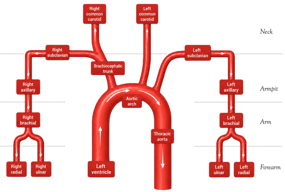
A lesion obstructs the brachiocephalic trunk before it divides. Blood flow would be directly affected to which TWO arteries?
A. Left common carotid + left subclavian
B. Right common carotid + right subclavian
C. Right radial + left radial
D. Left brachial + left common carotid
E. Right ulnar + left subclavian
Answer: B — Right common carotid + right subclavian
Explanation: The brachiocephalic trunk branches into the right common carotid and right subclavian arteries. The left common carotid and left subclavian arise directly from the aortic arch in Abbott's diagram.
300
A substance is traveling from an alveolus into pulmonary blood. Which sequence correctly traces its movement across the air-blood barrier and toward the pulmonary vein?
A. Alveolus → endothelium → pneumocyte → capillary → pulmonary artery
B. Alveolus → pneumocyte → basement membranes → endothelium → capillary → venule
C. Alveolus → basement membrane → pneumocyte → arteriole → capillary
D. Pneumocyte → alveolus → endothelium → capillary → venule
E. Alveolus → pneumocyte → endothelium → basement membrane → pulmonary artery
Answer: B
Explanation: Abbott gives the O₂ pathway as alveolus → pneumocyte → basement membranes → endothelial cell → capillary → venule → pulmonary vein.
300
A substance passes successfully through the glomerular filtration barrier and enters the space where filtered substances collect before potentially becoming urine. Where is it now located?
A. Efferent arteriole
B. Urinary space
C. Portal venule
D. Hepatic sinusoid
E. Pericardial cavity
Answer: B — Urinary space
Explanation: Abbott defines the urinary space as the space containing substances able to pass through the filtration barrier; these substances may eventually become urine.
300
A patient has a pleural effusion while upright. Fluid begins accumulating in the most dependent pleural recess shown in Abbott's review. Where would the fluid first collect?
A. Cervical pleura
B. Cardiac notch
C. Costodiaphragmatic recess
D. Mediastinal pleura
E. Lung apex
Answer: C — Costodiaphragmatic recess
Explanation: The costodiaphragmatic recess, or costophrenic angle, lies between the costal and diaphragmatic pleura and is the first part of the pleural cavity filled by a pleural effusion
400
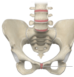
Imaging of the indicated joint shows no joint cavity. The bones are united by cartilage and permit only limited movement. Which structural classification applies?
A. Fibrous
B. Synovial
C. Cartilaginous
D. Diarthrosis
E. Syndesmosis
Answer: C — Cartilaginous
Explanation: Cartilaginous joints have no joint cavity, are held together by cartilage, and are slightly movable. Abbott uses a symphysis as the example.
400
Excess fluid accumulates in the space between the parietal and visceral layers of the serous pericardium. As the accumulation worsens, cardiac function becomes compromised. Which combination correctly identifies the location and complication?
A. Fibrous pericardium — pleural effusion
B. Pericardial cavity — cardiac tamponade
C. Visceral pericardium — pneumothorax
D. Superior mediastinum — cardiac tamponade
E. Pericardial cavity — pleural effusion
Answer: B — Pericardial cavity; cardiac tamponade
Explanation: The pericardial cavity containing serous fluid lies between the parietal and visceral serous layers. Excess fluid is a pericardial effusion, which Abbott states can lead to cardiac tamponade
400
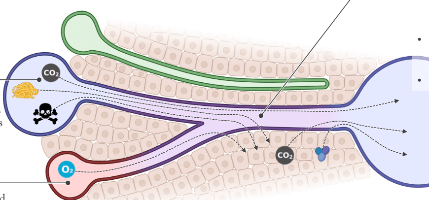
Two vessels empty into the indicated hepatic sinusoid. One contains deoxygenated blood, nutrients, and toxins from the GI tract; the other contains oxygenated blood from the heart. Which pair supplies the sinusoid?
A. Hepatic venule + hepatic vein
B. Portal venule + hepatic arteriole
C. Portal venule + hepatic venule
D. Hepatic arteriole + hepatic vein
E. Portal vein + IVC
Answer: B — Portal venule + hepatic arteriole
Explanation: Abbott's hepatic sinusoid receives blood from both the portal venule and hepatic arteriole. The sinusoid is discontinuous, has an incomplete basement membrane, and is surrounded by hepatocytes.
400
A patient's glomerular basement membrane is damaged. Laboratory analysis now detects substances in the filtrate that should appear with glomerular injury. Which finding is expected?
A. Only water and urea
B. RBCs and protein
C. Only glucose and K⁺
D. Albumin production by podocytes
E. Loss of all small ions from filtrate
Answer: B — RBCs and protein
Explanation: Abbott states that glomerular damage—including basement-membrane damage in Goodpasture syndrome—results in RBCs and protein being filtered.
400
Emergency access to the exposed heart and pericardium is required. The physician uses the indicated indentation of the anterior border of the left superior lobe. Which structure is being used?
A. Lingula
B. Oblique fissure
C. Cardiac notch
D. Costodiaphragmatic recess
E. Cardiac impression
Answer: C — Cardiac notch
Explanation: The cardiac notch is an indentation of the anterior border of the left superior lobe caused by the heart and pericardium. Abbott states that it provides access to the exposed heart/pericardium and lists intracardiac drug administration, pericardiocentesis, and echocardiographic visualization.
500
A malignant process begins in a superficial region of an extremity. During evaluation, the physician identifies a thin fibrous layer covering the deeper regions that can restrict spread of the disease. Immediately superficial to this layer is tissue rich in fat, vessels, and nerves. Which sequence correctly identifies these two layers from superficial → deep?
A. Deep fascia → superficial fascia
B. Dermis → epidermis
C. Superficial fascia → deep fascia
D. Epidermis → superficial fascia
E. Deep fascia → muscle → superficial fascia
Answer: C — Superficial fascia → deep fascia
Explanation: Abbott gives the order epidermis → dermis → superficial fascia → deep fascia → muscle. Superficial fascia contains abundant fat, vessels, and nerves; deep fascia is a thin fibrous covering that can limit spread of infection and malignant disease.
500
A thrombus is located in the left popliteal vein. If it travels proximally following the venous pathway shown in Abbott's review, which sequence does it encounter?
A. Femoral → external iliac → common iliac → IVC
B. Femoral → internal iliac → common iliac → SVC
C. Anterior tibial → femoral → external iliac → IVC
D. External iliac → femoral → common iliac → IVC
E. Femoral → common iliac → external iliac → IVC
Answer: A — Femoral → external iliac → common iliac → IVC
Explanation: Abbott's lower-body venous diagram follows tibial veins → popliteal → femoral → external iliac → common iliac → inferior vena cava.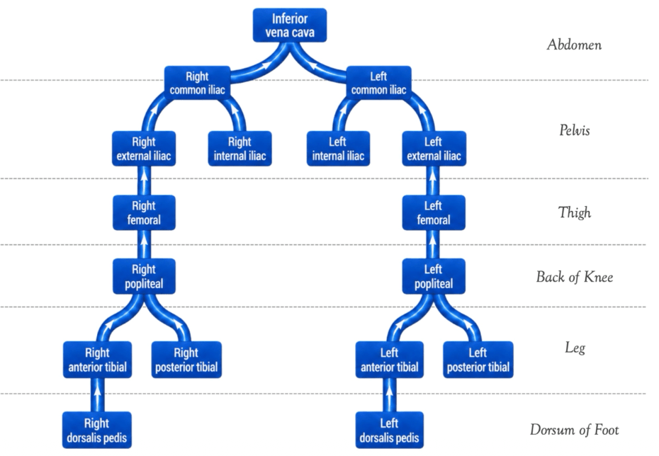
500
A molecule absorbed from the GI tract enters intestinal capillaries and must pass through the liver before returning to the systemic circulation. Which feature makes this arrangement a portal system?
A. Blood passes through one capillary bed before reaching an artery
B. Two arteries connect two capillary beds
C. Blood passes through two capillary beds in series connected by a portal vein
D. Blood passes directly from an artery to a vein without capillaries
E. The hepatic artery supplies both capillary beds
Answer: C
Explanation: Abbott defines a portal system as blood passing through two capillary beds in series before returning to the heart, with a portal vein connecting the two capillary beds. In this system, the hepatic capillaries form the secondary capillary bed.
500
A researcher compares two capillary beds. Bed X has endothelial pores and a complete basement membrane and participates in renal filtration. Bed Y has endothelial pores but an incomplete basement membrane and receives blood from a portal venule and hepatic arteriole. Which pairing correctly identifies X and Y?
A. X continuous; Y fenestrated
B. X sinusoidal; Y continuous
C. X fenestrated glomerulus; Y discontinuous hepatic sinusoid
D. X discontinuous glomerulus; Y fenestrated hepatic sinusoid
E. X pulmonary capillary; Y glomerulus
Answer: C — Fenestrated glomerulus; discontinuous hepatic sinusoid
Explanation: This integrates Abbott's capillary material: the glomerulus is fenestrated with pores but a complete basement membrane. The hepatic sinusoid is discontinuous with an incomplete basement membrane.
500
A child accidentally aspirates a small object while standing upright. Bronchoscopy confirms that the object entered the bronchial tree on the side anatomically favored for aspiration. Which combination best predicts the pathway and final location?
A. Left bronchus because it is wider → left superior lobe
B. Right bronchus because it is shorter, wider, and more vertical → right inferior lobe
C. Right bronchus because it is longer and narrower → right middle lobe
D. Left bronchus because it is more vertical → left inferior lobe
E. Right bronchus because it is more horizontal → right superior lobe
Answer: B — Right bronchus → right inferior lobe
Explanation: Abbott specifically states that the right bronchus is shorter, wider, and more vertical, making it more prone to foreign-body aspiration. When aspiration occurs upright, foreign bodies commonly lodge in the right inferior lobe.