You’ve Got Some Nerve
Head, Shoulders,
Knees and Toes...
Knees and Toes...
Ear We Go Again
Bugging Out
Dealer’s Choice
100
A 28-year-old male presents to the emergency department following an accident in which he slipped on ice and fell down, striking the base of his skull on the arm rail of a flight of stairs. The injury is suspected to have affected one of the parasympathetic nuclei within the brainstem. Which of the following cranial nerves could be spared during his neurological examination?
A. CN III
B. CN VII
C. CN VIII
D. CN IX
E. CN X
C. CN VIII
100
When attempting to pierce the tip of her tongue, a 15-year-old damages the indicated structures in the figure.
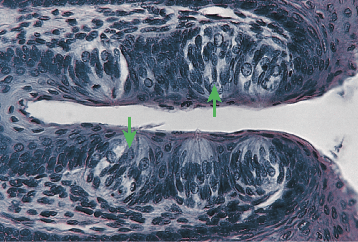
Primary afferents from the indicated structures travel through which of the following cranial nerves?
A. V, trigeminal
B. VII, facial
C. IX, glossopharyngeal
D. X, vagus
E. XII, hypoglossal
Correct: VII, facial (B)
The arrows indicate taste buds (identified by shape and component cells [sensory, supporting, and basal]). Taste sensation from the tip of the tongue is carried by the chorda tympani, a branch of the facial nerve. The glossopharyngeal (C) nerve conveys taste fibers from the posterior third of the tongue (including vallate papillae), and the vagus (D) nerve carries such fibers from the epiglottis, vallecula, and soft palate. Trigeminal (A) or hypoglossal (E) nerves are not involved with taste sensation. The chorda tympani nerve hitchhikes with the lingual nerve (branch of mandibular division, V3, of trigeminal) to reach the tongue. This does not mean that the taste fibers are carried by the
100
A 17-year-old girl is taken to the emergency department after being hit in the side of the head by a softball during a high-school game. Upon examination, it is determined that the incus bone within the right middle ear has been broken as a result of the head trauma, resulting in substantial conductive hearing loss. Which area in the presented image corresponds to the broken bone?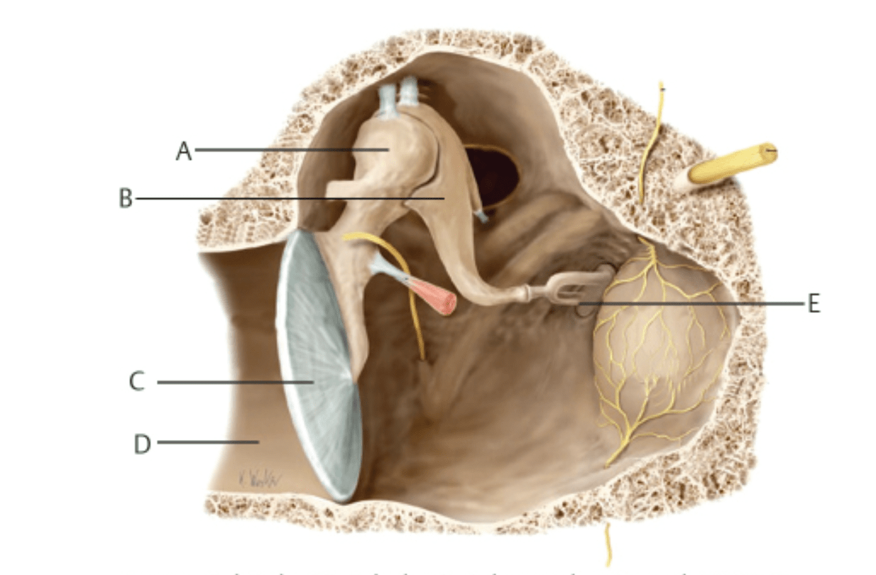
B
100
A 17-month-old child who recently recovered from an upper respiratory tract infection becomes irritable, has a slight fever (37.8°C, 100.4°F), and frequently pulls at his left ear. Suspecting an infectious etiology, the pediatrician prescribes a 7-day course of amoxicillin. Four days later, the child’s mother reports no improvement in symptoms and is very troubled because the boy seems to be less responsive to sound coming from his left side. The pediatrician made which of the following medical errors.
Failure to:
A. Differentiate acute otitis media from otitis media with effusion
B. Perform a myringotomy on the initial visit
C. Perform a proper review of the child’s immunization history
D. Prescribe the appropriate antibiotic
E. Refer the child to a hearing specialist
(A) This is most likely a case of otitis media with effusion (OME). Acute otitis media (AOM) is a common bacterial infection in the pediatric population, with clinical findings including ear pain and systemic symptoms such as fever and malaise. Purulent drainage may occur in AOM following perforation of the tympanic membrane. AOM is usually bacterial in etiology and may be treated with antibiotics such as amoxicillin or amoxicillin–clavulanate. OME occurs due to blockage of the eustachian tube and accumulation of fluid in the middle ear. It may occur after an episode of AOM or a viral upper respiratory infection. Most often, the only sign or symptom of OME is impaired hearing, which may be mild to moderate; fever, pain, and other systemic symptoms are usually absent. OME is generally self-limiting, so a “wait and watch” approach is recommended, and antibiotics are not warranted. (B) Myringotomy is a surgical incision of the tympanic membrane with tube placement, although newer methods may use laser incision without the need for tube placement. While this is a treatment for chronic OME, it would not have been appropriate for the pediatrician to perform this procedure at the initial visit. (C) Streptococcus pneumoniae and nontypeable Haemophilus influenzae are common bacterial causes of acute otitis media in children. Vaccination against S. pneumoniae and H. influenzae type b (Hib) reduces colonization and disease caused by vaccine-covered strains; however, the Hib vaccine does not affect colonization with nontypeable Haemophilus. Presumably, the child’s pediatrician would be aware of the vaccination history. (D) If this were a case of acute otitis media, amoxicillin would be an appropriate first-line antibiotic in many cases. S. pneumoniae is a common bacterial etiology, and β-lactam antibiotics can achieve high concentrations in middle-ear fluid. (E) Hearing loss is a common finding in OME. However, because most cases resolve spontaneously, immediate referral to a hearing specialist would generally be premature.
100
A proximal RCA occlusion causes an inferior MI with profound bradycardia.
What explains the bradycardia?
Answer: The RCA commonly supplies the SA and especially AV nodal regions; AV nodal ischemia may produce conduction abnormalities.
200
A 54-year-old man presents to a neurologist with weakness, pain, and tingling in his left arm and is subsequently diagnosed with neuropathy. A few weeks later, he presents to the emergency department with worsening pain and weakness in the affected arm, muscle twitching, difficulty swallowing and breathing, and increased salivation. He is admitted to the hospital and placed on mechanical ventilation. Two days after admission, he becomes disoriented and combative, and four days after admission, he progresses to a coma. The patient dies one week after admission. Autopsy examination of brain sections from the Ammon horn of the hippocampus demonstrates the findings shown in the image, confirming the diagnosis.
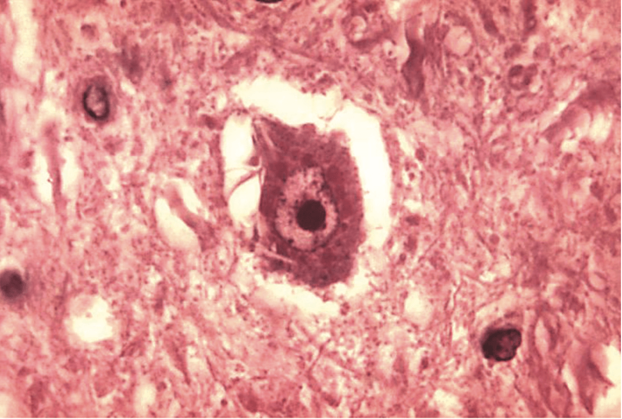
Contact with which of the following is most likely to be in this man’s history?
A. Bats
B. Birds
C. Mosquitos
D. Pigs
E. Ticks
(A) This is a case of rabies, caused by rabies virus, an enveloped, negative-sense, single-stranded RNA virus that is typically transmitted through the bite or saliva of an infected mammal. Important wildlife reservoirs in the United States include bats, raccoons, skunks, and foxes; globally, dogs remain an important source of human rabies. The image in the question stem shows Negri bodies, a classic histopathologic finding of rabies. Negri bodies are eosinophilic, round-to-oval cytoplasmic inclusions found in infected neurons, particularly pyramidal neurons of the hippocampus and Purkinje cells of the cerebellum. The incubation period is typically weeks to months but can vary considerably. Rabies initially produces a nonspecific prodrome and then progresses to neurologic disease. Encephalitic, or “furious,” rabies may present with hydrophobia, hypersalivation, agitation, autonomic instability, and seizures, whereas paralytic rabies produces progressive flaccid paralysis. Although the incubation period may be prolonged, once clinical neurologic symptoms develop, the disease typically progresses rapidly and is almost invariably fatal. (B, D) Birds and pigs can participate in the transmission of various infectious diseases but are not typical reservoirs or vectors for rabies. Rabies is primarily maintained and transmitted among mammals. (C, E) Mosquitoes and ticks transmit several pathogens capable of causing meningitis or encephalitis, but neither serves as a vector for rabies. Rabies transmission occurs primarily through exposure to the saliva of an infected mammal, most commonly through a bite.
200
A 39-year-old man comes to his physician because of a painful ulcer located in the tip of the organ depicted in the fi gure. The cranial nerve that transmits this patient’s pain is also involved in which of the following functions?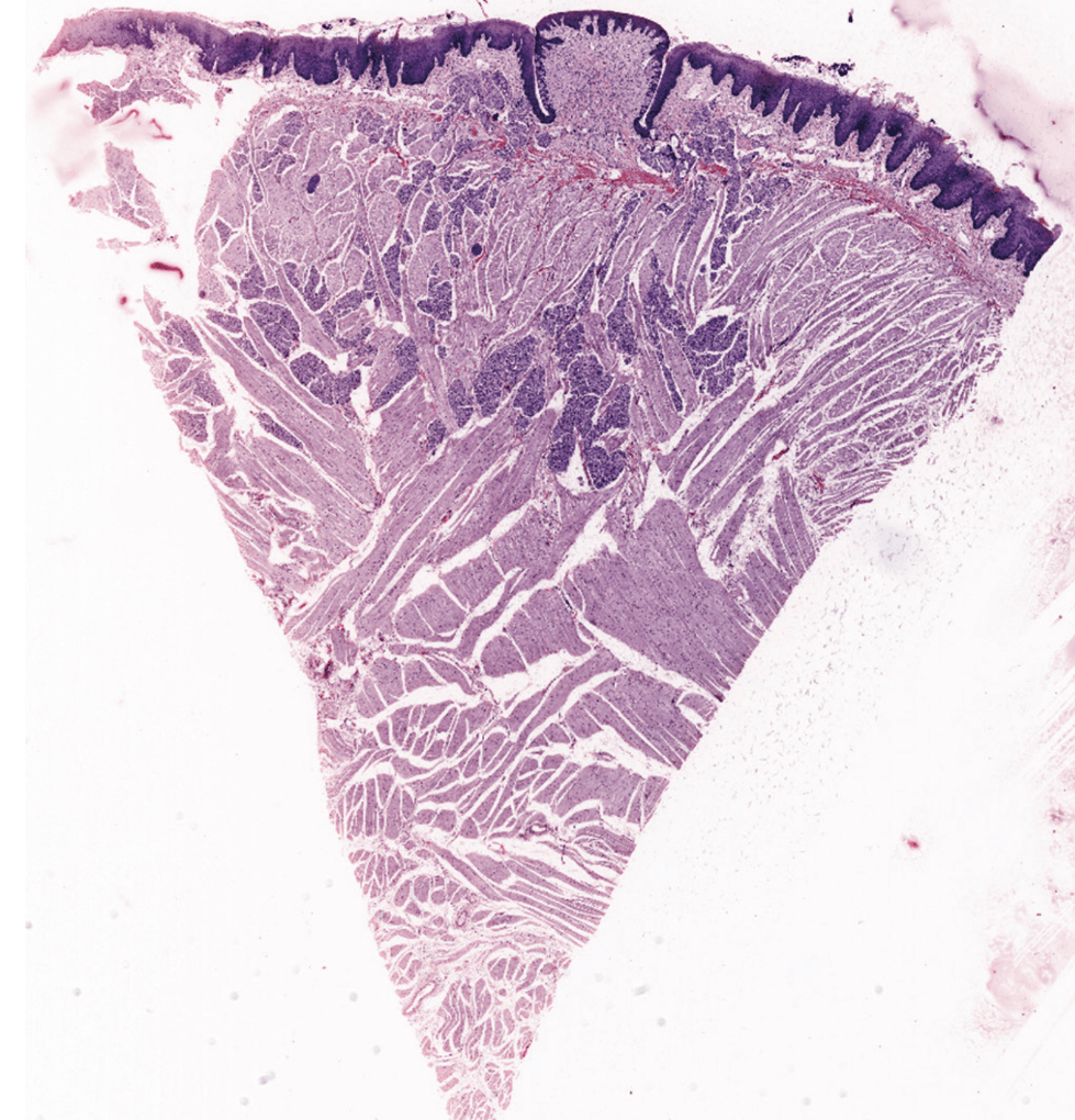
A. Lacrimation
B. Salivation
C. Mastication
D. Speech articulation
E. Tongue protrusion
The image can be identified as the tongue from the lining stratified squamous epithelium, papillae, and skeletal muscle fibers that run in three different planes (each arranged at right angles to the other two). Pain sensation from the tip of the tongue is carried by mandibular division of the trigeminal nerve, which also supplies motor fibers to the muscles of mastication. Lacrimation (A, facial nerve), salivation (B, facial and glossopharyngeal nerve), articulation (D, vagus nerve), and protrusion of the tongue (E, hypoglossal nerve) are not functions of the trigeminal nerve.
200
A 51-year-old Yemeni American develops fevers, rigors, nonproductive cough, and dyspnea 3 days after returning from Yemen to visit his relatives. His vital signs are: temperature, 38.2°C (101.8°F); blood pressure, 131/92 mm Hg; heart rate, 98 beats per minute; and O2 saturation, 94%. The patient is suspected of having Middle East respiratory syndrome (MERS) and is admitted to the hospital. PCR is pending. Which of the following describes the causative agent of MERS?
A. Enveloped, double-stranded DNA virus
B. Enveloped, double-stranded RNA virus
C. Enveloped, single-stranded DNA virus
D. Enveloped, single-stranded RNA virus
E. Nonenveloped, double-stranded DNA virus
F. Nonenveloped, single-stranded DNA virus
G. Nonenveloped, double-stranded RNA virus
H. Nonenveloped, single-stranded RNA virus
(D) Middle East respiratory syndrome (MERS) is caused by Middle East respiratory syndrome coronavirus (MERS-CoV). Coronaviruses are enveloped, positive-sense, single-stranded RNA viruses with a linear RNA genome. (A) Herpesviruses, including VZV, HSV-1, HSV-2, EBV, and CMV, are enveloped, double-stranded DNA viruses. (B) There are no medically significant examples of enveloped, double-stranded RNA viruses. (C) There are no medically significant examples of enveloped, single-stranded DNA viruses. (E) Adenoviruses are nonenveloped, double-stranded DNA viruses. (F) Parvoviruses are nonenveloped, single-stranded DNA viruses and are among the smallest DNA viruses. (G) Reoviruses are nonenveloped, double-stranded RNA viruses; rotavirus is a clinically important member of this family. (H) Picornaviruses, hepeviruses, and caliciviruses are nonenveloped, positive-sense, single-stranded RNA viruses.
200
A 16-year-old boy visits his primary care physician with a 4-day history of a sore throat, fever, and general malaise. He states that he is very tired and just wants to sleep. His mother notes that he has not been eating as much as he normally does. His medical history is unremarkable, he is up to date with vaccinations and is not on medication. On examination, notable pharyngeal inflammation and white tonsillar exudate is observed. In addition, he has marked posterior cervical lymphadenopathy. His temperature at the time of the examination is 38.8°C (101.8°F). A CBC demonstrates the presence of the cells shown below.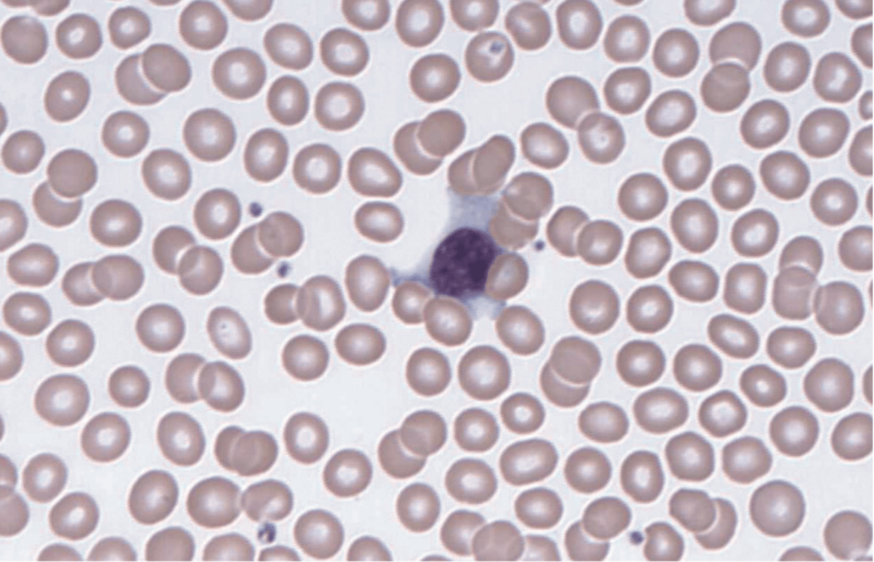
Which of the following would best confirm the diagnosis and identify the most likely agent of this infection?
A. β-Hemolytic growth on blood agar
B. Elek test positive
C. Growth on chocolate agar and not blood agar
D. Positive heterophile antibody
E. Positive DNase B antibody
(D) This boy likely has infectious mononucleosis caused by Epstein–Barr virus (EBV). The heterophile antibody test, or Monospot test, detects heterophile antibodies, which are nonspecific IgM antibodies produced as a result of EBV-induced B-cell activation. These antibodies do not react with EBV-specific antigens but instead recognize antigenic determinants on erythrocytes from certain animal species. In EBV-associated mononucleosis, the heterophile antibody test is typically positive, whereas CMV-associated mononucleosis is generally heterophile-negative. EBV is an enveloped, double-stranded DNA herpesvirus that may present with pharyngitis, severe fatigue, and posterior cervical lymphadenopathy. Atypical lymphocytes may also be present in the peripheral blood. EBV is additionally associated with several malignancies, including nasopharyngeal carcinoma and Burkitt lymphoma. (A) β-Hemolytic growth on blood agar is characteristic of several bacterial pathogens, notably Streptococcus pyogenes, which can cause acute pharyngitis with a similar presentation. However, the presence of atypical lymphocytes and other clinical findings makes EBV more likely. (B) The Elek test is an immunodiffusion test historically used to detect toxin production by Corynebacterium diphtheriae. Diphtheria may present with pharyngitis and formation of a pharyngeal pseudomembrane, but the findings in this case are more consistent with infectious mononucleosis. (C) Organisms that grow well on chocolate agar include Haemophilus and Neisseria species. H. influenzae can cause respiratory and head and neck infections, including pneumonia, otitis media, sinusitis, and epiglottitis; N. meningitidis commonly causes meningitis or meningococcemia, while N. gonorrhoeae is primarily a sexually transmitted pathogen. The clinical presentation and atypical lymphocytes make these organisms less likely than EBV. (E) Anti-DNase B antibodies are used as evidence of a preceding S. pyogenes infection, particularly when evaluating poststreptococcal complications. They are not used to diagnose acute streptococcal pharyngitis, which is evaluated with rapid antigen or molecular testing and/or throat culture, and they would not aid in the diagnosis of EBV infection.
200
A 15-day-old infant presents with dyspnea, stridor, and intercostal retractions. He was born post-term by a difficult forceps delivery. During a flexible laryngeal endoscopic examination, the right vocal cord was found to be paralyzed and in the paramedian line. Stretching of a specific nerve during forceps application was suggested to be the underlying cause.
Which of the following muscle groups, most likely, might suffer due to this iatrogenic injury?
A. Those derived from the 1st pharyngeal arch
B. Those derived from the 2nd pharyngeal arch
C. Those derived from the 3rd pharyngeal arch
D. Those derived from the 4th pharyngeal arch
E. Those derived from the 6th pharyngeal arch
Correct: Those derived from the 6th pharyngeal arch
(E) Congenital vocal cord paralysis is the second most common cause of congenital stridor, frequently consequent to birth trauma. The infant is most likely suffering from right recurrent laryngeal nerve palsy. The nerve supplies the 6th pharyngeal arch. Muscles of the first (A, by trigeminal nerve), second (B, by facial nerve), and third (C, by glossopharyngeal nerve) pharyngeal arches are not supplied by nerves related to the vocal cord. Muscles of the 4th pharyngeal arch (D) are supplied by the external laryngeal nerve. Damage to this nerve (supplying the cricothyroid) is not very common during childbirth and will cause hoarseness of voice due to uneven tension of the vocal cord.
300
A 13-year-old girl is brought to the emergency department with a 2-day history of fever, headache, nausea, and vomiting. On the second day of illness, her mother becomes concerned because the girl appears anxious and irritable, reports that her food tastes and smells “weird,” and has difficulty recalling recent events. Her temperature is 39.5°C (103.5°F), and her blood pressure is 95/60 mm Hg. She is oriented to time but not place. Physical examination reveals nuchal rigidity and a positive Kernig sign. Cerebrospinal fluid (CSF) Gram stain demonstrates numerous neutrophils but no organisms. A Wright–Giemsa stain of the CSF demonstrates the findings shown below.
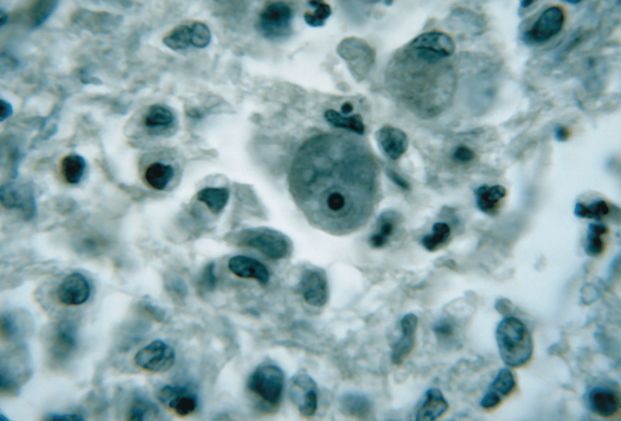
This girl is most likely to have a recent history of:
A. A mission trip to Haiti
B. Camping in the woods
C. Eating ice cream
D. Jet skiing in the ocean
E. Waterskiing in the lake
(E) The image in the question stem shows the trophozoite form of Naegleria fowleri, a free-living ameba found in warm freshwater. Transmission occurs when contaminated water enters the nasal cavity during activities such as waterskiing, swimming, diving, or jumping into warm freshwater. N. fowleri causes primary amebic meningoencephalitis (PAM), a rapidly progressive infection of the central nervous system. After entering the nasal cavity, the organism penetrates the olfactory mucosa and migrates along the olfactory nerves through the cribriform plate of the ethmoid bone to reach the brain. Involvement of the olfactory system may produce alterations in smell or taste. PAM progresses rapidly and is frequently fatal. (A) A mission trip to Haiti could increase exposure to several infectious diseases, including diarrheal and arthropod-borne infections, but the image and clinical presentation are more consistent with N. fowleri, which occurs in warm freshwater environments worldwide. (B) Camping may increase exposure to mosquito- or tick-borne pathogens capable of causing meningitis or encephalitis, but the clinical presentation and organism shown are more consistent with N. fowleri. (C) Listeria monocytogenes can be transmitted through contaminated foods, including dairy products, and may cause meningitis, particularly in neonates, older adults, pregnant individuals, and patients with impaired cell-mediated immunity. However, Listeria is a gram-positive rod and would not have the trophozoite morphology shown in the image. (D) N. fowleri is primarily associated with warm freshwater rather than saltwater. Saltwater recreational activities may instead expose individuals to other pathogens, including noncholera Vibrio species, which can cause gastrointestinal or skin and soft-tissue infections.
300
Early appendicitis typically produces referred pain around the umbilicus because visceral afferents enter at approximately:
T10
300
A 12-year-old boy wakes up at 2:00 a.m. with shooting pains in his right ear. The boy had returned from a 2-week summer camp near a local lake the previous weekend with daily swimming as one of the activities. He wakes his mother who notices that his ear canal is red and slightly swollen. A small amount of pus is visible on a cotton swab that she inserts into the boy’s ear. His mother drives the boy to an urgent care clinic that morning where the attending physician observes gram-negative rods in the pus and prescribes antibiotic drops along with an ear canal wick. What is the most likely etiology for the boy’s infection?
A. Candida albicans
B. Infl uenza H3N2
C. Pseudomonas aeruginosa
D. Staphylococcus aureus
E. Streptococcus pneumoniae
(C) This is a case of swimmer’s ear (otitis externa) caused by Pseudomonas aeruginosa. The boy most likely contracted the infection by swimming in contaminated water while at summer camp. P. aeruginosa is a common inhabitant of water and can act as an opportunistic pathogen. It can cause lung infections in patients with cystic fibrosis, eye infections in contact lens wearers, and skin infections such as hot tub folliculitis. Hot tub folliculitis is analogous to swimmer’s ear because both can result from exposure to contaminated water. P. aeruginosa produces several virulence factors, including an ADP-ribosylating toxin, alginate that contributes to biofilm formation, and elastase that can damage tissue. (A) Candida albicans is a common inhabitant of the skin and may cause skin and mucosal infections, such as diaper rash in infants or vaginitis. Considering the child’s history of swimming at summer camp and the presentation of the infection, C. albicans is an unlikely etiology. (B) Influenza H3N2 is not indicated in this case. A preceding viral respiratory infection may predispose a patient to otitis media, but this child presents with otitis externa. (D) Staphylococcus aureus may inhabit the skin or nares and can cause otitis externa. It can also cause infections of the eye, skin, respiratory tract, or bloodstream, particularly following disruption of normal barriers. S. aureus produces several virulence factors, including cytolytic toxins, superantigens, and protein A. However, it is a gram-positive coccus rather than a gram-negative rod, making this answer choice incorrect. (E) Streptococcus pneumoniae is a common cause of acute otitis media, or infection of the middle ear. Because S. pneumoniae commonly colonizes the upper respiratory tract, it may cause secondary bacterial infection following a viral respiratory infection, particularly when fluid accumulates in the middle ear. This case is more consistent with otitis externa, which is commonly associated with P. aeruginosa or S. aureus, rather than S. pneumoniae.
300
A 4-year-old girl presents to the emergency department with difficulty swallowing and difficulty breathing, and fever. Her mother states that the symptoms had just begun earlier that day. At the time of examination, the girl’s temperature is 40.1°C (104.2°F). On examination, the girl appears visibly ill and respiratory distress and stridor is observed. Additionally, while seated, she is leaning forward with her mouth open and drooling. Imaging findings are shown below.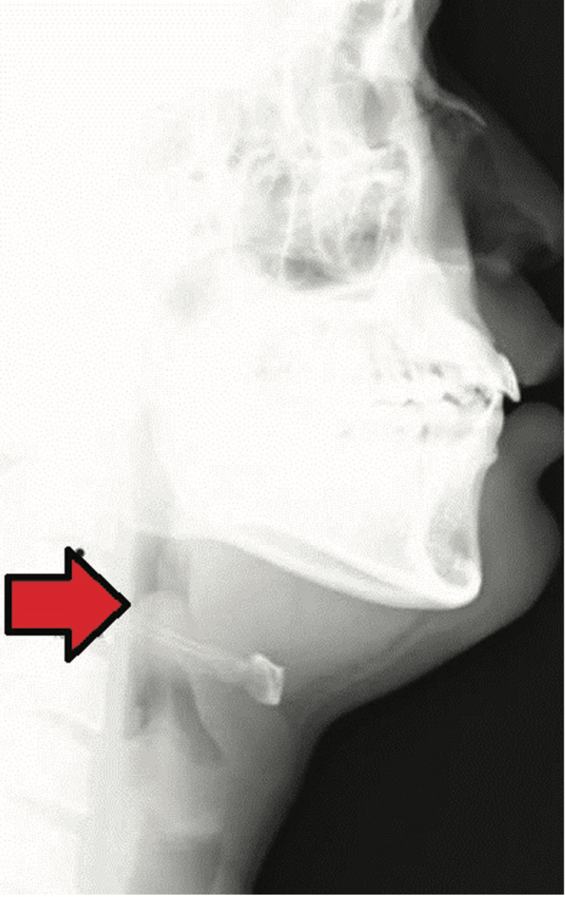
Which of the following virulence factors is produced by the likely causative agent of this infection?
A. Hemagglutinin-neuraminidase
B. Pili that can vary antigens
C. Polyribitol phosphate capsule
D. Toxin that inhibits protein synthesis
E. Toxin that interacts with Gi protein
(C) This child most likely has epiglottitis. Important bacterial causes of epiglottitis include Haemophilus influenzae type b (Hib), Streptococcus pneumoniae, and Streptococcus pyogenes. Key clinical features include difficulty breathing and swallowing, drooling, and stridor. Affected children may assume the tripod position, leaning forward to facilitate breathing. The polyribosylribitol phosphate (PRP) capsule is a major virulence factor of Hib, a gram-negative coccobacillus. Hib was historically the most common cause of pediatric epiglottitis, but widespread Hib vaccination has substantially reduced its incidence. (A) Hemagglutinin and neuraminidase are surface proteins associated with influenza virus; hemagglutinin facilitates viral attachment and entry, while neuraminidase promotes release of newly formed virions. Human parainfluenza viruses commonly cause laryngotracheobronchitis, or croup, rather than epiglottitis. (B) Pathogenic Neisseria species, including N. meningitidis and N. gonorrhoeae, possess pili that undergo antigenic variation, helping the organisms evade the host immune response. These organisms are not typical causes of epiglottitis. (D) Corynebacterium diphtheriae produces diphtheria toxin, which inhibits protein synthesis through inactivation of eukaryotic elongation factor 2 (eEF-2). This gram-positive rod can cause pharyngitis with formation of an adherent pseudomembrane but is not a typical cause of epiglottitis. (E) Bordetella pertussis produces pertussis toxin, which ADP-ribosylates the inhibitory G protein (Gi), resulting in increased intracellular cyclic AMP. B. pertussis causes pertussis, or whooping cough, rather than epiglottitis.
300
A woman brings her 67-year-old father to the physician because of increasing forgetfulness and paranoia. She states her father also seems to be more clumsy than usual, especially at night. The man has no prior psychiatric history. Physical examination reveals sensory ataxia. He is oriented to time and place, but has deficits in short-term memory. Laboratory results are shown below:
• CSF serology:
– VDRL: negative
– FTA-ABS: positive
– Toxoplasma gondii antibody, IgM: negative
– Toxoplasma gondii antibody, IgG: positive
– West Nile antibody, IgM: negative
– West Nile antibody, IgG: negative
Which of the following is the most likely diagnosis?
A. Guillain–Barré syndrome
B. Neurosyphilis
C. Rabies
D. Toxoplasmosis
E. West Nile encephalitis
(B) The patient has findings consistent with neurosyphilis, a manifestation of infection with Treponema palliduminvolving the central nervous system. Neurosyphilis can occur at any stage of syphilis, although some manifestations develop years to decades after the initial infection. Clinical presentations include meningitis, cognitive or psychiatric changes, general paresis, and tabes dorsalis. Because primary syphilis classically presents with a painless chancre that resolves spontaneously, patients may not recall the initial infection. The fluorescent treponemal antibody absorption (FTA-ABS) assay is a treponemal-specific test and supports prior or current infection with T. pallidum. Nontreponemal tests such as VDRL and RPR may have reduced sensitivity in some stages of syphilis, so diagnosis should incorporate clinical findings and appropriate treponemal and CSF testing. (A) Guillain–Barré syndrome is an important cause of acute ascending flaccid paralysis. It is typically an immune-mediated disorder that occurs following an infection, classically with Campylobacter jejuni, due in part to molecular mimicry between microbial antigens and peripheral nerve gangliosides. The clinical findings in this case are not consistent with Guillain–Barré syndrome. (C) Rabies is a viral infection that may have a prolonged incubation period. During the neurologic phase, patients may develop hydrophobia, pharyngeal spasms, agitation, autonomic dysfunction, and progressive encephalopathy. The clinical presentation and laboratory findings in this case make rabies less likely. (D) Toxoplasmosis is caused by the protozoan parasite Toxoplasma gondii and may cause fever, headache, confusion, and impaired coordination, particularly in immunocompromised patients. A negative IgM with positive IgG generally indicates previous exposure rather than a recent primary infection, making acute toxoplasmosis less likely in this case. (E) West Nile encephalitis is caused by West Nile virus, a mosquito-borne flavivirus. It may present with fever, headache, altered mental status, weakness, or flaccid paralysis. Negative West Nile virus serologic testing in this case makes this diagnosis less likely.
400
Gaze patterns obtained from five different patients from an ophthalmology clinic on a given day are presented below: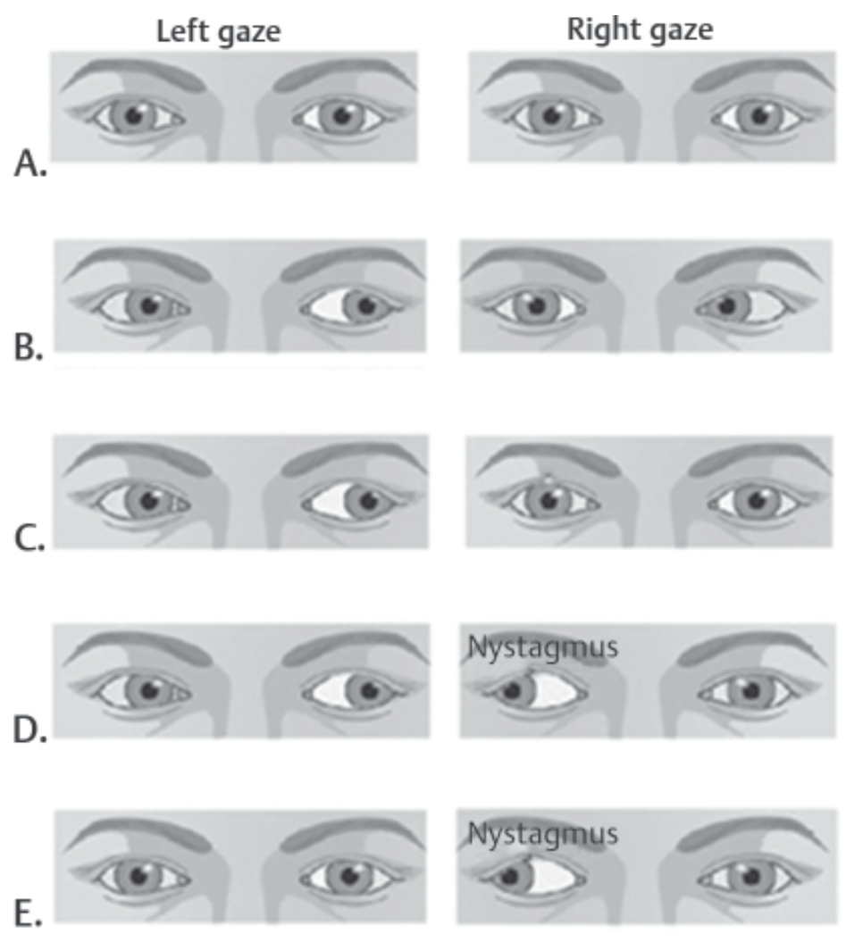
1. Which of the patients was suffering from right abducens nerve palsy?
2. Which of the patients was suffering from right abducens nucleus palsy?
3. Which of the patients was suffering from bilateral abducens nucleus palsy?
1 - B
2- C
3 - A
400
What are the three physiologic shunts present in the fetus and what is their purpose?
(1) Ductus venosus
(2) Patent Ductus Arteriosus,
(3) Foramen Ovale
Bypass liver, bypass lungs
400
A 36-year-old man presents with a tumor encroaching on the middle ear cavity and compressing the chorda tympani nerve. Which of the following structures is most likely to demonstrate impaired function?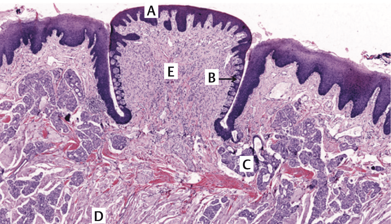
Correct: C (C)
Image key:
A – stratified squamous epithelium
B – taste bud
C – serous (von Ebner’s) glands
D – striated muscles
E – connective tissue
Identify the vallate papilla in the center of the slide. This is covered by stratified squamous epithelium that may be slightly keratinized. Each papilla is surrounded by a deep trench or cleft. Numerous taste buds are on the lateral walls of the papillae. The connective tissue near the papillae also contains several minor salivary serous (von Ebner’s) glands that open via ducts into the bottom of the trench. Secretomotor supply to these glands is by the facial nerve (through its chorda tympani branch). The lining epithelium (A) is nonnervous, and the connective tissue (E) will contain nerve endings responsible for general sensation (trigeminal nerve). Striated muscles (D) for the tongue are supplied by the hypoglossal nerve (with the exception of the palatoglossus). Taste buds (B) from vallate papillae are supplied by the glossopharyngeal nerve (this is an exception where taste buds anterior to sulcus terminalis are not supplied by the chorda tympani nerve).
400
A 5-year-old girl who recently emigrated from Eastern Europe is brought to the clinic complaining of throat pain. Symptoms started approximately 5 days ago and include low-grade fever, malaise, and sore throat without cough. The child denies any rhinorrhea, headache, or skin rash. Her mother states that the girl developed a “strange grating sound whenever she breathes in.” On examination, vitals include a temperature of 38.1°C (100.5°F), heart rate of 114 beats per minute, respiratory rate of 22 beats per minute, O2 saturation of 89% on room air and a normal blood pressure for her age. Prominent cervical lymphadenopathy is noted. On opening her mouth, a grayish-white, fibrinous, friable, coalescing membrane is visualized in an erythematous posterior oropharynx. Scraping this membrane with a tongue depressor causes minor bleeding and a diminished gag response. On auscultation, breath sounds are symmetrically reduced bilaterally and normal S1 and S2 heart sounds are heard. Electrocardiogram shows sinus tachycardia without other abnormalities.
What complication of this patient’s infection is most likely to contribute to her demise in the next 24 hours?
A. Cardiac dysfunction
B. Endophthalmitis
C. Neurological dysfunction
D. Skin ulcers
E. Suffocation
(E) This patient, who recently emigrated from a region where routine childhood vaccination may be less accessible, is most likely affected by respiratory diphtheria caused by the gram-positive rod Corynebacterium diphtheriae. The clinical presentation is characteristic of diphtheria, including fever, malaise, cervical lymphadenopathy—which can become severe enough to produce the classic “bull neck” appearance—and pseudomembranous pharyngitis. The characteristic diphtheritic pseudomembrane consists of fibrin, inflammatory cells, necrotic tissue, and bacteria. In this case, the pseudomembrane may extend beyond the oropharynx into the larynx or upper airway, producing stridor, respiratory distress, and potentially fatal airway obstruction. Thus, acute upper airway obstruction is the most immediate life-threatening complication. (A) Cardiac dysfunction is an important complication of diphtheria and may include myocarditis, conduction abnormalities, arrhythmias, and heart failure. However, a normal electrocardiogram and unremarkable cardiac examination make myocardial dysfunction a less likely immediate cause of death in this patient. (B) Endophthalmitis and other severe ocular complications are not typically associated with respiratory diphtheria. (C) Neurologic dysfunction is a recognized complication of diphtheria and may include cranial neuropathies, decreased gag reflex, impaired palatal movement, and peripheral neuropathy. These manifestations result from diphtheria toxin, which ADP-ribosylates eukaryotic elongation factor 2 (eEF-2), inhibiting host-cell protein synthesis. However, the presence of stridor and respiratory distress makes mechanical upper airway obstruction more likely than neurologic respiratory impairment as the immediate threat. (D) Cutaneous diphtheria may produce chronic, nonhealing ulcers covered by a gray membrane, but these dermatologic manifestations would not typically cause death within the next 24 hours.
400
Following a subdural hematoma, a 66-year-old male complains of headache, nausea, vomiting, and diplopia. Funduscopic examination reveals prominent papilledema in the left eye. Which area of the presented image is most likely to show earliest changes?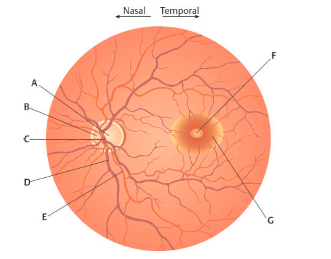
Area B
Papilledema is a noninflammatory congestion of the optic disc that is caused by increased ICP, such as what is often observed following subdural hematomas. Increased ICP causes the cerebrospinal fluid to compress the optic nerve within its sheath resulting in axoplasmic flow stasis and ischemia. Early papilledema causes a nerve fiber edema that surrounds the optic disc. Areas D (B) and E (C) represent the branches of the central retinal vein and artery, respectively. These are involved (obscured) in advanced stages of papilledema. Area F (D) represents the fovea centralis and area G (E) represents the macula lutea. None of these regions would be altered in papilledema.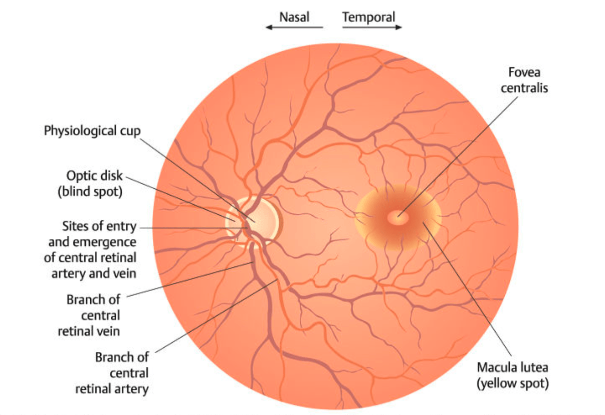
500
A patient is evaluated after a head injury. When the right cornea is lightly touched with a cotton wisp, neither eye blinks. When the left cornea is touched, both eyes blink normally.
Which nerve is most likely damaged?
A. Right oculomotor nerve
B. Left oculomotor nerve
C. Right ophthalmic nerve
D. Left ophthalmic nerve
E. Right facial nerve
F. Left facial nerve
C. Right ophthalmic nerve
500
What are the four features of tetralogy of Fallot?
Overriding Aorta
Ventricular Septal Defect
Subpulmonic (infundibular) stenosis
RV Hypertrophy
500
Indicate Common Nerve Injured and Nerve Roots Involved x10 points
1. Fibular neck injury →
2. Trendelenburg sign →
3. Humerus shaft →
4. Guyons Canal →
5. Absent knee-jerk reflex →
Bonus Question: What is the clinical condition characterized by injury to Option 1?
1. Fibular neck injury → Common fibular nerve (L4 - S2)
2. Trendelenburg sign → Superior gluteal nerve (L4 - S1)
3. Humerus shaft → Radial Nerve (C5 - T1)
4. Guyons Canal → Ulnar Nerve (C8 - T1)
5. Absent knee-jerk reflex → Femoral nerve (L2–L4)
Bonus - Foot Drop
500
A 22-year-old woman college student is preparing for final exams. During this time, she develops a blistering vesicular lesion on the border of her right upper lip. The genomic composition of the most likely causative agent of this infection is most similar to that of:
A. Hepatitis A virus (HAV)
B. Infl uenza virus
C. Measles virus
D. Parvovirus
E. West Nile virus
(A) The patient most likely has cystic fibrosis (CF). Burkholderia cepacia, a gram-negative rod, is an important cause of recurrent and chronic pulmonary infections in patients with CF and chronic granulomatous disease. It possesses several virulence mechanisms, including quorum sensing, motility, biofilm formation, siderophore-mediated iron acquisition, and secretion systems. B. cepacia is also highly resistant to many antimicrobial agents, in part because of multidrug efflux pumps that actively remove antibiotics from bacterial cells. Another gram-negative rod, Pseudomonas aeruginosa, is a major pulmonary pathogen in CF and shares several characteristics with B. cepacia, including biofilm formation and substantial antimicrobial resistance. Staphylococcus aureus, a gram-positive coccus, is another important cause of pulmonary infection in patients with CF. (B) Candida albicans is a yeast that can cause pulmonary infection, particularly in severely immunocompromised patients. Although patients with CF may become colonized with Candida, invasive pulmonary candidiasis is much less characteristic than bacterial pulmonary infections caused by organisms such as P. aeruginosa, B. cepacia, and S. aureus. (C) Klebsiella pneumoniae is a gram-negative rod that can cause severe pneumonia, particularly in patients with certain comorbidities or healthcare exposures. Its prominent polysaccharide capsule produces mucoid colonies and has classically been associated with thick, blood-tinged sputum. However, K. pneumoniae is not as characteristically associated with chronic CF pulmonary infections as P. aeruginosa, B. cepacia, and S. aureus. (D) Legionella pneumophila is a gram-negative bacillus that causes Legionnaires’ disease, often following exposure to contaminated aerosolized water systems. Risk factors include older age, smoking, chronic lung disease, and immunosuppression. It is not a characteristic cause of recurrent pulmonary infection in CF. (E) Streptococcus pneumoniaeis a gram-positive, lancet-shaped diplococcus and a common cause of community-acquired pneumonia. It is α-hemolytic on blood agar, producing a green discoloration around colonies. However, it is not as strongly associated with chronic pulmonary infection in CF as P. aeruginosa, B. cepacia, and S. aureus.
500
A 26-year-old soccer player sustained a head injury while contesting a ball in the air. He transiently lost his consciousness and remained dizzy for about an hour. A CT scan revealed a fracture line passing through the stylomastoid foramen. Which of the following might be found in him?
A. Paralysis of all muscles that develop from the 1st pharyngeal arch
B. Paralysis of all muscles that develop from the 2nd pharyngeal arch
C. Loss of taste sensation from the tip of the tongue
D. Loss of taste sensation from the vallate papillae
E. Loss of general sensation over a small part of the external acoustic meatus
Correct: Loss of general sensation over a small part of the external acoustic meatus
(E) The facial nerve exits the skull through the stylomastoid foramen. As the nerve exits the stylomastoid foramen, it gives off a sensory branch that supplies part of the external acoustic meatus and tympanic membrane. It then gives off motor branches that supply muscles of facial expression. Muscles that develop from the 1st pharyngeal arch (A) are supplied by the trigeminal nerve. The stapedius is a muscle derived from the 2nd pharyngeal arch. It is supplied by a branch of the facial nerve given off from its mastoid segment (course from pyramidal eminence to stylomastoid foramen), which is proximal to the stylomastoid foramen. Therefore, the stapedius will not be affected by the fracture (B). Taste fibers for the tip of the tongue (C) are supplied by the chorda tympani branch of the facial nerve, which is also given off from its mastoid segment and hence will be unaffected by the fracture. Taste fibers for the vallate papillae (D) are supplied by the glossopharyngeal nerve. It exits the skull through the jugular, but not the stylomastoid, foramen.
600
A 32-year-old patient sustains a penetrating injury that produces a right hemisection of the spinal cord at T8. Several weeks later, examination reveals right lower-extremity weakness with an upper motor neuron pattern. Which additional finding is most likely?
A. Left loss of vibration with right loss of pain below the lesion
B. Right loss of vibration with left loss of pain below the lesion
C. Bilateral loss of vibration with preserved pain below the lesion
D. Bilateral loss of pain with preserved vibration below the lesion
E. Right loss of vibration with right loss of pain below the lesion
F. Left loss of vibration with left loss of pain below the lesion
Answer: B. Right loss of vibration with left loss of pain below the lesion
Rationale: A right hemisection damages the right dorsal column, causing ipsilateral loss of vibration and proprioception below the lesion. The spinothalamic fibers have already crossed before ascending, so damage to the right spinothalamic tract causes contralateral loss of pain and temperature. Thus, the patient has right-sided vibration/proprioception loss and left-sided pain/temperature loss below the lesion.
600
A pudendal nerve block is commonly performed near which landmark?
Ischial spine
600
Discuss the neonatal process that results in closure of the ductus arteriosus and interventions used to keep it open?
The ductus arteriosus closes due to a rise in oxygen tension and decreased prostaglandin levels. PGE-1 is used to keep it open.
600
A 72-year-old patient develops a left homonymous hemianopia following an ischemic stroke. Visual field testing demonstrates preservation of central vision. Collateral blood flow from which artery most likely accounts for the preserved central vision?
Middle cerebral artery
Rationale: The primary visual cortex is supplied predominantly by the posterior cerebral artery (PCA). The occipital pole, where the macula is represented, may also receive collateral supply from branches of the middle cerebral artery (MCA). Thus, a PCA infarct can cause a contralateral homonymous hemianopia with macular sparing when MCA-supplied cortex remains perfused.
700
The AV node is located near which major anatomical landmark?
Triangle of Koch in the right atrium.
700
A patient undergoes surgical repair of an aortic coarctation and later develops lower-extremity neurologic deficits.
What vascular anatomy explains the complication?
Answer: Interruption of segmental blood supply to the spinal cord, particularly vessels contributing to the anterior spinal artery, including the artery of Adamkiewicz.
700
The posterior descending artery determines:
Coronary dominance.
700
What structures fuse and close the atrioventricular canals?
Endocardial cushions