RA
Heme/Onc/Endo
GI
Renal/Cardio/Pulm
Monoclonal Gammopathy
HIV
100
What serum lab finding is associated with these skin symptoms?
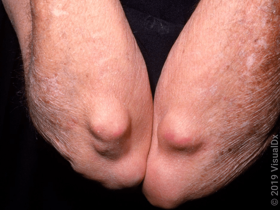
Rheumatoid nodules
• 20% of RA patients
• Firm, on periarticular locations
• Associated with high RF titers
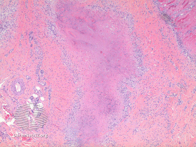
• Path with central eosinophilic fibrin + palisaded histiocytes
100
Which disease is associated with these skin findings:
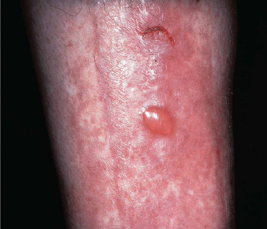
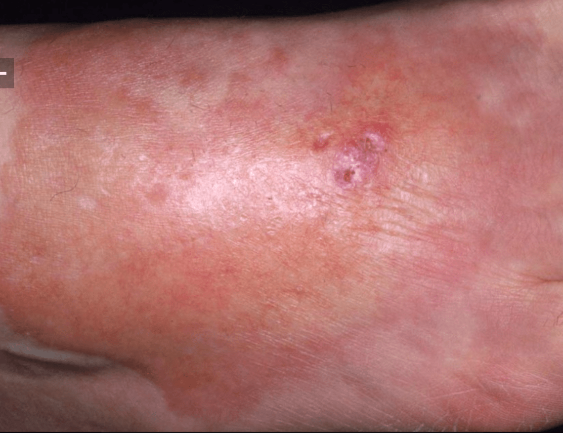
Photos show:
1) diabetic bullae
2) necrobiosis lipoidica
Other dermatologic associations with DM:
- acanthosis nigricans
- acral dry gangrene
- acral erythema
- carotenoderma
- charcot foot (neuropathic osteoarthropathy)
- diabetic cheiroarthropathy
- diabetic dermopathy
- disseminated GA
- eruptive xanthoma
- hemochromatosis
- neuropathic ulcers
- perforating disorders
- rubeosis
- cleredema
100
What systemic disease is responsible for these skin lesions?
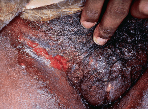
Crohn's disease
Erythematous plaques, lymphedema (common & stricking), knife-like ulcerations (usu intertriginous areas), most commonly in anogenital regions. 20% of patients without preceding diagnosis of Crohn's.
Part of spectrum of mucocutaneous findings of Crohn (20-45%)
- Specific lesions – metastatic Crohn, contiguous perianal (fistulas), oral disease (aphthous ulcers, pyostomatitis vegetans),
- Non-specific/reactive – EN, PG, PAN
- Nutritional skin changes – acrodermatitis enteropathica (zinc)
- Treatment-related side effects
Histo with non-caseating granulomas; combination lichenoid + granulomatous inflam & granulomatous perivasculitis.
DDx: sarcoid, mycobacterial infx, deep fungal, foreign body rxn, actinomycosis.
Tx (severity not related to control of intestinal disease): tCS, calcineurin inhibitors, systemic CS, sulfasalazine, azathioprine, TNF-a inhib
100
What is this acute form of sarcoidosis?
Löfgren syndrome (sarcoidosis variant)
• Fever, arthritis, hilar LAD, EN
Systemic granulomatous disorder unknown etiology. Most commonly involves lungs (90%). Cutaneous manifestations in up to 1/3 (can be only manifestation). Bimodal age distribution, F>M.
Red-brown violaceous papules + plaques on face (nose), upper back & extremities. "Apple jelly" under pressure. Can also present within scars & tattoos.
Variants include
• Löfgren syndrome – fever, arthritis, hilar LAD, EN; often need systemic CS
• Darier-Roussy – painless firm nodules without epidermal changes (limited to panniculus)
• Lupus pernio – papulonodules on nose + cheeks; a/w lung (75%) & upper resp tract (50%)
EN can be a/w acute form of sarcoidosis. Also with LAD (esp hilar), uveitis, liver & heart involvement.
Histo with non-caseating naked granulomas ± Schaumann bodies (rounded laminated basophilic inclusions). ACE level elevated (2-3x normal).
Dx of exclusion: can be drug-induced (TNFα inhibitors, IFNα) or infx (r/o acid fast, fungus).
DDx GA, necrobiosis lipoidica, cutaneous Crohn.
Tx = corticosteroids, HCQ, minocycline, MTX, TNFα inhibitors
100
What is this?
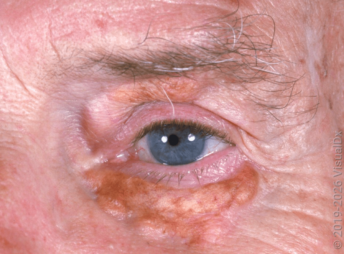
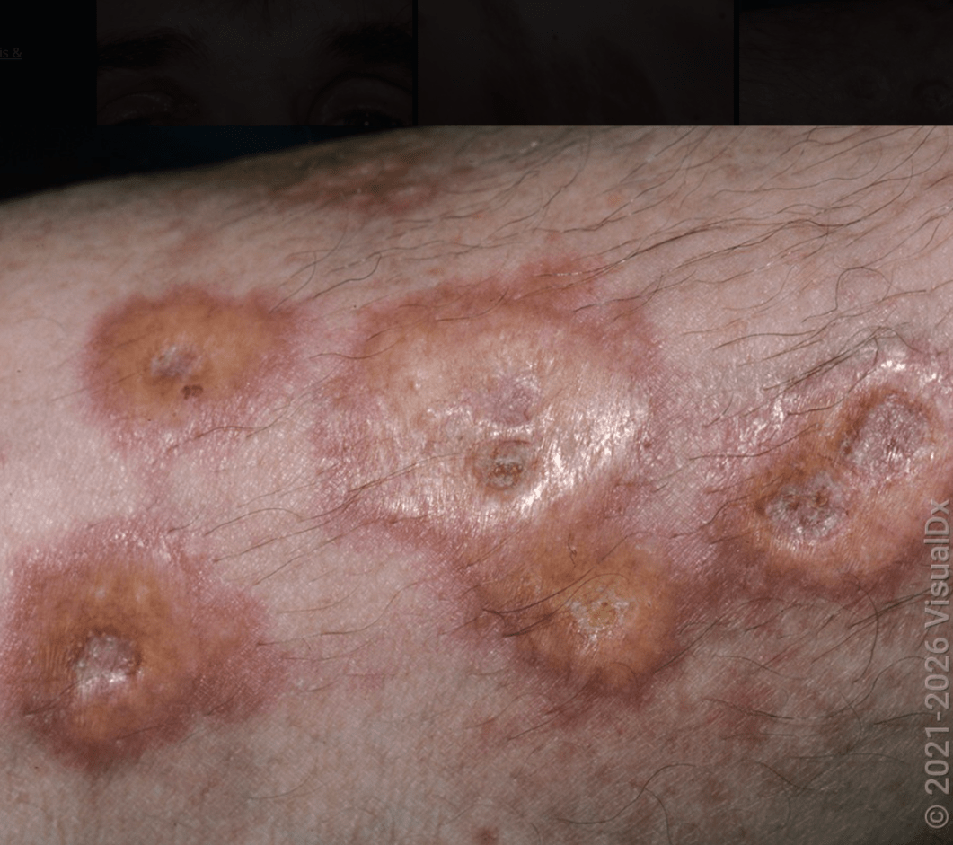
Necrobiotic xanthogranuloma
• IgG monoclonal gammopathy κ light chain
Rare (less than 100 cases), generally adults >50 yo. Asymptomatic, firm, yellowish plaques or papulonodules, classically periorbital. 50% with ophthalmic manifestations (orbital masses, ectropion, ptosis, conjunctival lesions).
A/w IgG monoclonal gammopathy κ light chain (80% - order SPEP), MM or other lymphoproliferative disorder rarely
Histo with palisading xanthogranuloma in mid dermis to panniculus. Cholesterol clefts, giant Touton cells & large, bizarre foreign body giant cells.
Differential includes necrobiosis lipoidica, normolipemic plane xanthomas, xanthelasma, JXG, sarcoidosis.
No good treatment, low dose CS and chlorambucil
100
What are these lesions?
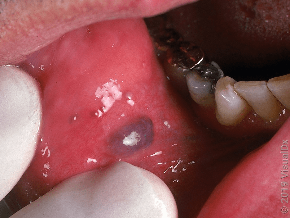
Kaposi sarcoma
Classical form (older men of Mediterranean descent)
African endemic form (men & young children)
Iatrogenic immune suppression-associated form (solid organ transplant recipients)
AIDS-related epidemic form (MSM)
HHV-8 infects endothelial cells of lymphatics & blood vessels → mixed vascular phenotype. Red, brown to violaceous papules, plaques & nodules. Classic form on legs, slow progression vs AIDS-related (more disseminated, can involve internal organs).
Histo: dermal proliferation of small, angulated vessels, promontory sign (small vessel protruding into an abnormal vascular space); patch → plaque → nodular stage (proliferation of spindled & epithelioid cells admixed with extravasated erythrocytes); latency-associated nuclear antigen (LANA-1).
DDx: capillary changes (e.g. venous stasis), KHE & spindle cell hemangioma when nodular stage.
Tx: cryotherapy, laser surgery, PDT, topical alitretinoin gel & radiation therapy; systemic chemo for visceral involvement
200
Name a medication that this patient might be receiving to explain this skin finding?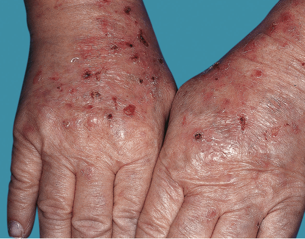
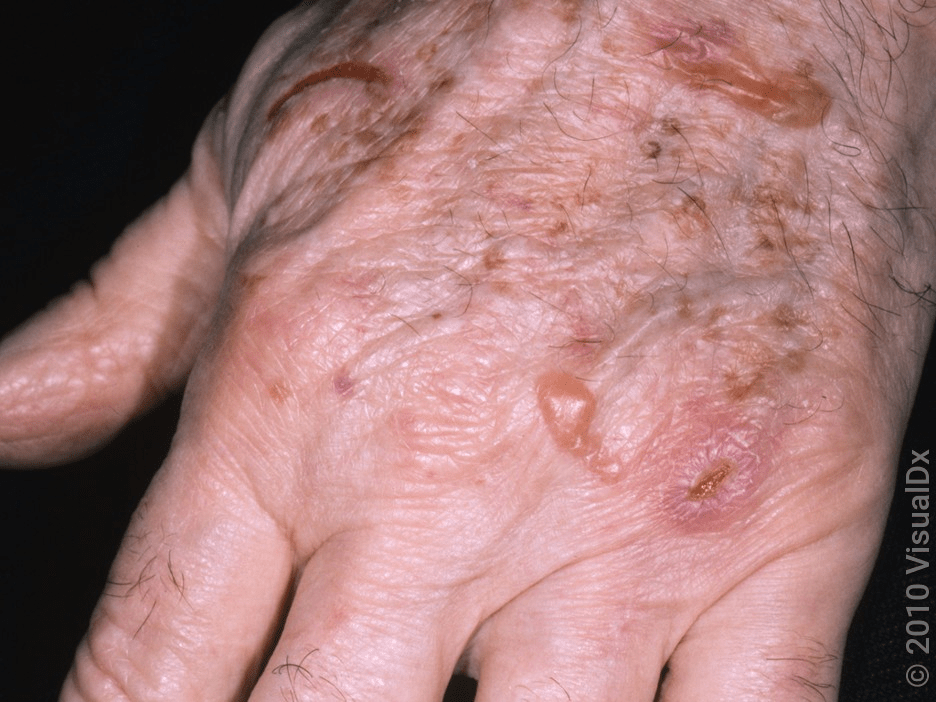
Pseudoporphyria secondary to NSAID use.
Common culprit medications:
NSAIDs (especially naproxen), nalidixic acid, thiazides, furosemide, tetracyclines, ciprofloxacin, voriconazole, and imatinib
No biochemical abnormalities in porphyrin metabolism.
Most commonly seen in patients with CKD4-5 or ESRD.
200
What is 1 systemic condition that could explain the hyperpigmentation?
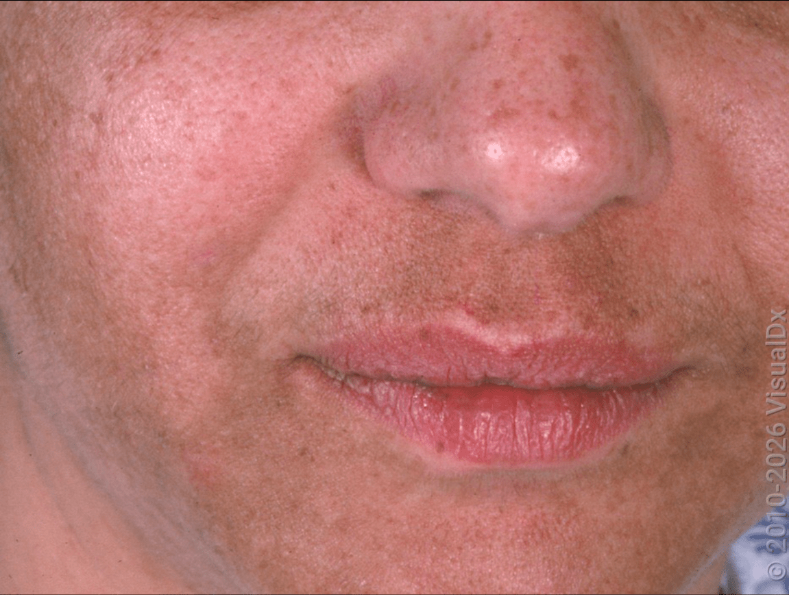
Addison disease – diffuse hyperpigmentation from MSH-like effect of ACTH. Accentuation in sun-exposed skin or sites of trauma. Loss of androgen-stimulated hair in post-pubertal women.
Hemochromatosis – AR mutation in HFE (impaired transferrin receptor function). Fe build-up in organs incl skin. Generalized bronzing (esp face), cirrhosis/HCC risk, cardiomyopathy, DM (bronze diabetes). Histo with increased melanin and hemosiderin granules.
200
What is one potentially fatal complication associated with this finding?
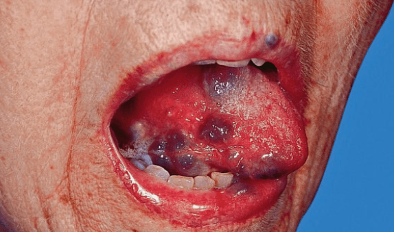
Blue rubber bleb nevus syndrome
• GI hemorrhage
Sporadic, usually present at birth. TEK mutations. Blue-violaceous compressible papules & nodules (venous malformations). Involve trunk/extremities, mucosa, GI tract (melena or hemorrhage).
Histo with ectatic vascular spaces surrounded by fibrous tissue in deep dermis/subcutis
200
What the are the 2 general locations for cutaneous ulcerations in this autoimmune condition?
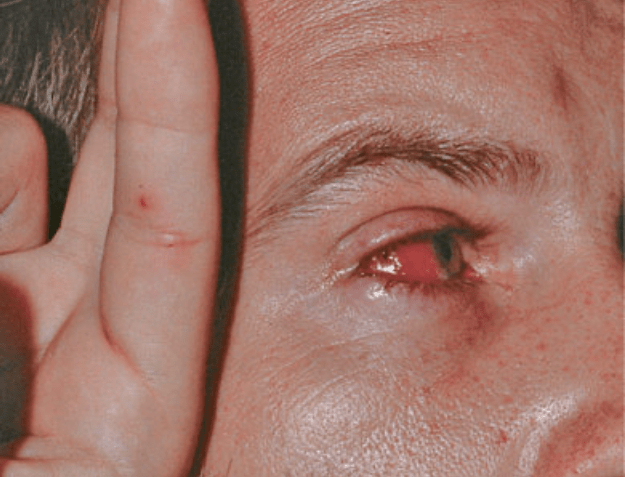
Behçet's disease
• Aphthous & genital ulcers
Young adult males, Middle East (Turkey) and Japan genetic ancestry. Associated with HLA-B51
Recurrent painful aphthous ulcers (#1) at least 3x in 1 yr + 2 of following:
• Recurrent genital ulcerations
• Ocular lesions – uveitis, conjunctivitis
• Cutaneous lesions – acral/facial papulopustules, purpura, EN-like nodules. +pathergy
Affects other organs. Ocular (90%, #1 cause of morbidity), vascular (superficial migratory thrombophlebitis), joints, neuro.
Histo with neutrophilic infiltrate + LCV
Treatment: colchicine, daspone, thalidomide, TNFi, prednisone, MMF, IVIg
200
Name an associated plasma cell dyscrasias
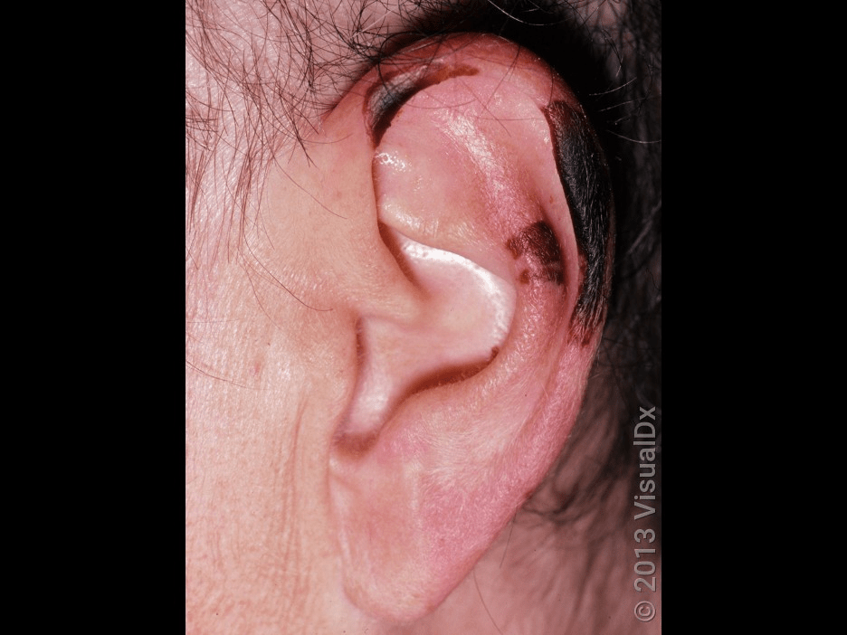
Type I cryoglobulinemia
• MM and Waldenström macroglobulinemia
Presence of cryoglobulins (Ig which reversibly precipitate at temp <37°C) in serum.
Type I: monoclonal IgM (rarely IgG or IgA). Pathology is vascular occlusion. A/w B cell lymphoproliferative disorders. Purpuric or necrotic lesions, often retiform at acral sites (or cold exposure), livedo reticularis, Raynaud's phenom.
Histo with bland eosinophilic hyaline occlusion of blood vessels.
DDx incl cholesterol emboli, chilblains/pernio.
Tx is preventive, treat underlying disorder
200
What is this condition?
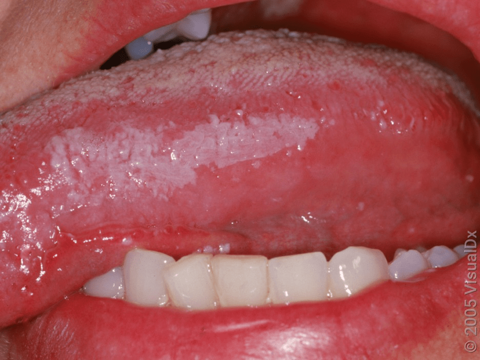
Oral hairy leukoplakia
Benign mucosal disorder caused by EBV.
Painless, corrugated, white plaques on lateral borders of tongue. A/w immunocompromise (HIV, transplant). Lesions do not scrape off (≠ candida). Superimposed candidal infection common.
Histo with irregular parakeratosis, focal balloon cell degeneration. +EBER (Epstein-Barr virus-encoded RNA).
DDx includes candidiasis, LP, hairy tongue.
Tx: regresses with ART
300
What histological pattern would you find associated with these tender lesions?
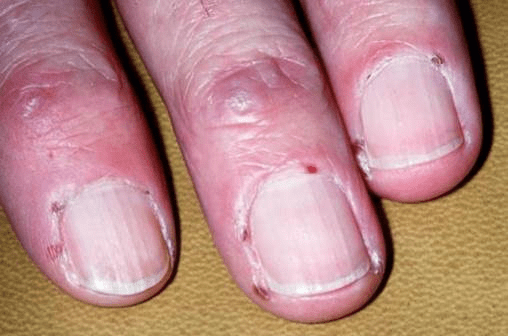
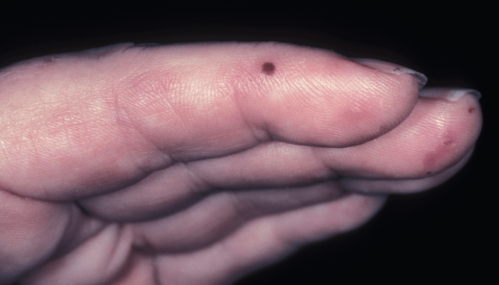
Bywaters lesions due to cutaneous small vessel vasculitis
• Leukocytoclastic vasculitis
Punctuate purpuric papules on the distal digits d/t cutaneous small vessel vasculitis.
300
Most commonly associated malignancy with this condition?
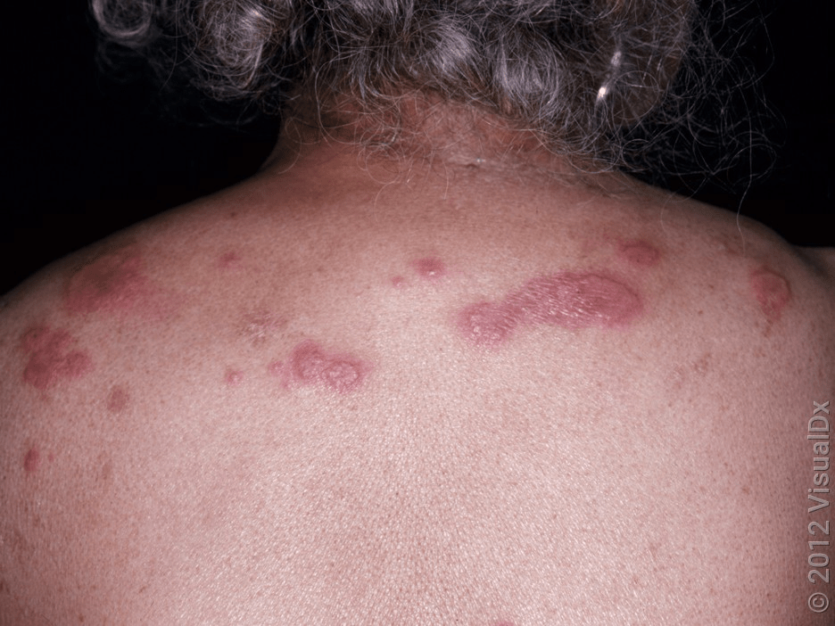
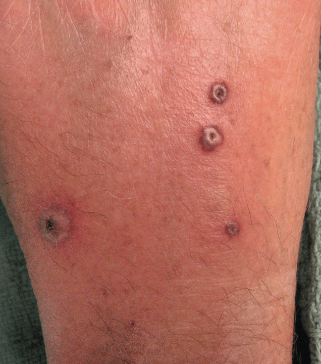
Sweet syndrome (acute febrile neutrophilic dermatosis)
• AML
4:1 female predominance. Fever, leukocytosis common; other systemic manifestations less (athralgias, myalgias, ocular involvement). 50% pts with associated disorder or trigger: 15–30% malignancy, (hematologic ≫ solid organ), ~25% infx (URI), ~10% drug (abx, antihypertensive, antineoplastics e.g. iplimumab, vemurafetinib), autoimmune disorders (AI-CTD, sarcoidosis, Behçet).
Tender, non-pruritic erythematous papules/plaques, pronounced edema (can vesiculate). Favor H&N + upper extremities. Can see pathergy. Usually resolve spontaneously within 5-12 wks (but 30% have recurrence).
Histo with diffuse nodular & perivascular neutrophilic infiltrate without evidence of vasculitis. Epidermal changes usually not significant.
Diagnostic criteria = 2 major (abrupt onset of typical lesions + histo c/w Sweets) + 2 minor (a/w infx/malignancy/inflammatory disorders/drug OR fever + constitutional sx OR leukocytosis OR excellent response to systemic CS).
DDx includes PG, neutrophilic eccrine hidradenitis, autoinflam syndromes, vasculidities, granulomatous diseases, infx, leukemia cutis.
Tx = address underlying condition + PO pred (0.5-1 mg/kg) x2-6 wks (alternatives include potassium iodide, dapsone, colchicine
300
What is the diagnosis and/or mutation associated?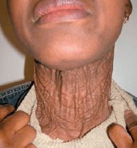
Pseudoxanthoma elasticum
• ABCC6
Autosomal recessive disorder, LoF in ABCC6. The absence of functional ABCC6 in PXE patients
results in reduced ATP release from hepatocytes.
Clumped, distorted & calcified elastic fibers (skin, eyes, CV). Yellowish papules, “cobblestoning” or “plucked chicken skin,” redundant folds in flexural areas. Calcification of elastic fibers in walls of medium-sized arteries can lead to luminal narrowing (claudication, renovascular HTN, angina, MI). Ocular angioid streaks reflect breaks in calcified elastic lamina of Bruch membrane; choroidal neovascularization. GI hemorrhage is another possible complication.
Histo: distorted, fragmented elastic fibers (purple clumps) in mid + deep reticular dermis.
DDx: actinic elastosis, perforating calcific elastosis, late-onset focal dermal elastosis, elastoderma, perforating calcific elastosis, white fibrous papulosis of the neck.
Tx: no specific treatment for skin, referral for ophtho, cards
300
What is an important lab to order to monitor for a complication for patients with this condition?
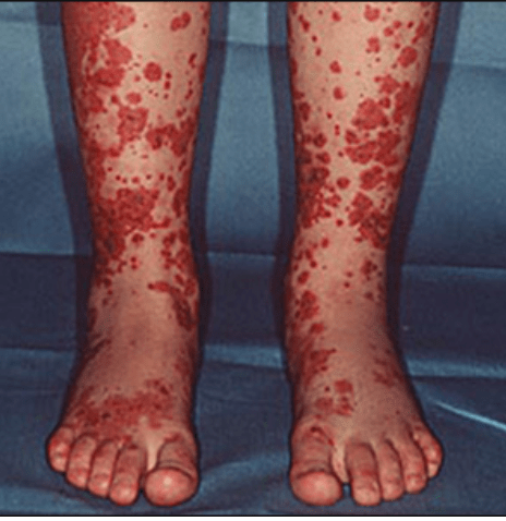
Urinalysis
IgA vasculitis or (Henoch–Schönlein
purpura) notable for palpable purpura of the distal extremities.
Most common in children but can occur in adults. Renal complication = IgA-associated glomerulonephritis. Systemic steroids especially helpful for nephritis.
In addition to skin findings, can have Arthritis, GI vasculitis with abdominal pain, GI bleeding, and/or vomiting.
300
Name the diagnosis and/or associated plasma cell dyscrasia
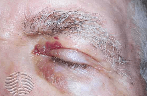
Primary systemic amyloidosis
• 15% in multiple myeloma
Acquired amyloidosis. Waxy, translucent or purpuric papules; periorbital and pinch purpura; macroglossia. Wide spectrum of organ involvement incl carpal tunnel.
A/w plasma cell dyscrasia incl MM (15%)
Histo with amyloid deposits (amorphous, eosinophilic deposits with fissures) in dermis + subcutis. Can also see deposits in vessel walls.
DDx includes papular mucinoses, nodular amyloidosis, lipoid proteinosis.
Poor prognosis, supportive measures, HSCT
300
What is the differential for these lesions in HIV+ patient? (4)
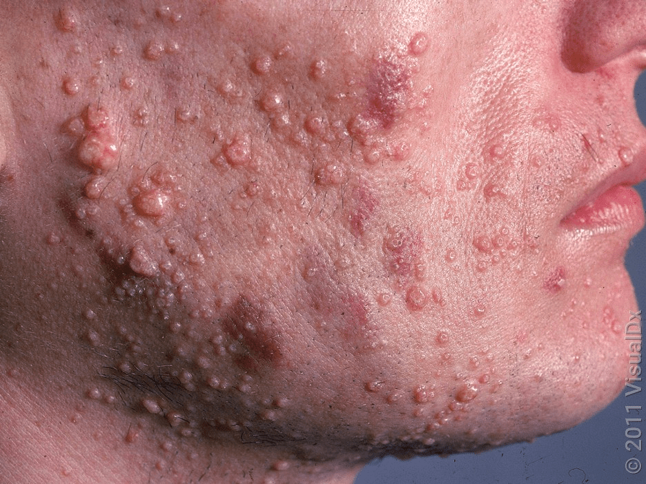
Molluscum-like lesions
• Cryptococcus
• Coccidioides
• Histoplasmosis
• Penicillium
400
What is this condition? (Hint: it is a specific disease within a broader, unifying condition)
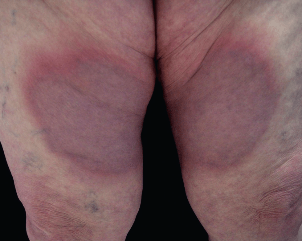
Interstitial granulomatous dermatitis (IGD) with arthritis
Secondary process, classically seen in RA or seronegative arthritis. Also seen with medications (ACEi, CCB, BBs, lipid-lowering agents, antihistamines, anticonvulsants, antidepressants, TNFα blockers)
Symmetric annular plaques, linear cords (rope sign)
Histology with rosettes of palisading histiocytes surrounding tiny foci of degenerative collagen + neutrophils without vasculitis
400
Name commonly associated malignancies with these cutaneous findings
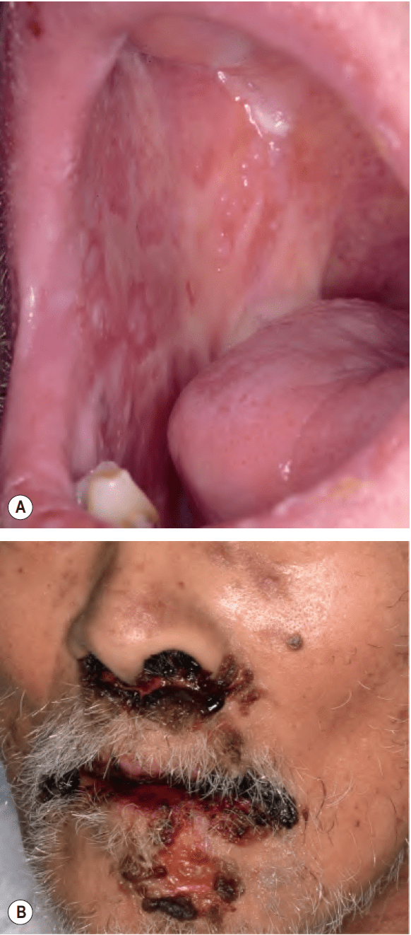
Paraneoplastic pemphigus
• NHL, CLL, Castleman (peds)
Intractable stomatitis (affecting all surfaces of oropharynx & extending on to vermilion lip). Blisters & erythema multiforme-like lesions on palms and soles. Severe pseudomembranous conjunctivitis also common. Polymorphic cutaneous findings.
Bronchiolitis obliterans is serious internal manifestation.
A/w Non-Hodgkin lymphoma & chronic lymphocytic leukemia (2/3 of pts), Castleman disease (otherwise very rare) for children; malignant & benign thymomas, sarcomas, Waldenstrom macroglobulinemia.
Can affect palms and soles vs. unusual involvement for pemphigus vulgaris.
400
What GI surgery did this patient likely have in the past?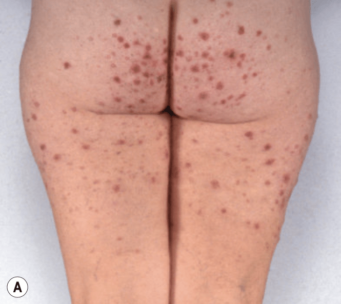
Bowel-associated dermatosis arthritis syndrome (BADAS)
• Bowel bypass
10-20% of intestinal bypass surgery patients.
Recurrent erythematous macules and patches with overlying papulovesicles and pustules on proximal upper extremities and trunk
Serum-sickness like symptoms, diarrhea
Possible bowel bacterial overgrowth leading to complement activation and antibody complexes deposition
Histo with perivascular neutrophilic infiltrate, dermal edema (similar to Sweets)
DDx includes urticarial vasculitis (papular phase) and CSVV, septic vasculitis, Behçet.
Tx: prednisone, PO abx, colchicine, dapsone; consider surgical revision of bowel bypass if severe
400
Which 2 organ systems requires screening?
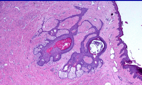
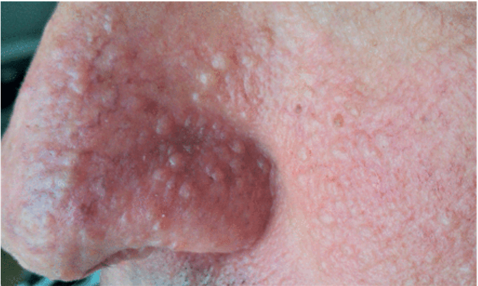
Birt-Hogg-Dubé
• RCC, pulm cyst & pneumothorax
AD mutation in FLCN (folliculin)
Fibrofolliculoma + acrochordon + trichodiscoma (FAT Hogg)
Pulmonary cysts (most common; up to 90%) lead to spontaneous pneumothorax (30%). Increased risk of renal cell carcinoma. Also see colonic polyps and connective tissue nevi
400
What is the associated monoclonal gammopathy
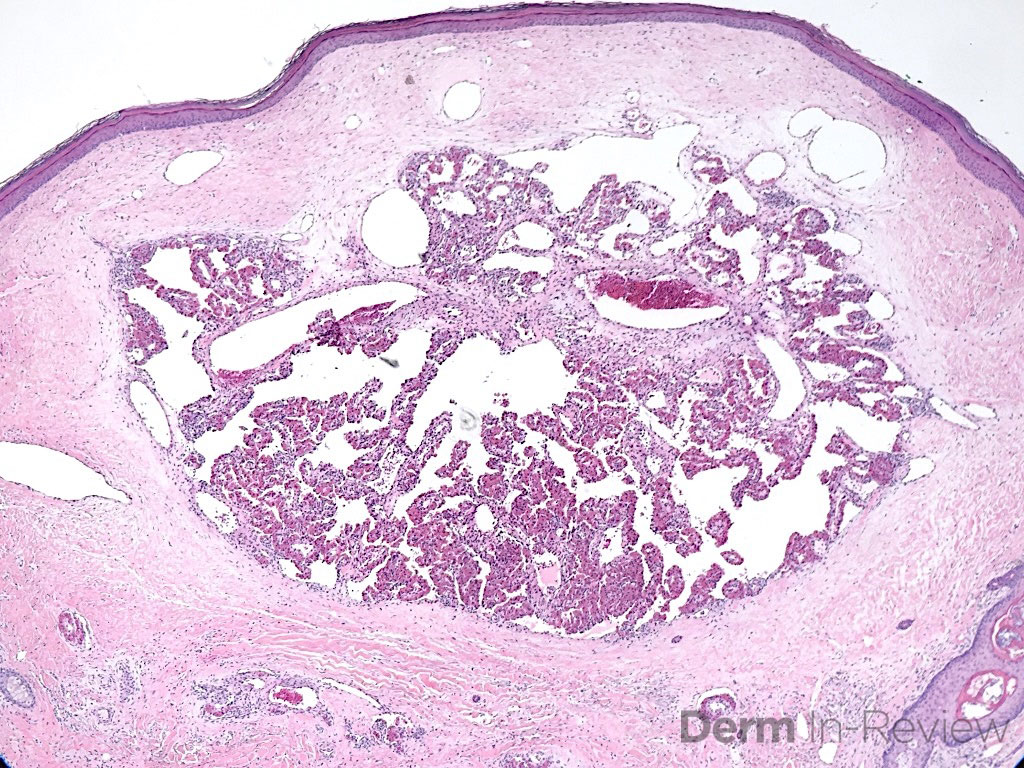
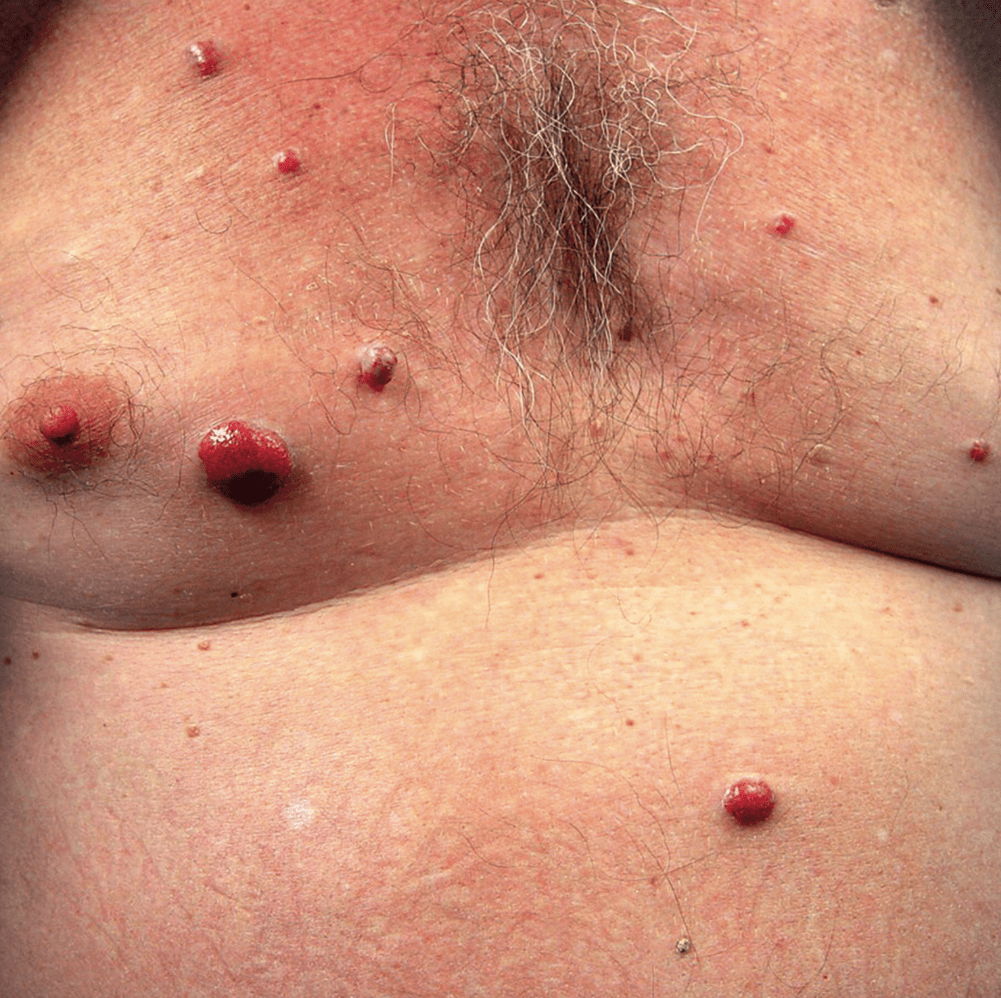
Glomeruloid hemangioma
• IgG lambda
Pathognomonic for POEMS syndrome (type of monoclonal gammopathy) =
Polyneuropathy (necessary)
Organomegaly
Endocrinopathy – hypogonadism
Monoclonal plasma cell prolif – IgG lambda
Skin changes – cherry angiomas, hyperpig, hypertrichosis, sclerotic skin, acrocyanosis
Sclerotic bone lesions
Histo with numerous dermal ectatic vascular spaces lined by flat endothelial cells with conglomeration of capillaries lined by plump, swollen endothelial cells
DDx incl MM, MGUS, Waldernström macroglobulinemia, primary amyloidosis
Treatment = heme/on
400
This is a sequelae to what in HIV?
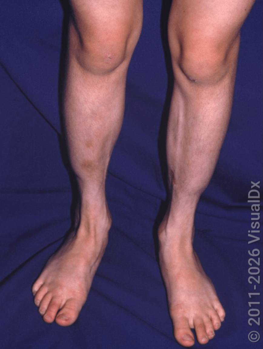
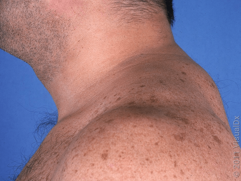
Antiretroviral-associated lipodystrophy
• Caused by protease inhibitors, nucleoside reverse transcriptase inhibitors, and to lesser extent, nonnucleoside reverse transcriptase inhibitors. ≤2 yrs of starting therapy
• Lipoatrophy (loss of fat in face, extremities, and buttocks) or lipohypertrophy (accumulation and redistribution of fat to upper back, neck, or abdomen)
• A/w metabolic abnormalities (hyperlipidemia and insulin resistance)
• Tx: poly-L-lactic acid & calcium hydroxylapatite
500
What are some considerations for management of this treatment complication for a patient with RA?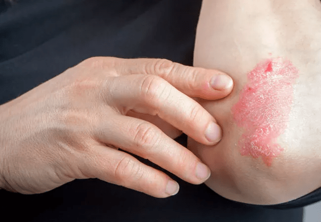
TNFi-induced psoriasis
• Occurs in 2-5% of patients on these medications
• Plaque most common presentation but also high % of scalp, inverse and pustular Pso
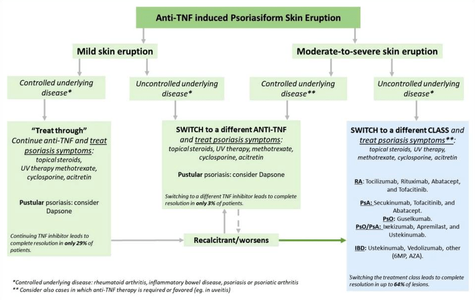
• Review from JAMA Derm showed improvement with topicals in 60% of patients. Switching TNFi only led to improvement in 36% whereas discontinuation was effective in 60%
• PMID: 30576759, 31093599
500
What is this syndrome?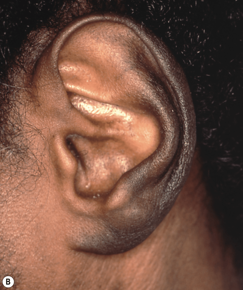
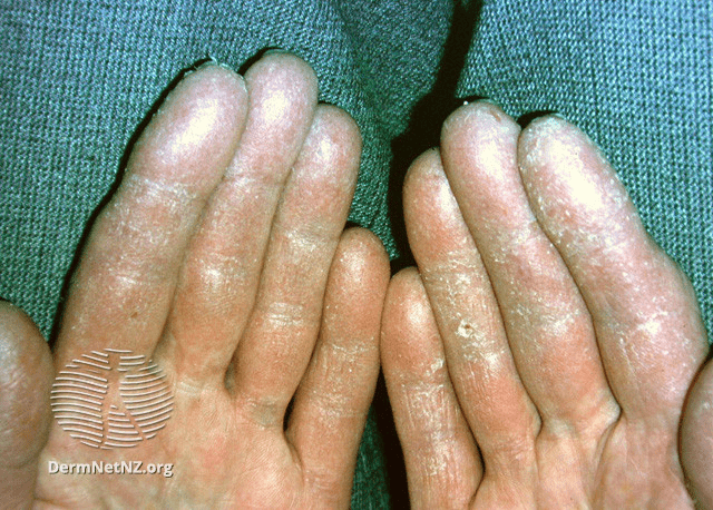
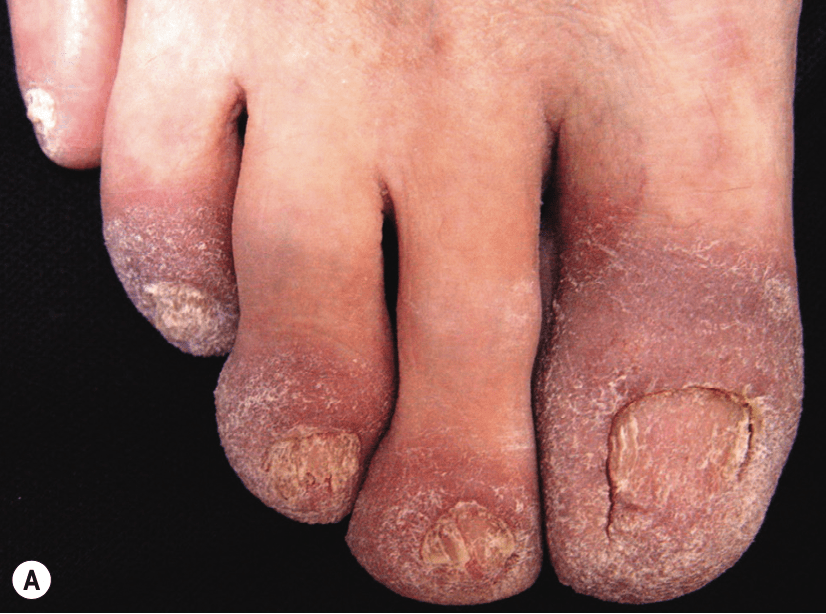
Bazex syndrome (acrokeratosis paraneoplastica)
Symmetric erythematous psoriasiform plaques on distal extremities, nasal bridge & helices. 75% with horizontal + longitudinal nail ridging
A/w upper bronchial/GI tract carcinoma (SCC of pharynx, larynx or esophagus). Often precedes diagnosis
500
What is this condition and/or what GI infection is it associated with?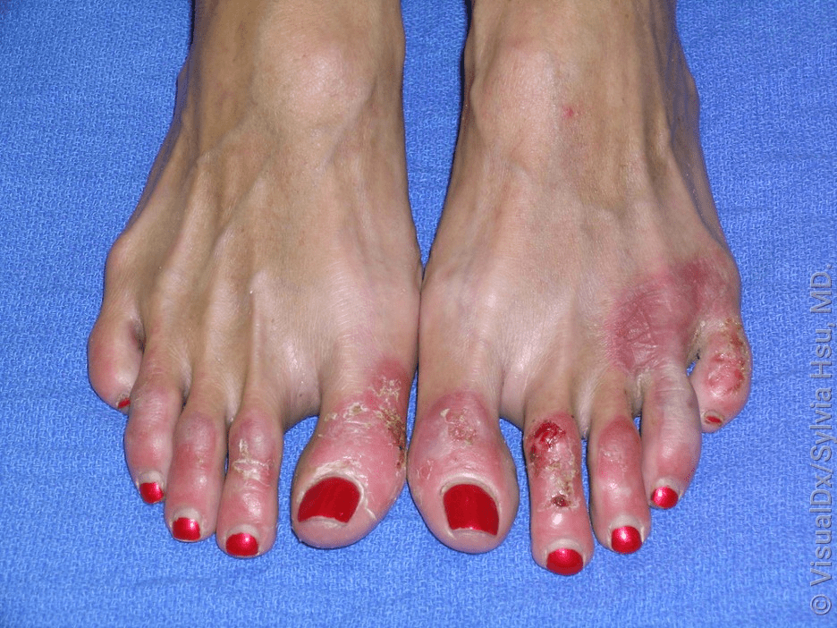
Necrolytic acral erythema
• HCV infection
Rare cutaneous disorder a/w HCV. Often advanced disease.
Pronounced erythematous papules + occasionally vesicles progressing to well-demarcated dusky red/purple plaques. Most often dorsal acral surfaces, sparing palms/soles.
DDx includes necrolytic migratory erythema, acrodermatitis enteropathica, pellagra
Treatment of underlying HCV
500
What is the most common associated cardiac anomaly?
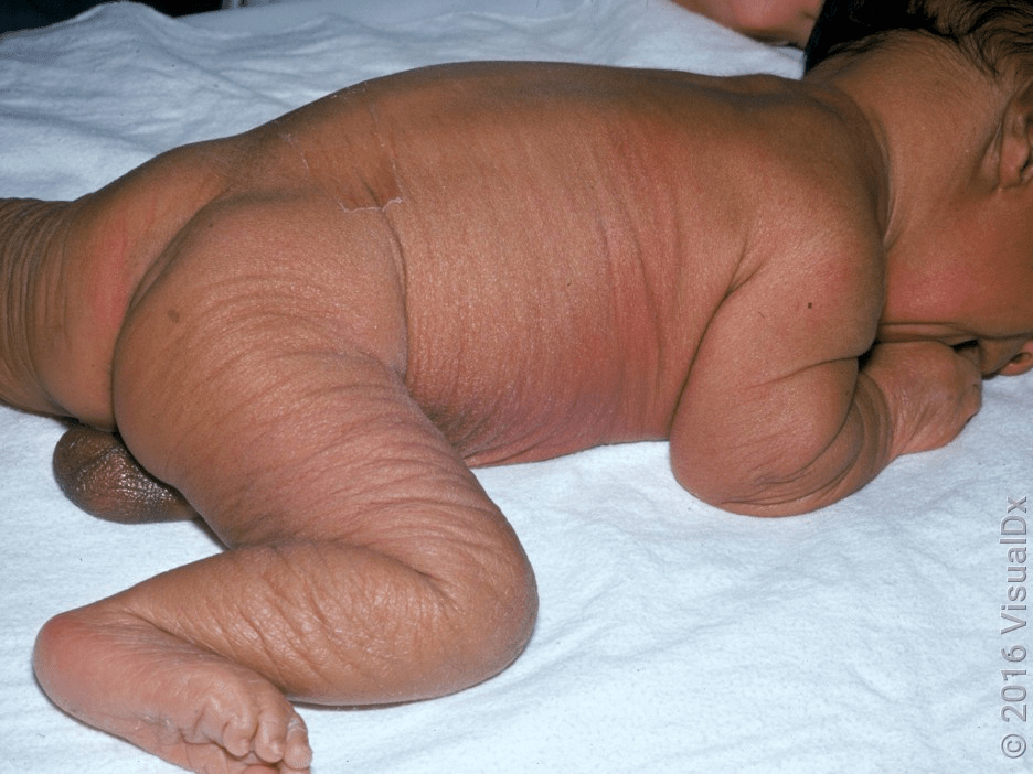
Cutis laxa
• Aortic dilation & rupture
Sparse & fragmented elastic fibers. Multiple modes of inheritance and multiple forms. Generally, AR most common and most severe with systemic involvement. Fibulin-5 (FBLN5) commonly mutated. Occasionally acquired (penicillamine, systemic disease e.g. RA, SLE)
Loose, pendulous skin of face (esp. periocular and cheeks→ “bloodhound facies”), neck, axillae, and thighs; skin lacks elastic recoil.
Depending on type can see hypoplastic lungs, emphysema, GI/GU diverticula, corneal clouding/cataracts
Treatment is multidisciplinary
500
What is the asssociated monoclonal gammopathy with this presentation?
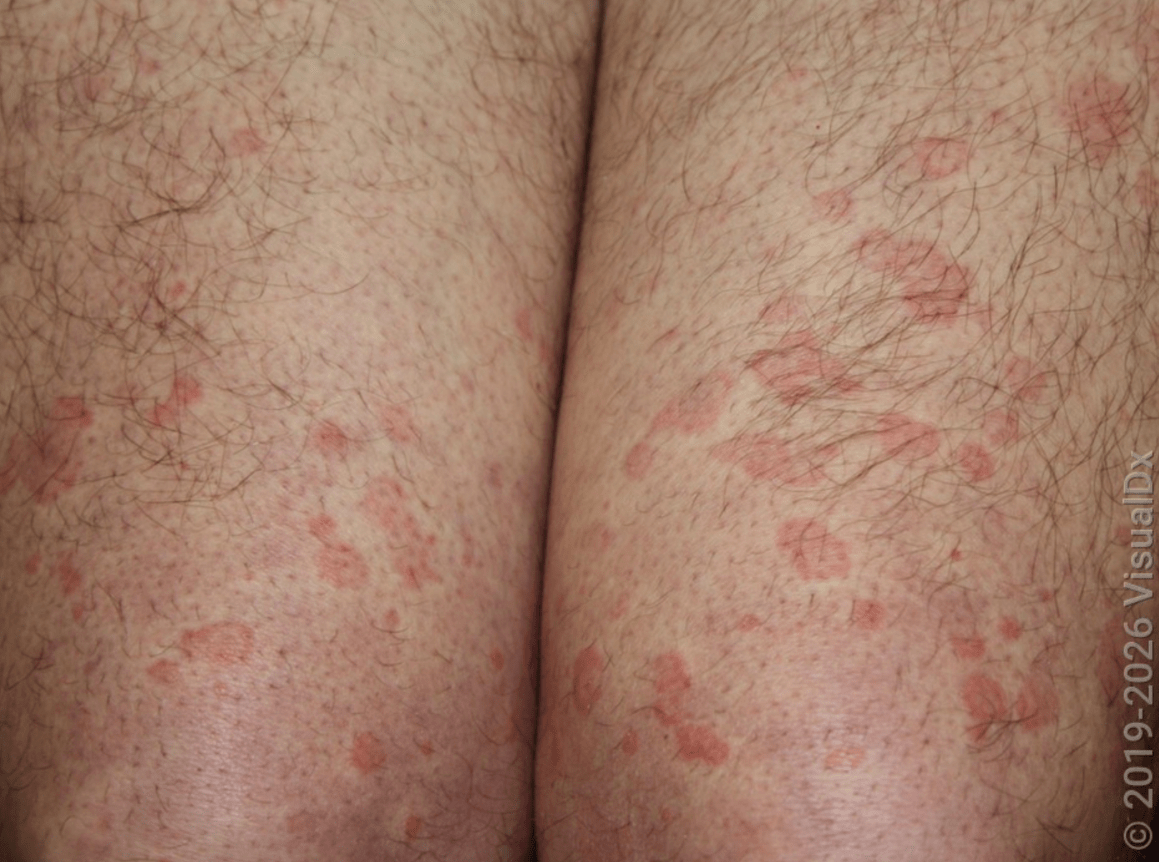
Schnitzler syndrome
• IgM κ
Rare autoinflammatory disorder a/w IgM monoclonal gammopathy. Chronic, recurrent urticarial lesions, fever, arthralgias & bone pain. Can also see LAD + HSM, elevated ESR. May progress to lymphoprolif disorder (incl. Waldeström macroglobulinemia).
Histo with neutrophilic-rich dermal infitrate.
DDx adult-onset Still, RA, urticarial vasculitis.
Tx: heme/onc referral, IL-1 inhibition
500
What HIV medication is associated with these nail findings?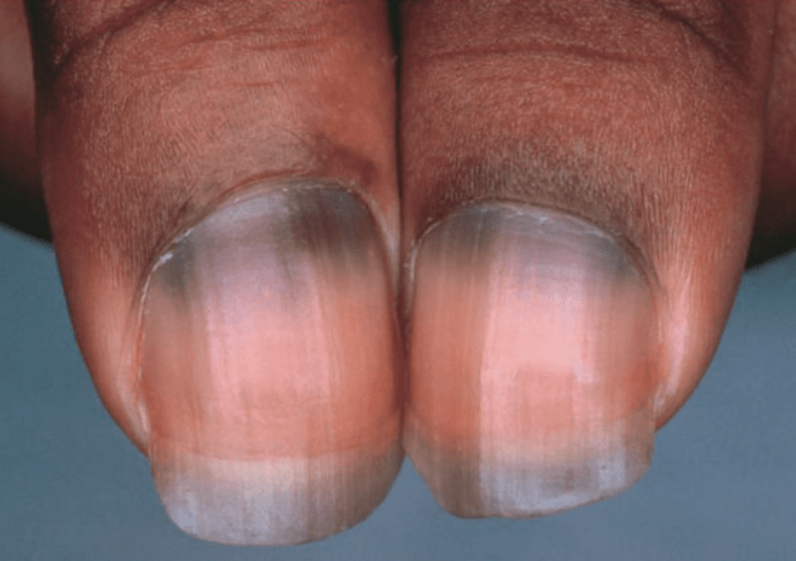
Zidovudine-associated melanonychia
Patients receiving zidovudine may develop longitudinal streaks, horizontal bands, and diffuse hyperpigmentation.
Other common HIV nail findings (2/3 of patients): onychomycosis, onychoschizia, onycholysis, paronychia (from proteinase inhibitors)