Transducer Placement
What's that Image?
The Pelvic Cavity
The Liver
100
What plane is the transducer placed in, and what is the large, dark structure?
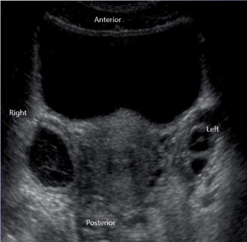
Transverse! The Bladder.
100
What is this an image of, and what are direction are each side of the image (transverse)?
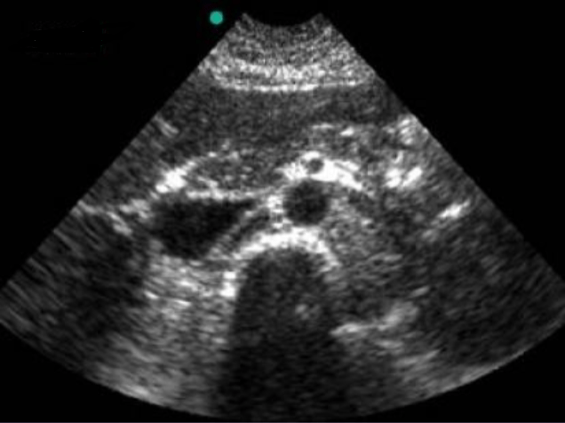
The Aorta!
Top: Anterior
Bottom: Posterior
Right: Left
Left: Right
100
Here we can see the "uretral jets" at work. What exactly is happening?
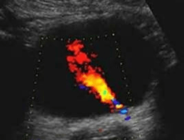
The ureters are emptying urine into the bladder.
100
In this ultrasound, we can visualize the liver lobes (supposedly) well. Which lobes are which?
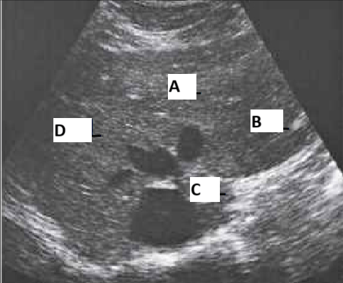
A. Quadrate
B. Left
C. Caudate
D. Right
200
What plane was the transducer placed in for this image?
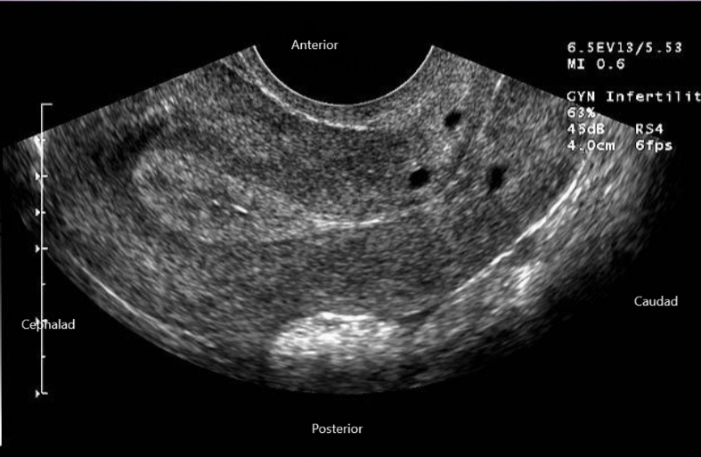
Sagittal via transvaginal approach.
200
What is the structure in the image, and what direction does each side represent (sagittal)?
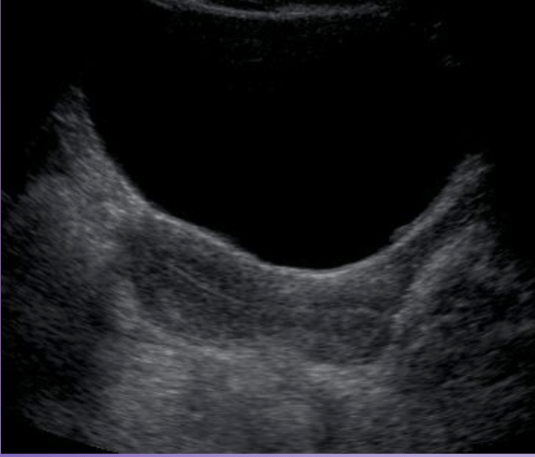
The Bladder (top) and Uterus!
Top: Anterior
Bottom: Posterior
Left: Cephalad
Right: Caudad
200
What is this an image of, and why do the walls look thicker than normal?
Hint: this organ has transitional epithelium.
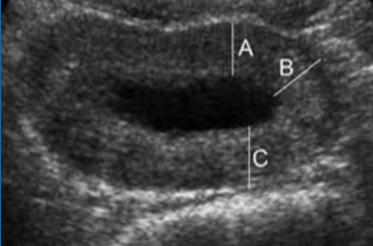
The bladder, and it's mostly empty! The walls get thinner as it expands.
200
Which liver ligament is represented in the photo?
Hint: Divides the left lobe into medial and lateral aspects and attached liver to the anterior wall.
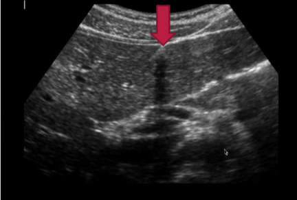
Falciform ligament.
300
What plane was the transducer placed in for this image?
Sagittal.
300
What is the yellow arrow is pointing at?
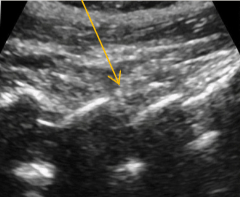
The lumbar vertebrae!
300
What is the structure in this image?
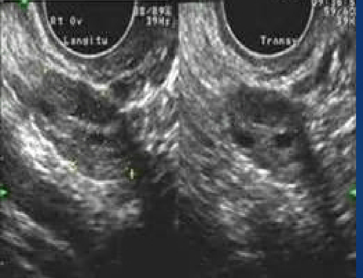
The ovary!
300
Which liver ligament is represented below?
Hint: Once the umbilical vein, now located in the left lobe.
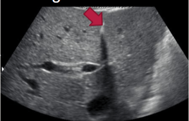
Ligamentum teres.
400
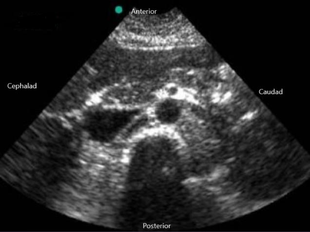
What plane was the transducer placed in for this image? (It’s actually right/left not cephalad/caudad sorryyyyyyyyy)
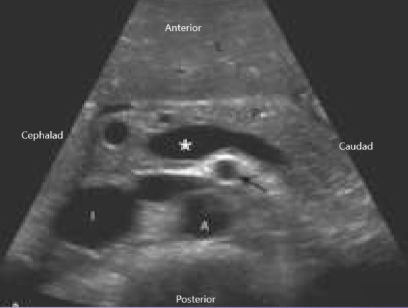
Transverse!
400
What is this structure, and what direction does each side of the image represent (sagittal)?
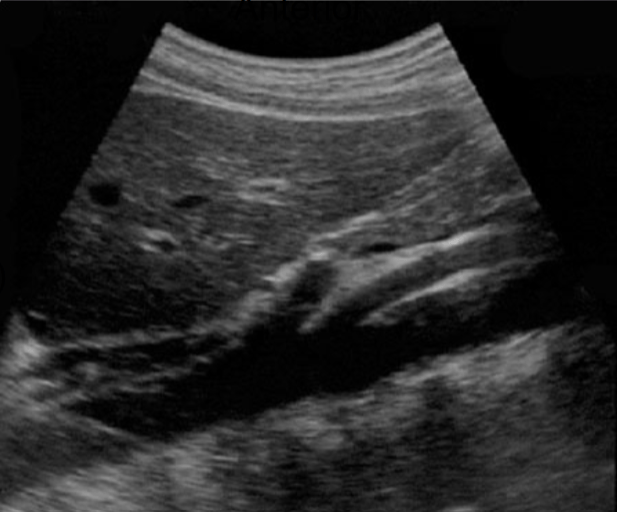
The SMA (Superior Mesenteric Artery)
Top: Anterior
Bottom: Posterior
Right: Cephalad
Left: Caudad
400
Sagittal is also called ________.
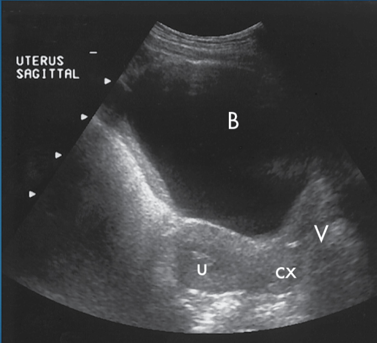
Longitudinal.
400
Which liver ligament is represented in the photo?
Hint: it separates the caudate lobe from the left lobe.
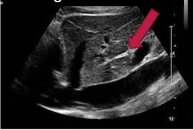
Ligamentum venosum.
500
Here is the dreaded ultrasound. What plane was the transducer placed in for this image?
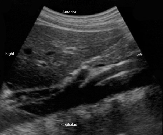
Sagittal.
500
What is this an image of, and what directions does each side of the image represent (sagittal)?
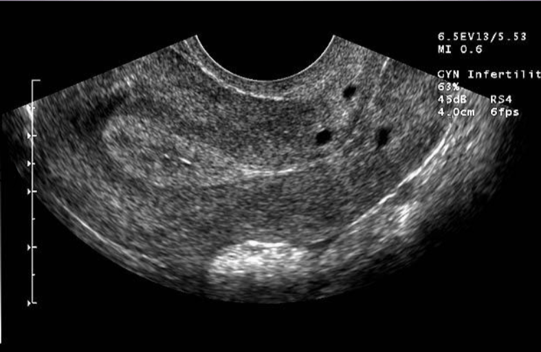
The uterus!
Top: Anterior
Bottom: Posterior
Right: Caudad
Left: Cephalad
500
Identify the structures in this image.
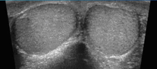
Testes.
500
Here we have an image of the hepatic veins (no idea what plane though, not in the powerpoints). Label A, B, & C.
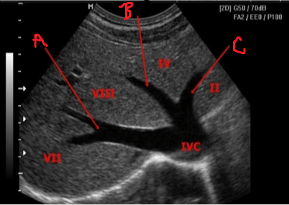
A. Right Hepatic Vein (branches into anterior and posterior)
B. Main Hepatic Vein
C. Left Hepatic Vein
600
Which plane is the transducer placed in for both of these images?
Bonus: what is this structure?
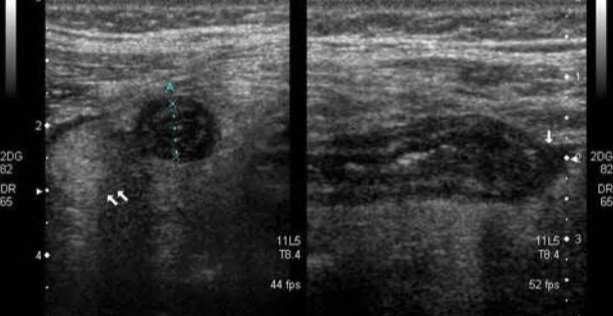
Left: transverse
Right: sagittal
The appendix!
600
What is the white line in the photo?
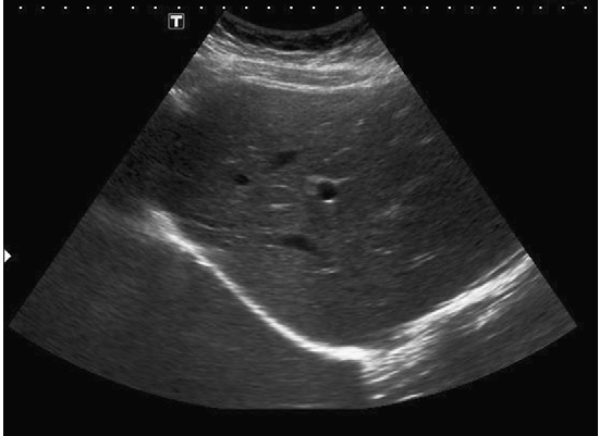
The diaphragm.
600
What do the starred structures represent?
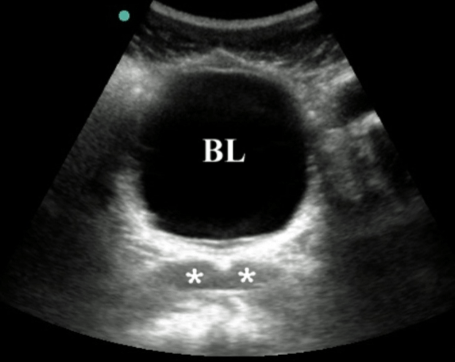
Seminal vesicles.
600
What is this structure, and are A, B, and C, and what plane was the transducer placed in to get this image?
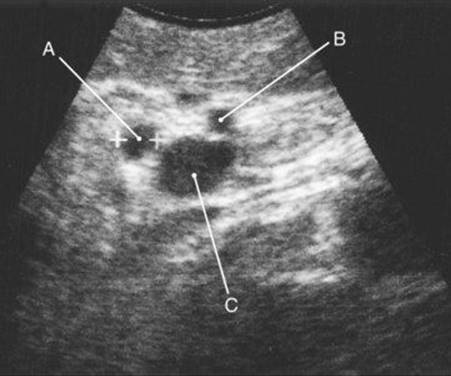
Porta Hepatis!
A. Common bile duct
B. Hepatic artery
C. Main Portal Vein
Transverse!
700
Here we have the left kidney and the spleen. It looks a lot like the liver and kidney, but okay. What plane is the transducer placed in to get this image?
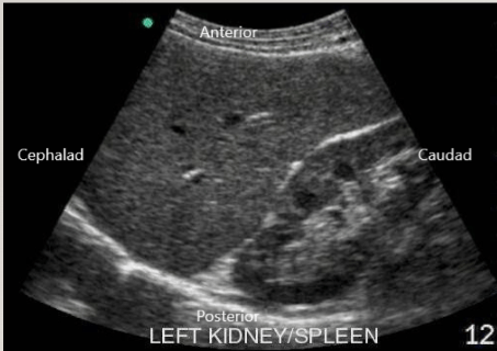
Sagittal!
700
What is this structure, and what direction does each side of the image represent?
Hint: This organ can only be seen when it's distended, and it's only distended when a patient has not eaten that day.
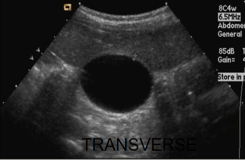
Gallbladder.
Top: Anterior
Bottom: Posterior
Left: Right
Right: Left
700
What is the structure the arrows are pointing at, and what direction does each side of the image represent?
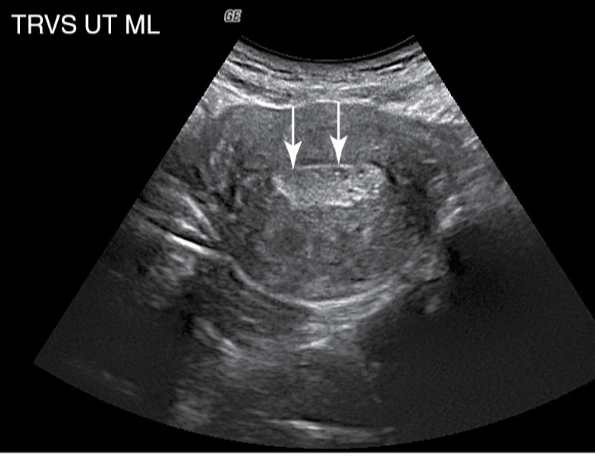
The endometrium.
Top: Anterior
Bottom: Posterior
Left: Right
Right: Left
700
Here we have an image of the right lobe of the liver. What plane was the transducer placed in to get this image?
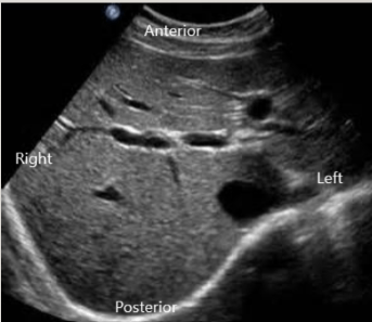
Transverse.
800
Here we have another picture of the kidney. What plane was the transducer placed in for this image?
Hint: when we view in one plane, it's very long. In this plane it looks more squished.
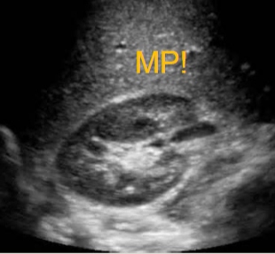
Transverse!
800
What is the structure(half) circled on the image?
Hint: it nestles the pancreatic head.
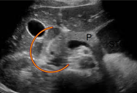
Duodenum (Small Intestine)
800
This organ in the male pelvis sits just below the urethra, and is often scanned by a urologist if there are concerns. What is it, and what symptom is often reported as concerning?
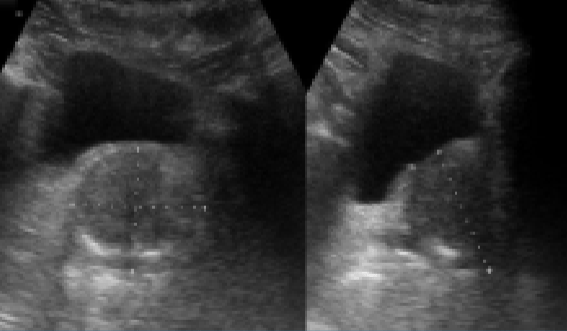
The prostate, and frequent urination is a warning sign.
800
What plane is the transducer placed in for this image, and what two structures can you see on it?
Hint: one is right there haha
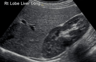
Right Kidney and Right Lobe of Liver
Sagittal (Longitudinal)
900
Here we have an image of the gallbladder. It is taken in the sagittal view, meaning the transducer is placed in a ________ direction.
Hint: Latitude is ---, but what is l?
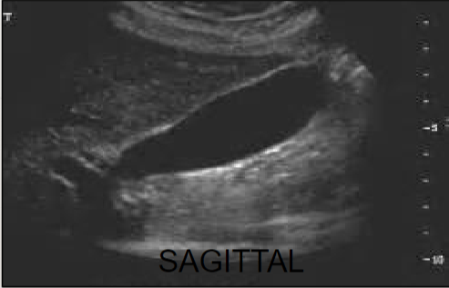
Longitudinal!
900
Unfortunately I think we're just going to have to study this one, I can't make sense of it.
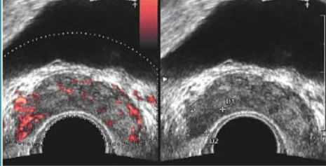
Seminal vesicles.
900
What is the structure the yellow arrow is pointing at?
Hint: it appears as an elongated tube around the testes in the sagittal view
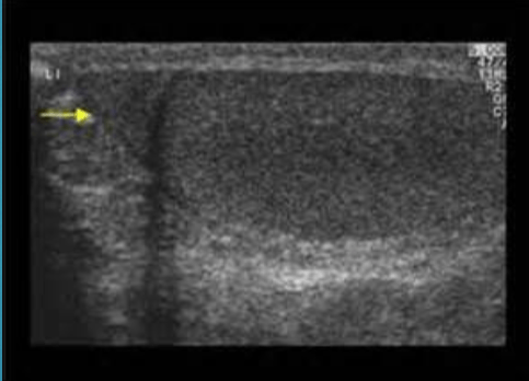
Epididymis
900
What plane is the transducer placed in the photo?
Hint: This is the porta hepatis!
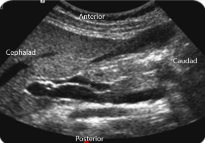
Sagittal!