Leukocytes
RBC/Anemias
Hemoglobin
Case Studies
Blood Cell Inclusions, Morphology & Physiology
Random
100
Which is true of specific (secondary) granules of the neutrophilic granulocyte?
a. Contain esterase
b. Are derived from azurophil (primary) granules
c. Appear first at the myelocyte stage
d. Are formed on the mitochondria
Appear first at the myelocyte stage
(Secondary granules first appear at the myelocyte stage, though primary granules are still retained)
100
In which of the following conditions would you expect to find Howell-Jolly bodies?
a. Iron deficiency anemia
b. Splenomegaly
c. Diabetes
d. Megaloblastic anemia
Megaloblastic anemia
(Howell-Jolly bodies are associated with megaloblastic anemia as well as in other conditions where normal mitosis may be interrupted and may be observed on a peripheral blood smear from a patient who has a hypo-functioning spleen)
100
Hemoglobin (g/100mL) x 10 / RBC count (millions/mm3) is the formula for calculating:
a. MCHC
b. MCV
c. MCH
d. RDW
MCH
(stands for “mean corpuscular hemoglobin” and is a measure of the average hemoglobin content of the RBC. MCH = (Hgb x 10) / RBC count = pg)
100
In a patient with an increased red cell mass into the 99th percentile and serum erythropoietin level below reference range for normal, which of the following criteria confirms a diagnosis of polycythemia vera?
a. Bone marrow panmyeloysis
b. BCR/ABL1 translocation
c. inv(16) mutation
d. JAK2 V617F mutation
JAK2 V617F mutation
(In 95-100% of polycythemia vera patients the JAK2 V617 mutation is present)
100
New methylene blue and brilliant cresyl blue are used to stain and identify which type of blood cell?
a. Blasts
b. Nucleated RBC
c. Reticulocytes
d. Neutrophils
Reticulocytes
(These stains are primarily used as they stain the residual RNA present in the cell)
100
A platelet determination is performed on an automated instrument and a very low value is obtained. The platelets appear adequate when estimated from the stained blood film. The best explanation for this discrepancy is:
a. The blood sample is hemolyzed
b. White cell fragments are present in the blood
c. Red cell fragments are present in the blood
d. Many platelets are abnormally large
Many platelets are abnormally large
(The instrument counts particles within defined size limits, the upper limit separates large platelets from erythrocytes.)
200
A total leukocyte count is 10.0 x 10^3/uL and 25 NRBCs are seen per 100 leukocytes on the differential. What is the corrected leukocyte count?
a. 12,000/uL (12.0 x 10^9/L)
b. 10,000/uL (10.0 x 10^9/L)
c. 8,000/uL (8.0 x 10^9/L)
d. 2,000/uL (2.0 x 10^9/L)
8,000/uL (8.0 x 10^9/L)
The WBC count can be corrected for NRBCs using the formula
(uncorrected WBC count x 100) / (# of NRBCs per 100 WBCs + 100)
(10,000 x 100) / (25+100)
1,000,000 / 125 = 8,000/ uL
200
Blood collected in EDTA undergoes which of the following changes if kept at room temperature (22°) for 6-24 hours?
a. Increased MCHC and MCV
b. Increased hematocrit and MCV
c. Increased ESR and MCV
d. Decreased reticulocyte count and hematocrit
Increased hematocrit and MCV
(Tubes that remain at room temperature for >5 hours have unacceptable blood cell artifacts. This will affect the HCT and the MCV)
200
A screening procedure for detecting hemoglobin F is the:
a. Fluorescent spot test
b. Dithionite solubility test
c. Kleihauer-betke test
d. Heat stability test
Kleihauer-betke test
(This procedure is commonly used as a screening test to determine the amount of fetal blood that has mixed with maternal blood)
200
A patient has a congenital nonspherocytic hemolytic anemia. After exposure to anti-malarial drugs, the patient experiences a severe hemolytic episode. This episode is characterized by red cell inclusions caused by hemoglobin denaturation. Which of the following conditions is most consistent with these findings?
a. Thalassemia major
b. G6PD deficiency
c. Paroxysmal nocturnal hemoglobinuria
d. Pyruvate kinase deficiency
G6PD deficiency
(G6PD deficiency RBCs are sensitive to the anti-malarial drug primaquine and oxidized and denatured hemoglobin can precipitate as Heinz bodies)
200
Which RBC inclusions are composed of iron containing granules and can be seen on Wright’s stain but confirmed with a Prussian blue stain?
a. Howell-Jolly bodies
b. Basophilic Stippling
c. Pappenheimer bodies
d. Heinz body
Pappenheimer bodies
(The prussian blue stains non-heme iron that makes up these inclusions)
200
In an electronic or laser particle cell counter, clumped platelets may interfere with which of the following parameters?
a. Red blood cell count
b. Hematocrit
c. White blood cell count
d. Hemoglobin
White blood cell count
(The large clumps of platelets will be mistaken for and counted as WBC due to their increased size)
300
Which of the following stains is closely associated with the lysosomal enzyme in primary (azurophilic) granules?
a. Methylene blue
b. Myeloperoxidase
c. Prussian blue
d. Nonspecific esterase
Myeloperoxidase
(Myeloperoxidase is an enzyme found in the primary granules of granulocytic cells)
300
All of the following are present in a normal bone marrow except what?
a. Siderocyte
b. Sideroblast
c. Ringed sideroblast
d. Hemosiderin
Ringed sideroblast
(These are formed from an accumulation of non-ferritin iron in the mitochondria that encircle the erythroblast nucleus and are not present in the normal bone marrow. This morphology is associated with sideroblastic anemia and myelodysplasia)
300
Which of the following curves in this figure represent the production of gamma polypeptide chains of hemoglobin?
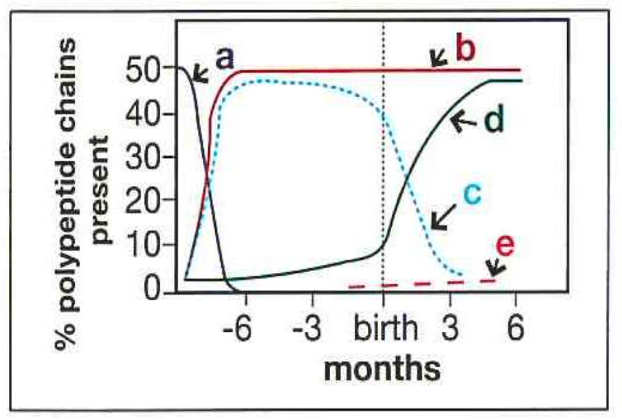
a. Curve e
b. Curve b
c. Curve c
d. Curve d
Curve c
(Gamma chains are a component of fetal hemoglobin that are synthesized during liver and bone marrow erythropoiesis in the fetus, with production that decreased after birth)
300
A 20-year-old woman with sickle cell anemia, whose usual hemoglobin concentration is 8 g/dL, develops a fever, increased weakness and malaise. The hemoglobin concentration is 4 g/dL and the reticulocyte count is 0.1%. The most likely explanation for her clinical picture is:
a. Occult blood loss
b. Thrombotic crisis
c. Aplastic crisis
d. Increased hemolysis due to hyperplenism
Aplastic crisis
(Aplastic crisis is a severe complication of sickle cell disease, causing a temporary pause in RBC production, which dramatically worsens the anemia)
300
Which of the following statements about macrocytes is true?
a. MCV is within normal limits
b. The appearance of the cell is due to exaggerated flattening
c. The cells are typically larger than a resting lymphocyte
d. Macrocytic cells are generally hypochromic
The cells are typically larger than a resting lymphocyte
(This is correct as size wise, they will typically appear larger than a normal lymphocyte)
300
Which of these is a diagnostic hallmark of Hodgkin's lymphoma?
a. Hairy cells
b. Rouleux
c. Reed Sternberg
d. Heinz bodies
Reed Sternberg
(The presence of these cells in a lymph node biopsy can be used to diagnose Hodgkin’s lymphoma)
400
The mediator cell type that binds to IgE antibodies is the:
a. Eosinophil
b. Polymorphonuclear neutrophil (PMN)
c. Macrophage
d. Basophil
Basophil
(Basophils have high concentrations of heparin and histamine in their granules which play an important role in the acute system, hypersensitivity reactions. Degranulation occurs when an antigen such as pollen binds to two adjacent immunoglobulin E (IgE) antibody molecules located on the surface of mast cells)
400
Peripheral blood smears from patients with untreated pernicious anemia will be characterized by:
a. Pancytopenia and microcytosis
b. Leukocytosis and elliptocytes
c. Leukocytosis and ovalocytosis
d. Pancytopenia and macrocytosis
Pancytopenia and macrocytosis
(The impaired DNA synthesis associated with pernicious anemia causes decreased production of all blood cells as well as abnormally large, macrocytic RBCs)
400
The following polypeptide chains are found in normal adult hemoglobin A:
a. Alpha and gamma
b. Alpha and beta
c. Alpha and delta
d. Beta and delta
Alpha and beta
(A normal hemoglobin molecule is a tetramer made up of two alpha and two beta chains)
400
In patients who present with bleeding disorders caused by platelets, the most common type of bleeding is:
a. Mucosal bleeding
b. Hemarthrosis
c. Delayed bleeding
d. Deep hematomas
Mucosal bleeding
(Certain characteristics of platelet-type bleeding disorders include mucosal bleeding, petechiae, and a female predominance.)
400
The light blue-gray inclusions observed in the cytoplasm of many of the bands and segmented neutrophils of a burn patient are most likely?
a. Auer rods
b. Toxic granules
c. Dohle bodies
d. May-Hegglin bodies
Dohle bodies
(These are associated with the peripheral smears of burn patients, severe infections, and cancer as a result of toxic drugs used on the patients)
400
What principle(s) of flow cytometry is employed when performing immuno-phenotyping?
a. Diffraction grating
b. Impedance
c. Diffraction grating and impedance
d. Fluorescent antibody tagging and light scatter
Fluorescent antibody tagging and light scatter
(Flow cytometry employs a combination of fluorescent antibody tagging of the cells and analysis with laser light scatter)
500
After briefly circulating in the peripheral blood, which of the following cells becomes a tissue macrophage (or histocyte) whose main function is phagocytosis?
a. Monocyte
b. Basophil
c. Plasma cell
d. Neutrophil
Monocyte
(Monocytes typically circulate in the peripheral blood between one and three days before entering into the tissues in a process called diapedesis and transforming into a tissue macrophage)
500
What is the mechanism that causes megaloblastic anemia?
a. Interruption of DNA synthesis
b. Interruption of development of precursor cells
c. Interruption of hemoglobin production
d. Defective stem cell production
Interruption of DNA synthesis
(DNA synthesis that is being interrupted results in the development of a megaloblastic anemia)
500
The abnormal cells indicated by the arrows are indicative of which of the following?
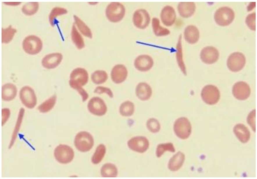
a. Sickle cell disease
b. Vitamin B12 deficiency
c. Severe burns
d. Iron deficiency
Sickle cell disease
(Variable anisocytosis and poikilocytosis with drepanocytes and target cells are shown which are characteristic of sickle cell disease)
500
Acquired Hemophilia A (not classic hemophilia A) may result from which of the following conditions?
a. Anit-factor VIII inhibitor
b. Factor VIII deficiency
c. Warfarin therapy
d. Thrombosis complications
Anit-factor VIII inhibitor
(Patients with acurired hemophilia A are suffering from a coagulation facotr inhibitor, specifically against factor VIII)
500
Which of the following RBC inclusions are seen in sideroblastic anemia and contain high amounts of iron?
a. Howell-Jolly bodies
b. Cabot rings
c. Heinz bodies
d. Pappenheimer bodies
Pappenheimer bodies
(RBCs in sideroblastic anemia can contain iron deposits known as pappenheimer bodies. These are irregular clusters of small light to dark granules found near the periphery of the red blood cell using wright stain or with prussian blue for iron)
500
Which of these characteristics is not associated with Lymphoproliferative disorders?
a. Increased risk of myelofibrosis
b. Slow progressing
c. Lymphadenopathy
d. Primarily seen affecting the elderly
Increased risk of myelofibrosis
(Though it can be seen it is rare and more closely associated to myeloproliferative disorders)
600
Disease associated with a dysfunction of polymorphonuclear neutrophils (PMNs) include all of the following with the exception of:
a. Chediak-Higashi syndrome
b. Chronic granulomatous diseases
c. Gaucher’s Disease
d. Myeloperoxidase deficiency
Gaucher’s Disease
(This is a monocyte-macrophage disorder caused by a rare genetic defect. There is a high incidence of this disease in Ashkenazi Jews. Gaucher cells, typically a large cell with one to three eccentric nuclei and a characteristically wrinkled cystoplasm, are found in the bone marrow, spleen, and other organs.)
600
Giant, vacuolated, multinucleated erythroid precursors are present in which of the following?
Primary myelofribrosis
Chronic myelocytic leukemia
Acute myelocytic leukemia
Erythroleukemia
Erythroleukemia
(RBC precursors in erythroleukemia display several dysplastic characteristics)
600
In the erythrocyte, the main function of the hexose monophosphate shunt is to:
a. Prevent the reduction of heme iron
b. Regulate the level of 2,3-DPG
c. Provide energy for membrane maintenance
d. Provide reduced glutathione to prevent hemoglobin oxidation
Provide reduced glutathione to prevent hemoglobin oxidation
(The hexose monophosphate shunt prevents hemoglobin degradation by producing reduced glutathione)
600
A 40-year-old caucasian male is admitted to the hospital for treatment of anemia and presents with symptoms of lassitude, weight loss, and loss of libido. Admission laboratory data are shown in the table:
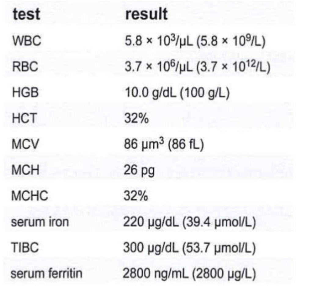
Examination of the bone marrow reveals erythroid hyperplasia with a shift to the left of erythroid precursors. Prussian blue staining reveals markedly elevated iron stores noted with occasional sideroblasts seen. This data is most consistent with which of the following conditions?
a. Iron deficiency anemia
b. Anemia of chronic inflammation
c. Hemochromatosis
d. Acute blood loss
Hemochromatosis
(Hemochromatosis is iron overload. Primary, as in hereditary hemochromatosis, or secondary, due to chronic anemia or treatments such as multiple transfusions. Secondary hemochromatosis is associated with anemia and abnormally high serum iron studies)
600
Which of the following statements characterize a reticulocyte?
a. Reticulum visible with a supravital stain, such as new methylene blue
b. Reticulum visible with Wright stain
c. Pyknotic nucleus still present in the cell
d. Contains some organelles used for hemoglobin production
Reticulum visible with a supravital stain, such as new methylene blue
(Reticulocytes must be stained with a supravital stain when performing a manual reticulocyte count as its the only stains that will color the residual RNA)
600
Patients with Waldenstrom’s macroglobulinemia frequently encounter thrombotic complications because of:
a. Increased platelet count
b. Increased megakaryocytes in the bone marrow
c. Increased plasma cells
d. Coating of platelets and clotting by increased IgM
Increased plasma cells
(Plasma cells are involved in the overproduction of monoclonal IgM proteins which can cause thrombotic complications)
700
A manual white blood cell count was performed by the hematology tech. The cell counts for both sides were 152 and 164 respectively. All nine large squares were counted on each side. The dilution for this kit was pre-measured at 1:100. What should the technologist report as the white cell count?
a. 177.5 x 10⁹/L
b. 175 x 109/L
c. 17.5 x 10⁹/L
d. 1.75 x 10⁹/L
17.5 x 10⁹/L
The formula used will be:
(The average cells counted of both sides) x (dilution factor) / (# of squares counted) x (area of each square) x (depth factor)
So the problem will be:
158 x 100 / 9 x 1 x 0.1 mm = 17555.55 / mm3
There are 1,000,000 mm3 in a liter so it would be 17555.55 x 1,000,000 = 17.5 x 10⁹/L
700
Which one of the following sets of laboratory results is consistent with hemolytic anemia?
a. Increased concentration of haptoglobin; negative hemoglobinuria
b. Decreased erythrocyte survival; increased catabolism of heme; decreased haptoglobin levels
c. Decreased serum LDH activity; normal catabolism of heme
d. Normal concentration of haptoglobin; marked hemoglobinuria
Decreased erythrocyte survival; increased catabolism of heme; decreased haptoglobin levels
(Hemolysis is the process of RBC destruction which leads to a decrease in erythrocyte survival. Increased catabolism of heme is also associated with the RBC destruction)
700
Which of the following forms of hemoglobin cannot be detected by routine hemoglobin methods?
a. Carboxyhemoglobin
b. Sulfhemoglobin
c. Oxyhemoglobin
d. Reduced hemoglobin
Sulfhemoglobin
(Sulfhemoglobin cannot combine with oxygen. Routine hemoglobin measurements are modified versions of the Cyanmethemoglobin method. Sulfhemoglobin is not readily converted to hemoglobincyanide which is a required for detection using this method)
700
A 2-year-old girl, who had been treated with upper respiratory tract infection a few weeks ago, showed multiple bruises and had the following laboratory findings:
Hgb = 13.5 g/dL
RBC - 3.9 x 10¹² /L
WBC = 8.0 x 10⁹ /L
Platelets = 5 x 10⁹ /L
Bone marrow M:E ratio - 3:1
Megakaryocytes = increased
These results are most suggestive of which condition?
a. Glanzmann thrombasthenia
b. Wiskott-Aldrich syndrome
c. Immune (idiopathic) thrombocytopenic purpura
d. Thrombotic thrombocytopenic purpura
Immune (idiopathic) thrombocytopenic purpura
(A characteristic lab finding of ITP is very low platelet count with elevated megakaryocytes in the bone marrow)
700
Many microsphereocytes, schistocytes and spherocytes with budding cytoplasm can be seen on peripheral blood smears of patients with:
a. Thalassemia
b. Pyruvate kinase deficiency
c. Hereditary spherocytosis
d. Extensive burns
Extensive burns
(Patients who have suffered burns to >15% of their body generally show evidence of intravascular hemolysis.
700
Which pair of malignancies represent different clinical manifestations of the same disease?
a. Chronic lymphocytic leukemia and adult T-cell leukemia
b. Chronic lymphocytic leukemia and small lymphocytic lymphoma
c. Hairy cell leukemia and hodgkins lymphoma
d. Sezary syndrome and prolymphocytic leukemia
Chronic lymphocytic leukemia and small lymphocytic lymphoma
(CLL and SLL represent different clinical presentation of the same disease)
800
What cytokines are most responsible for eosinophil differentiation and release from the bone marrow?
a. IL-5
b. IL-4
c. IL-2
d. IL-1
IL-5
(IL-5 released by T helper cells, mast cells, eosinophils and other lymphocytes has lineage specificity for eosinophils and is the major cytokine required for production and differentiation of eosinophils)
800
All of the following are known causes of aplastic anemia except:
a. Viral infections
b. Chemical agents
c. Enzyme deficiencies
d. Ionizing radiation
Enzyme deficiencies
(Enzyme deficiencies are typically associated with hemolytic anemias, not aplastic)
800
Which of the following alpha thalassemia syndromes has the genotype -a/aa?
a. Silent carrier
b. Alpha thalassemia minor
c. Hemoglobin H disease
d. Alpha thalassemia major
Silent carrier
(Only one of the four genes is missing. Individuals who are silent carriers show no clinical disease and demonstrate normal results during routine laboratory testing)
800
What would be the logical first step to obtain correct values when the following results were obtained on an electronic particle counter in hematology for an alcoholic patient?
WBC 6,500/cu. mm
RBC 4,550,000/cu. mm
HGB 18.0 g/dL
HCT 41.5%
MCV 90.1 fL
MCH 39.6 pg
MCHC 43.4%
a. Perform a microhematocrit
b. Check and correct for lipemia
c. Dilute the blood
d. Replace the lysing agent
Check and correct for lipemia
(The patient is an alcoholic and the possibility of lipemia can cause falsely increased hemoglobin results on automated hematology analyzers since the hemoglobin measurement is performed spectrophotometrically. To correct it would be beneficial to perform a saline replacement to resolve the discrepancy)
800
All of the following are sites of active hematopoiesis in the adult except:
Skull
Ribs
Liver
Pelvis
Liver
(The liver is not an active site of hematopoiesis as the bone marrow becomes the dominant site of hematopoiesis in adults)
800
Which of the following causes schistocyte formation in disseminated intravascular coagulation (DIC)?
a. Degranulation of promyelocytes
b. Excessive fever in patients with DIC
c. Microthrombi disposition in small vessels
d. Excessive platelet size
Microthrombi disposition in small vessels
(Schistocyte are the result of erythrocytic membrane damage caused by shearing of red blood cells as they pass through a fibrin mesh of clot formation occurring in the blood vessels. This intravascular clot formation process occurs in DIC)
900
Which of the following leukemias can be excluded as a possible diagnosis if Auer rods are seen in blasts on the peripheral blood smear?
a. Acute myelomonocytic leukemia
b. Acute lymphoblastic leukemia
c. Acute myeloid leukemia without maturation
d. Acute promyelocytic leukemia
Acute lymphoblastic leukemia (ALL)
(This is correct as auer rods are not seen in acute lymphoblastic leukemia and most commonly found in myeloid cells)
900
Which anemia is formed due to the inability to incorporate iron into the protoporphyrin ring?
a. Iron deficiency anemia
b. Sideroblastic anemia
c. Anemia of chronic inflammation
d. Thalassemia
Sideroblastic anemia
(Sideroblastic anemia occurs due to lead poisoning or porphyrias. The iron is unstable to be incorporated into the protoporphyrin ring upon the development of heme resulting in anemia)
900
How is beta thalassemia defined?
a. A decrease in the rate of production of beta chains due to a partial or total deletion of loci from chromosome 11 that code for the beta chain.
b. A decrease in the rate of production of Hb A due to a substitution of an amino acid in the beta chain.
c. The production of an abnormal form of hemoglobin due to the substitution of an amino acid in the beta chain.
d. A decrease in the rate of production of beta chains due to a partial or total deletion of loci from chromosome 16 that code for the beta chain.
A decrease in the rate of production of beta chains due to a partial or total deletion of loci from chromosome 11 that code for the beta chain.
(Beta thalassemia demonstrates problems with the beta-globin chain production. One of two loci that code for the beat chain may be detected from chromosome 11. The greater number of loci detected or inactivated, the greater the severity of the anemia that develops)
900
A patient has anemia with an elevated mean corpuscular volume (MCV) of 104 fL (Reference range: 80-100 fL). All of the following could be likely causes except:
a. Vitamin B12 deficiency
b. Alcoholism
c. Anemia of chronic inflammation
d. Liver disease
Anemia of chronic inflammation
(ACI is typically a normocytic normochromic anemia and may be microcytic in certain conditions)
900
The WBC anomaly indicated by the arrow in this illustration is:
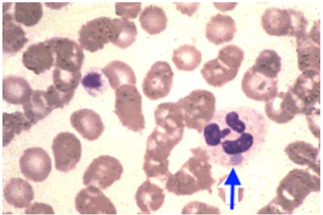
a. Alder-Reilly anomaly
b. Pelger-Huet anomaly
c. May-Hegglin anomaly
d. Toxic granulation
May-Hegglin anomaly
(May-Hegglin anomaly is a rare autosomal dominant conditions where patients are at risk for bleeding and infections and is characterized by large Dohle body-like inclusions within neutrophils, eosinophils, basophils and monocytes)
900
If a patient has a white blood count of 40,000/uL, what would be the MOST useful staining procedure to distinguish between bacterial infection and chronic myelogenous leukemia (CML)?
a. Wright’s stain
b. Leukocyte alkaline phosphatase (LAP)
c. Peroxidase (i.e. Myeloperoxidase)
d. Alpha-naphthy acetate
Leukocyte alkaline phosphatase (LAP)
(This stain is used to determine if an increase of cells is due to chronic myelogenous leukemia or a leukemoid reaction due to infection or similar conditions. Cells from a noncancerous reaction stain are positive with many intense blue granules)
1000
Bone marrow examination reveals a hypercellular marrow consisting of probable lymphoblasts. The cells are positive for TdT, CD3 and CD7; however, the lymphoblasts are negative for surface immunogobulins, CD19, and CD10 (CALLA). The most likely diagnosis is:
a. Chronic lymphocytic leukemia (CLL)
b. Large granular lymphocytosis
c. T-cell leukemia (T-ALL)
d. Hairy cell leukemia
T-cell leukemia (T-ALL)
(T-cell markers include CD34, CD2, CD3, CD4, CD5, CD7, CD8 and TDT)
1000
All of the following are associated with HELLP syndrome except:
a. Hemolysis
b. Elevated liver enzymes
c. Low platelet count
d. Elevated lipid levels
Elevated lipid levels
(HELLP syndrome is a severe pregnancy complication involving the breakdown of red blood cells, impaired blood clotting and liver dysfunction. Elevated lipid levels are not associated with HELLP syndrome)
1000
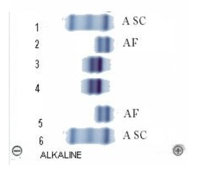
The alkaline hemoglobin electrophoresis patterns shown on the right include controls for A, S and C; and A and F above and below the patient results. (NOTE: ASC and AF are labeled for controls and do not indicate the migration order) The patient was tested in duplicate and the results are in lanes 3 and 4. The patient lanes (3 and 4) displayed in these hemoglobin electrophoresis patterns are consistent with what diagnosis?
a. HbSA
b. HbSC
c. HbSD
d. HbS/HPFH
HbS/HPFH
(Nearly equal bands are shown in the “S” and “F” positions. In HbS/HPFH, there would be two bands on the alkaline electrophoresis, one would be in the S position. Hereditary Persistence of Fetal Hemoglobin will result in a band in the F position)
1000
A 90-year-old patient is admitted to the hospital with the following laboratory data:
WBC: 9,000/ mm3
PLT: 190,000 / mm3
Hgb: 6.1 g/dL
Differential:
11% Neutrophils
40% Lymphocytes
4% Monocytes
45% Myelobasts
45 NRBC’s/100 WBC
Bone Marrow: 45% Myeloblasts and 55% Megaloblastoid Erthroblasts
Serum Vitamin B12 and Folic Acid: Normal
The most likely diagnosis is:
a. Pernicious anemia
b. Polycythemia vera
c. Erythroleukemia
d. Myelomonocytic leukemia
Erythroleukemia
(Erythroleukemia is also known as Acute Myelogenous Leukemia type M6. This type is associated with either the presence of both erythroid and myeloid precursors or strictly erythroid precursors.)
1000
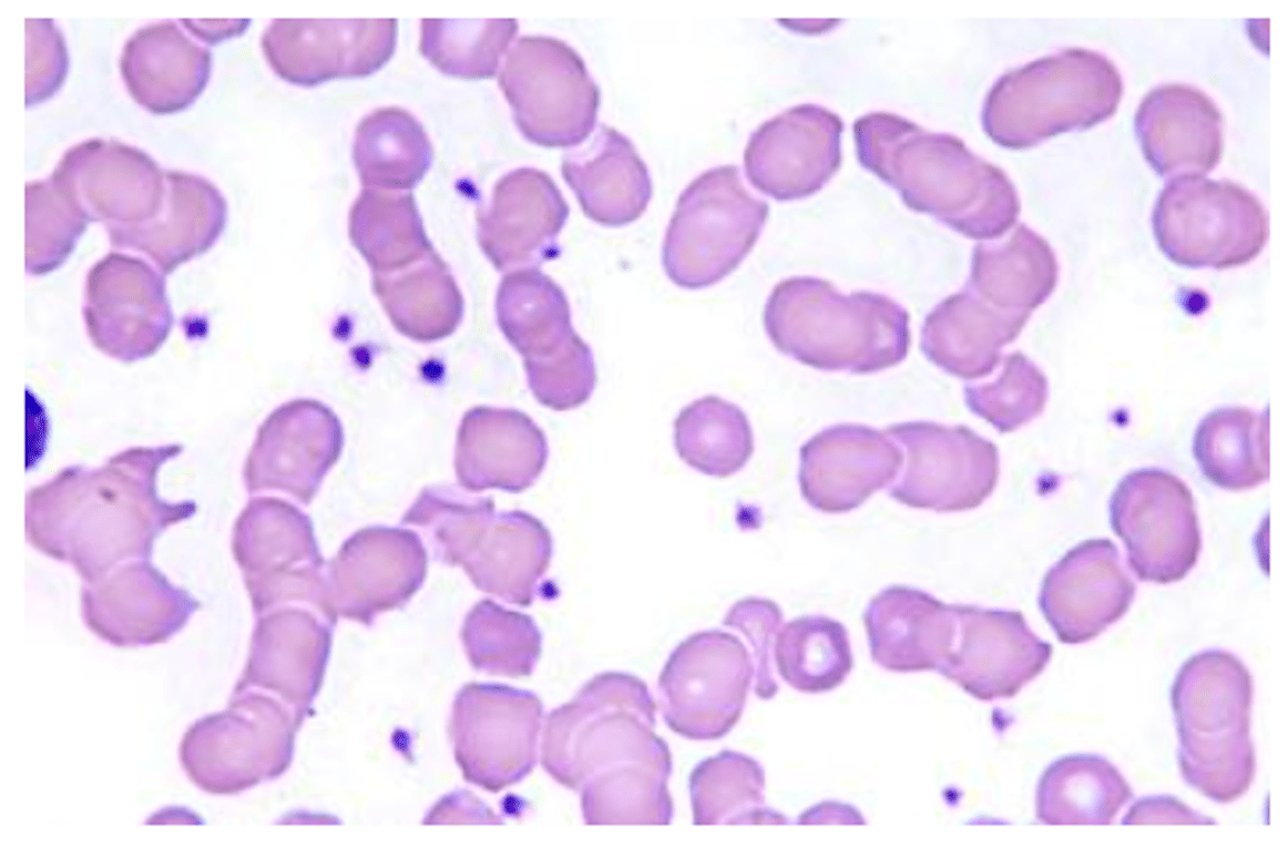
The arrangement of erythrocytes on this peripheral blood smear can be associated with each of the following conditions except:
a. Multiple myeloma
b. Cold agglutinin disorder
c. Chronic inflammatory disorders
d. Lymphoplasmacytic lymphoma
Cold agglutinin disorder
(Erythrocyte agglutination rather than rouleaux formation appears in these disorders when cold reactive antibodies, most commonly IgM, are circulating in the plasma)
1000
Which potent inhibitor of platelet aggregation is released by endothelial cells?
a. Epinephrine
b. Ristocetin
c. Thromboxane A2
d. Prostacyclin
Prostacyclin
(Prostacyclin is produced by endothelial cells and prevents the formation of the platelet plug involved in primary hemostasis by inhibiting platelet activation and aggregation)