you make me nervous
Happy studying
A
B
100
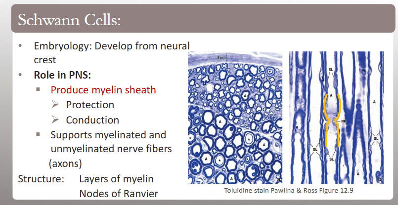
What is A and NR?
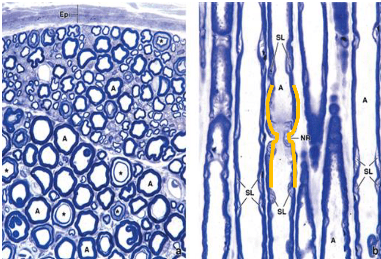
A = Axon
NR = Nodes of Ranvier
100
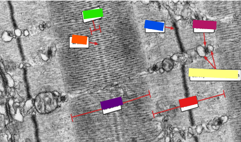
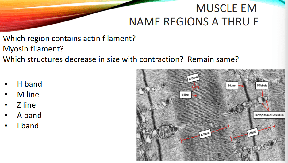
label this image
100
Patient B has osteoporosis. Where is loss of bone mass predominant?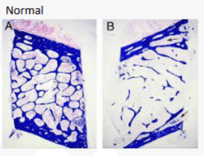
Cancellous (trabecular/spongy) bone
In image B, notice how the internal trabeculae are much thinner and more widely separated compared with normal bone.
Why cancellous bone? It has a larger surface area and undergoes more active remodeling, so increased bone resorption becomes apparent there earlier/more prominently.
RECALL: In osteoporosis:
Osteoclast resorption > osteoblast formation → net ↓ bone mass
100
where are these tissues commonly found?
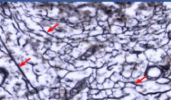
this is reticular tissue and is commonly found in the lymph nodes, spleen, and bone marrow.
200
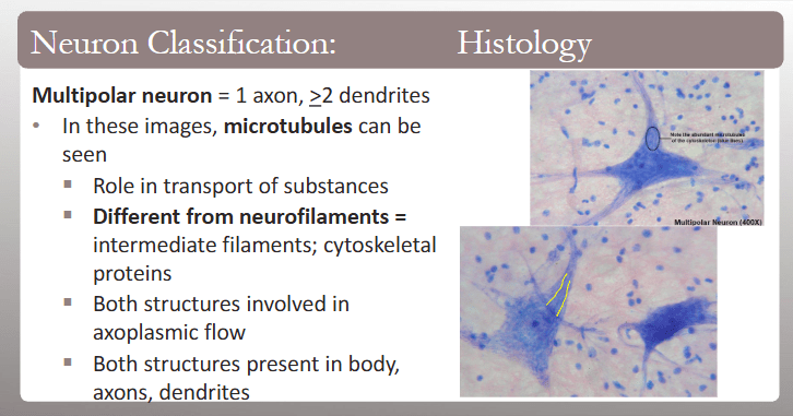
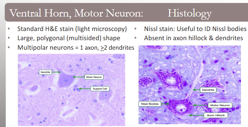
fill in the blanks!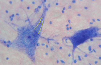 this is an example of a ________ neuron which contains ___ axon and ____ dendrites
this is an example of a multipolar neuron which contains 1 axon and >2 dendrites
200
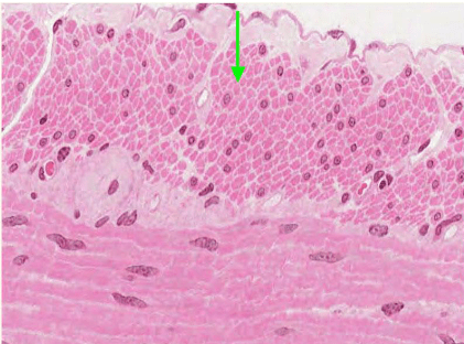 Name 3 locations this type of muscle is in.
Locations:
- esophagus
- small and large intestines
- ducts of glands
- bladder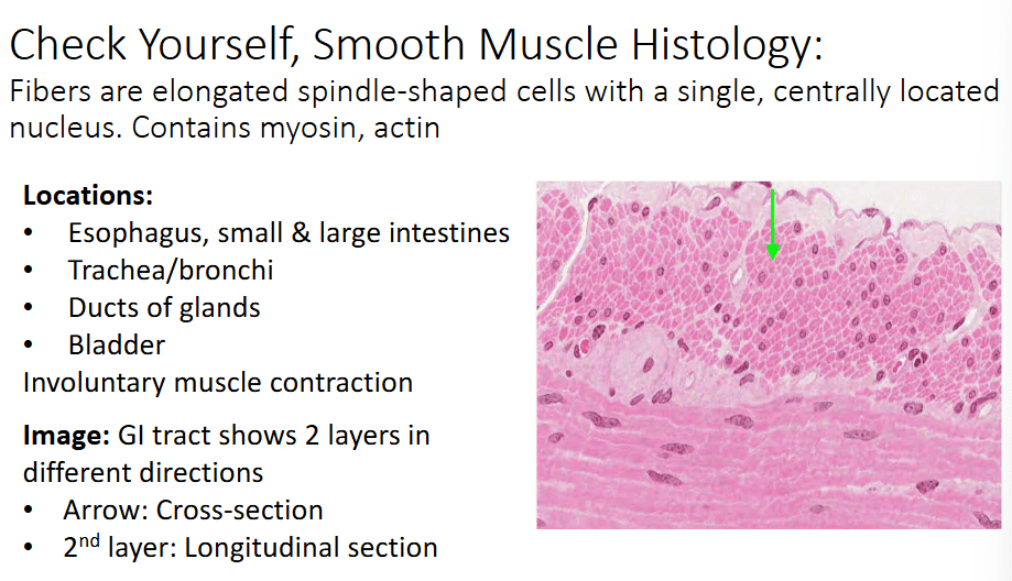
200
label the two boxes and explain the difference between the two
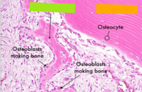
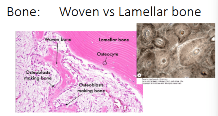
Woven: immature, random/ disorganized collagen, weak strength, rapid formation, seen in fetal development and early fracture repair, becomes remodeled into lamellar bone
Lamellar bone: mature, organized into layers (lamellae), stronger, slower formation, seen in in normal adult bone.
Fracture
→ soft fibrocartilaginous callus
→ hard bony callus containing woven bone
→ remodeling
→ lamellar bone
200
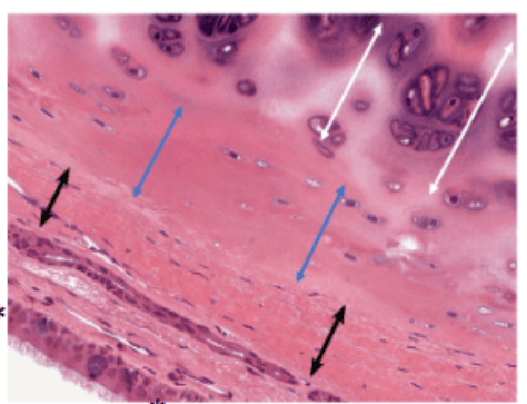
what is the function/why is the blue arrow section important?
The blue arrows span the connective tissue immediately surrounding the cartilage → perichondrium.
This is important because cartilage is avascular, so the perichondrium provides vascular/nutritional support for the cartilage by diffusion.
300
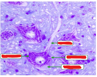
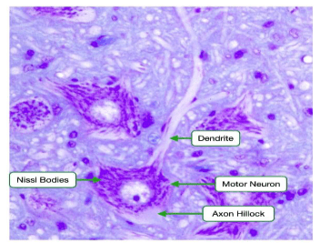
identify the structure in the red circle and its function
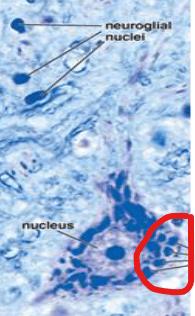
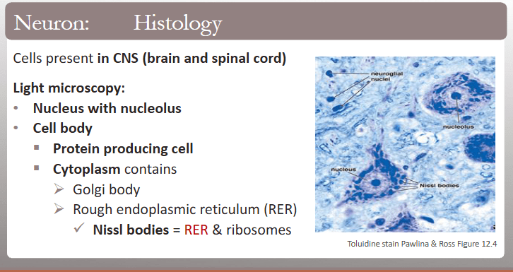
Nissl bodies are clumps of rough endoplasmic reticulum (RER) and free ribosomes found in neurons.
They are responsible for protein synthesis, producing proteins needed for neuronal maintenance and function.
300
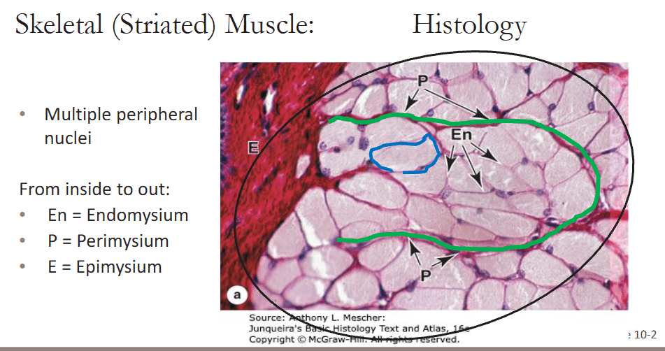
name the structure in green
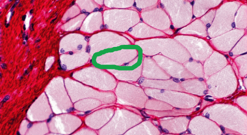
endomysium
300
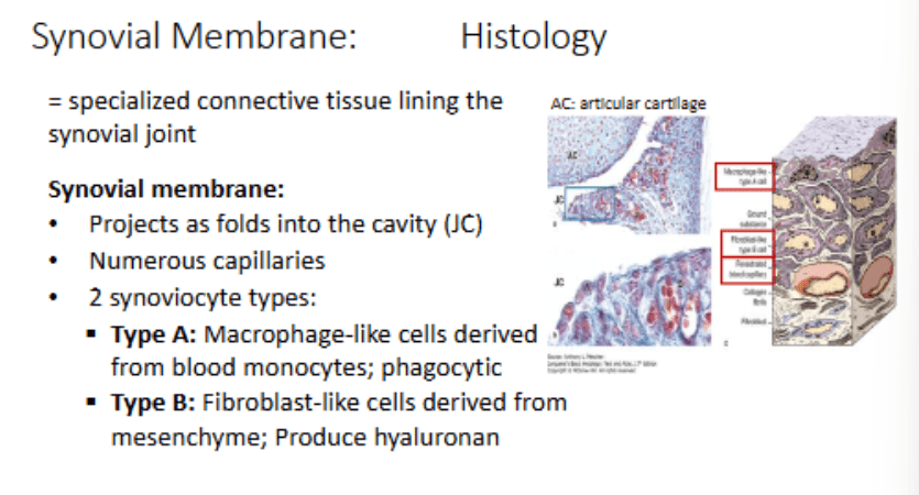
identify the yellow box and its function
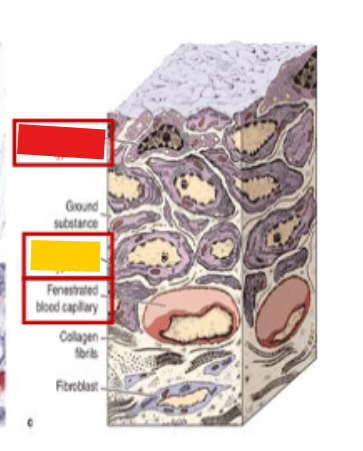
Fibroblast-like type B synoviocyte (derived from mesenchyme): Produces components of synovial fluid/ECM, especially hyaluronan
300
what type of collagen is this tissue type?
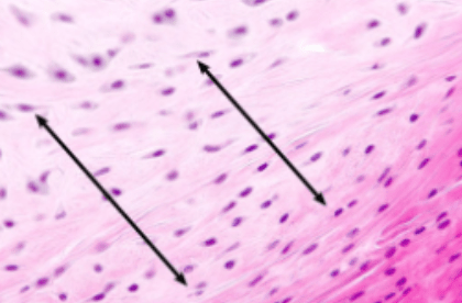
Fibrocartilage = Type I because it needs to be TOUGH → handles both tension and compression.
hyaline + elastic = Type II collagen
400
label ALL
400
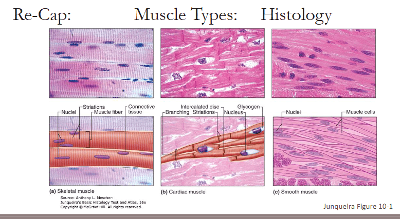
identify this muscle and explain how they regenerate?
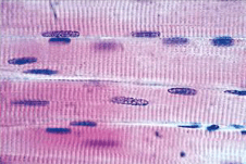
skeletal muscle!
Mature skeletal muscle fibers are highly differentiated and generally don't undergo mitosis themselves.
Instead, regeneration depends on satellite cells.
Muscle injury → satellite cells activated → proliferate → differentiate into myoblasts → fuse with/repair muscle fibers
Satellite cells are therefore the stem/progenitor cell population of skeletal muscle.
400
A bone biopsy shows mature cells embedded within mineralized matrix. In which structure is the cell body located?
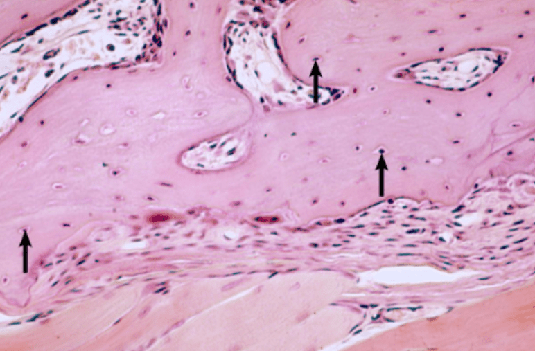
Lacunae!
Recall:
Lacuna → osteocyte location
Canaliculi → osteocyte processes travel through them
Connexins → gap junctions → cell-to-cell communication
Fibronectin → cell-to-ECM adhesion
400
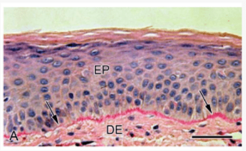
A skin biopsy shows separation of the stratified squamous epithelium from the underlying connective tissue due to damage at their junction. Which structure has most likely been disrupted?
Basement membrane
500
these cells line the ventricles of the brain and help with _______
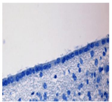
These are ependymal cells. Their apical cilia help circulate/move CSF along the ventricular system and central canal.
500
name 2 examples for this type of cartilage
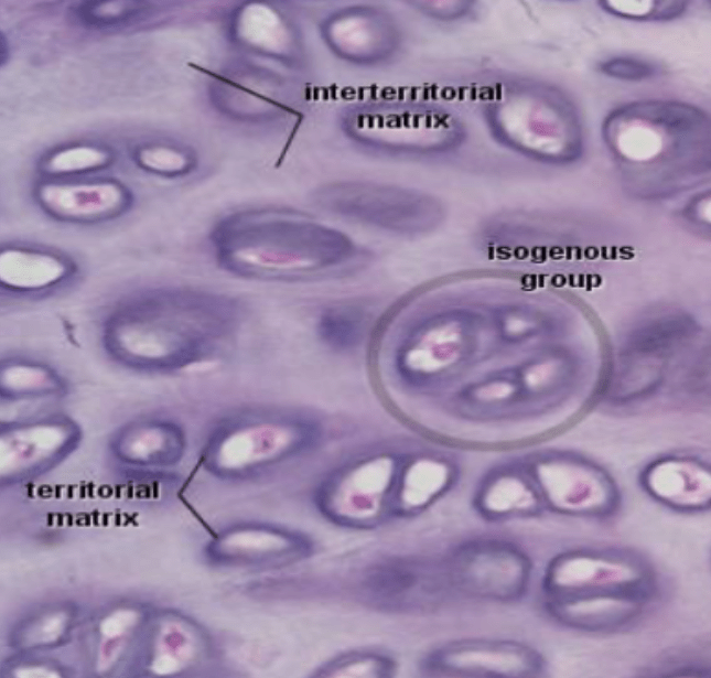
1. Articular cartilage of joints
2. Nose
3. Trachea/ Bronchial airways
4. epiphyseal plate
5. embryonic skeleton
500
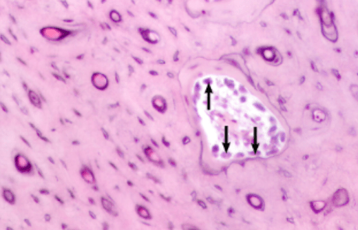
______ activates this structure (see pointed arrow)
RANKL drives osteoclast differentiation, survival, and activation → ↑ bone resorption → Ca²⁺ released from bone
500
what type of neuron cell bodies are located here? (green arrow)
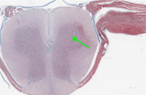
large multipolar lower motor neuron (LMN) cell bodies