Cerebrovascular and Demyelinating Pathologies
Infectious Pathologies
Neurocutaneous I
Neurocutaneous II
100
Red dead neurons in the acute stage and activated macrophages in the subacute.
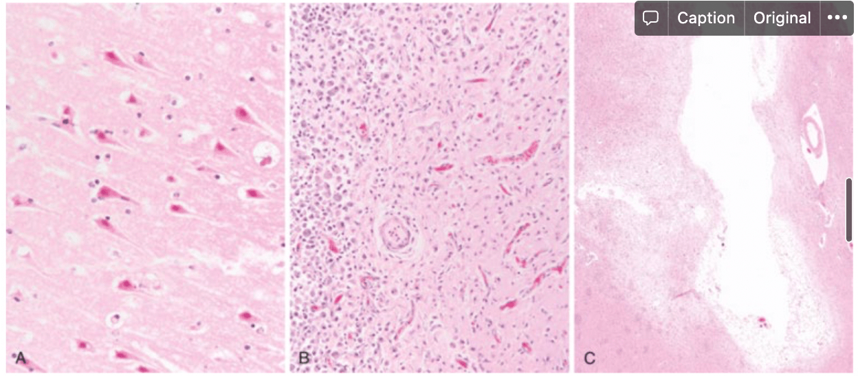
Focal Cerebral Ischemia
100
A 84-yo presents to clinic with high fever, confusion, a stiff neck, and a skin rash on the lower left leg. You order a lumbar puncture which finds increased neutrophils and low glucose levels. With these results you suspect this organism as the primary infectious agent.
Pneumococcus (Streptococcus pneumoniae)
Notes:
Neonates - E. coli and group B strep
Young adults - Meningococcus (Neisseria meningitidis)
100
The gene product that plays a role in the RAS pathway, which plays a role in Von Recklinghausen’s Disease.
neurofibromin
100
The gene products involved in the mTOR pathway, which when unchecked leads to increased tumor production.
hamartin (TSC1) and tuberin (TSC2)
200
Presentation: sudden headache and stiff neck; bloody CSF upon lumbar puncture
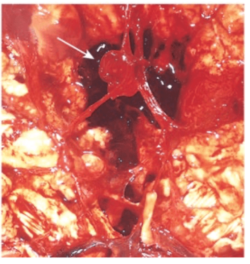
SAH
200
You're a pathologist doing an autopsy of 2 week old infant who passed from encephalitis. In this process you note that there is necrosis near the ventricles. Histology shows cytomegaly and owl's eye inclusions. From these findings, you determine this infectious agent as the primary cause of the meningoencephalitis.
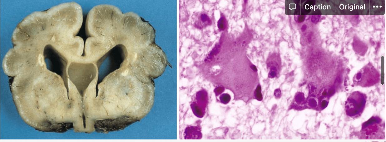
CMV
200
Condition with autosomal dominant inheritance caused by mutations in the merlin gene.
Neurofibromatosis Type 2
200
A patient presents to clinic with increasing pain and motor dysfunction of the lower limbs. Axillary freckles, cafe au lait spots, and lisch nodules are noted on physical exam. Based on these findings, you decide to order imaging to look for tumors that could be causing the pain and motor dysfunction. These benign tumors could be observed in such imaging. (4 possible answers)
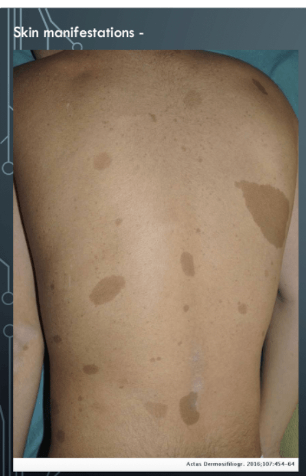
nerve sheath tumors, optic gliomas, schwannoma, neurofibroma
300
Tangled networks of worm-like vascular channels with high blood flow
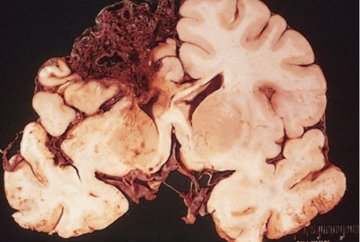
Arteriovenous Malformations (AVM)
300
You are a pathologist doing an autopsy of a 43-yo male you passed away from meningoencephalitis. The patient was also HIV+. Upon gross examinations, you observe a "soap bubble" appearance of the basal ganglia and a thick, white layer covering the brain. Histology shows histiocytes present. From these findings you determine this infectious agent to be the primary suspect of the meningoencephalitis.
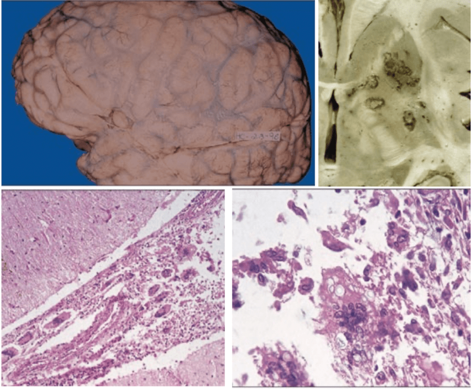
Cryptococcus
300
A 22-yo male presents to clinic with concerns over poor coordination, nausea, and blurred vision. Upon physical exam you notice shagreene patches and numerous ashleaf spots. You decide to order a MRI, suspecting this structure/pathology to show up to confirm your suspected diagnosis.
enlarged ventricles or subependymal giant cell astrocytoma (SEGA)
Note: This is Tuberous Sclerosis/Bourneville’s disease/Adeoma sebaceum
Could also see subependymal nodules and tubers
300
A 24-yo female presents to clinic due to concerns over recurrent nosebleeds. Additionally, she notes that she has been coughing up blood and has been having black and tarry stools. During physical exam, you also note multiple red spots on the face and limbs. Based on this presentation, you suspect a genetic disorder that caused formation of these structures (2 acceptable answers)
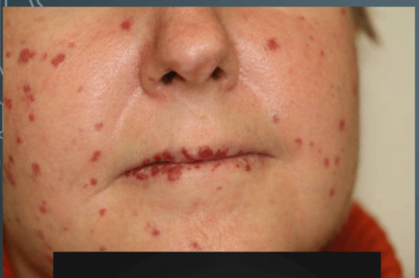
telangiectasias and arteriovenous malformations
400
Multifocal, white matter plaques that are localized to the periventricular area, as well as hypercellularity (mostly foamy macrophages) are sign's of a demyelinating disease process. The plaques can be visualized via this stain.
luxol fast blue
Note: Neurofilament stain used to visualize preserved axons
(Hallmark of MS: demyelination and preserved axons)
400
You are a pathologist doing an autopsy of a 48-yo female who died while recovering in the hospital from neurosurgery. During the recovery period, the patient was noted to have rapidly progressing dementia, ataxia, and myoclonus. Given this information, you expect to find this key finding in your autopsy of the patient's brain.
spongiform encephalopathy (vacuoles in the gray matter)
Note: This is CJD
400
A 9-yo male presents to clinic after experiencing recurrent seizures. His parents are concerned over weakness on the right side of his body and recent visual impairments of the left eye. You note an angiomatous nevus on the left upper face during the patient interview as well. You give the parents medication to manage the seizures and tell them that the nevus will need to be monitored and possibly managed by this procedure (2 possible answers).
resection or radiotherapy
Note: This is Sturge Weber Syndrome/Encephalotrigeminal Angiomatosis
400
A patient presents at clinic with bilateral hearing loss, tinnitus, and unsteadiness. The patient also notes a history of multiple benign tumors. Based on this presentation, you order a head CT to look for this type of tumor.
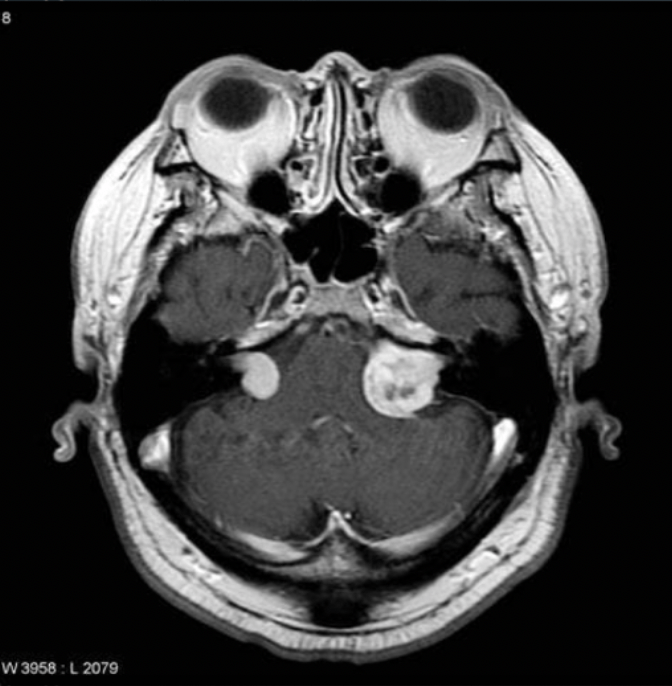
acoustic schwannoma/vestibular neuroma
Note: This is neurofibromatosis type 2, which also has meningiomas, ependymomas and gliomas as spinal and peripheral nerve tumors. Management includes surgical resection and hearing aid use.
500
A 24-yo male presented to clinic with headaches and lethargy. In his history, he notes that he had a URI one to two weeks prior. You give the patient medications for the headache and send them home. 1 week later, the patient goes into a coma and eventually dies. In the autopsy, histology shows perivascular inflammation, as well as myelin loss and preserved axons. The pathologist tells you these findings indicate this disease process.
Acute disseminated encephalomyelitis (ADEM)
500
Liquefactive necrosis surrounded by a rim of granulation and fibrous tissue. Sulfur granules present as well. This organism is suspected as the primary infectious agent.
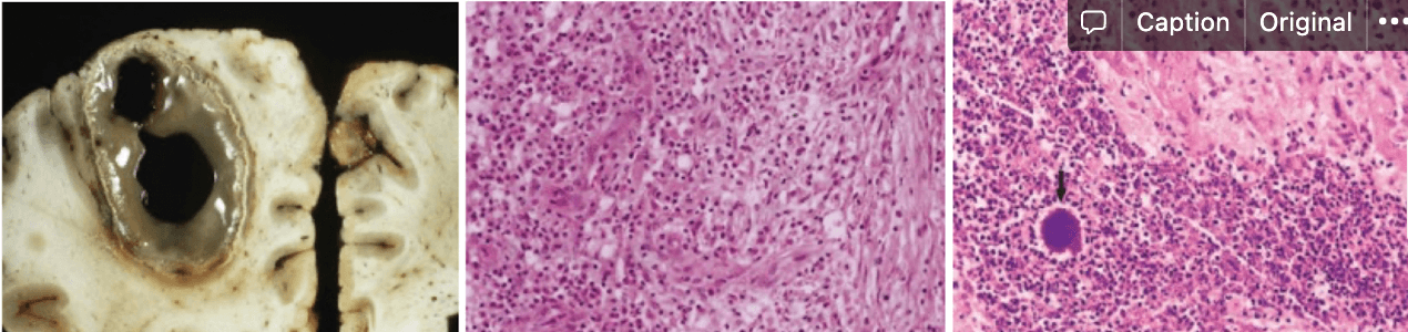
Actinomyces israelii
Note: This is a brain abscess
500
A 34-yo male presents with recurrent bloody noses and hemoptysis. Upon physical exam, you also note multiple telangiectasias. Based on the patient's condition, this could be included in their management plan. (3 possible answers)
Embolization, resective surgery and anti-angiogenesis drugs