Shoulder Complex
Elbow/Forearm
Wrist/Hand
Face/Neck
Misc.
100
A fracture at the surgical neck of the humerus leads to impaired shoulder abduction and loss of sensation over the lateral shoulder.
1. Which Nerve is involved?
2. Which muscles are supplied by this nerve?
1. The Axillary Nerve
2. Deltoid and Teres minor
100
What causes a hard end feel and limits extension ROM at the elbow?
hard end feel at the elbow that limits extension is caused by bony approximation—specifically the olecranon process of the ulna contacting the olecranon fossa of the humerus
100
1. The Hook of this carpal and the most "protruding" carpal bone on the palmar side are connected by a ligament that houses this nerve
2. What muscles are innervated by this Nerve?
1. The Ulnar Nerve
2. Flexor Carpi Ulnaris (FCU): Flexes and adducts the wrist.
- Flexor Digitorum Profundus (FDP - Medial/Ulnar Half): Flexes the distal interphalangeal joints of the ring and little fingers. [1, 2, 3]
Hand Muscles Supplied (Intrinsic)
Hypothenar Muscles (via deep branch):
- Abductor digiti minimi
- Flexor digiti minimi brevis
- Opponens digiti minimi
- Interossei Muscles (Deep branch):
- Four Dorsal Interossei: Abduct the fingers (DAB).
- Three Palmar Interossei: Adduct the fingers (PAD).
100
1. Name the 4 major muscles of mastication
2. Which is the primary and most powerful muscle of mastication?
3. Which passes under the zygomatic arch?
4. What is their innervation
1. M/L Pterygoid, Masseter, Temporalis
2. Masseter
3. Temporalis
4. Cranial N. 5(Trigeminal)
100
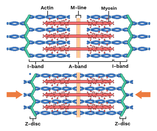
What is a sarcomere?
the fundamental, repeating contractile unit of striated muscle fibers (skeletal and cardiac), responsible for muscle contraction. Consists of actin and myosin filaments.
200
These are the osteokinematic motions needed at your right UE to pitch a baseball
Bonus: Give me a muscle that also does each!
Shoulder abduction
Shoulder ER --> IR
Elbow flexion
Forearm pronation
Wrist extension --> flexion
Finger Flexion
200
These are the osteokinematic motions needed at your right UE to punch straight forward
Bonus: Give me a muscle that also does each!
start in shoulder at rest (adducted/extended to 0 deg), elbow flexion, forearm neutral wrist neutral, finger flexion -->
Shoulder flexion, elbow extension, forearm pronation, wrist and fingers stay neutral
200
A patient presents with an injury to this tendon
 1. What is it?
2. What "region" of the body does this muscle create the border of ?
3. Which Carpal Bone can you palpate in this region?
1. Extensor Pollicis Longus
2. Anatomical Snuff Box
3. The Scaphoid
200
1. Which muscle is primarily responsible for elevating the corners of the mouth during smiling?
2. Which muscle makes the face of disgust?
1. zygomaticus major
2. levator labii superioris alaeque nasi.
Why this muscle?
- It raises the upper lip
- It flares the nostrils
- Creates that “wrinkled nose / sneer” look people make when something smells bad
200
The Axilla is _______ to the Cervical region (Provide anatomical descriptors)
Appropriate choices:
1. Inferior
2. Lateral
3. Caudal
4. Distal
300
A patient is recovering from shoulder surgery and is having difficulty with bringing their arm to the side. Every time that they do, the PTA can tell that the tendon of this muscle is hitting the acromion process and causing pain.
1. Which muscle is involved?
2. Which joint(s) is not moving properly?
The supraspinatus muscle. The tendon is getting impinged under the acromion because the Scapulothoracic joint is not _______ rotating. 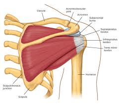
300
1. Which muscles are involved in supination?
2. Which of those is the most powerful when the elbow is flexed to 90 deg?
1. Supinator, Biceps Brachii, (and the brachioradialis assists)
2. The Biceps Brachii
300
Compression beneath the Flexor Retinaculum leads to sensory deficits in the Volar Side of the thumb, index, middle, and lateral half of the ring finger.
1. Which Nerve is Involved?
2. What is the area beneath the flexor retinaculum called? (The area that houses all the flexor tendons, nerves)
1. The Median Nerve
2. The Carpal Tunnel
300
Your patient is trying to look through a microscope while closing his right eye. What muscle is needed to do that?
Orbicularis oculi
300
1. What is myelin, what is it's purpose?
2. Each axon is surrounded by myelin, and then a connective tissue layer known as:____
1. Is an insulating layer, allowing electrical impulses to travel quickly, efficiently, and with high speed across nerve cells via ___________ conduction.
2. Endoneurium
400
Weak wrist extension with wrist drop indicates injury to this nerve.
Bonus: Injury to which part of the humerus could cause this?
The Radial Nerve
Bonus: Radial Groove of the Humerus
400
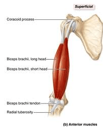
What muscle inserts here, and what nerve innervates it?
The Brachialis
The musculocutaneous N.
400
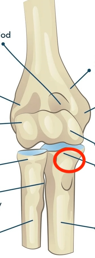
What is the structure marked #9?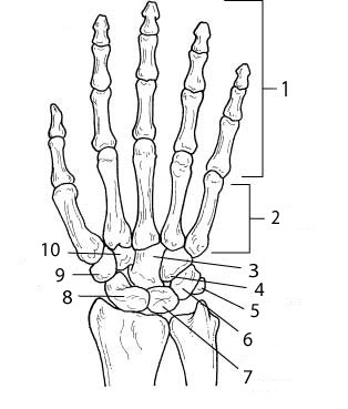
The Trapezium
400
1. What do the anterior scalenes do bilaterally?
2. What do all the scalenes ( anterior, middle, and posterior) do on one side (unilaterally)?
1. Cervical Flexion
2. Contralateral (opposite side) Cervical rotation and Ipsilateral (same side) lateral Cervical Flexion
400
What type or part of the bone consists of microscopic cylinders called osteons, which contain matrix and osteocytes deposited around central canals?
Compact Bone
500
This muscle originates on the infraglenoid tubercle, AND is an antagonist to the muscle that originates on the supraglenoid tubercle.
Triceps Brachii (Long head) and Biceps Brachii (Long head)
500
A patient falls and fractures their olecranon process. Which Action will be most affected and why?
Elbow extension; the Triceps insertion is on the olecranon
500
Name the muscle. How do you know which muscle this is?
This is the Flexor Carpi Radialis
- It is a flexor as it is originating on the medial epicondlye
-It only crosses the wrist joint ( not a finger muscle mover)
-Its inserting on the radial side
500
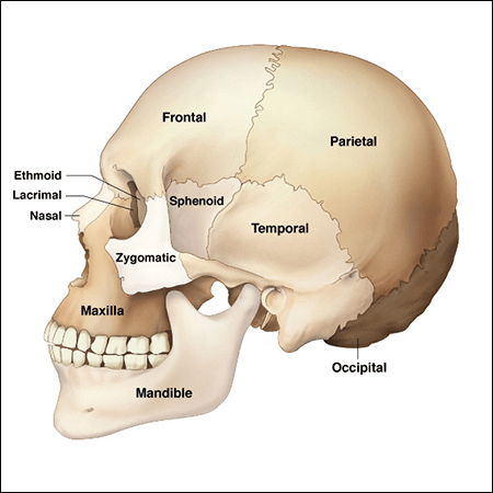
The mastoid process, the styloid process, the zygomatic arch, and mandibular fossa; are all parts of which bone?