1
2
3
4
5
100
One of every eight women will develop this disease.
→ breast cancer
100
Approximate incidence of breast cancer by location within the breast is highest in this quadrant.
→ upper lateral quadrant of the breast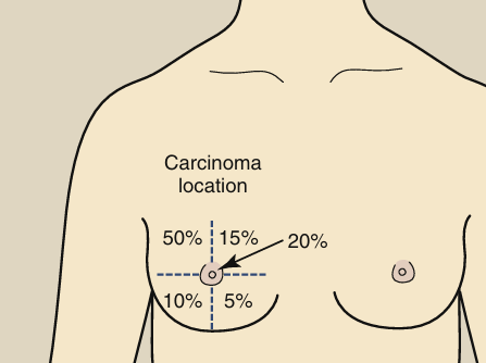
100
This is required in mammography to visualize tiny microcalcifications.
→ small focal spots
100
If a malignancy is present, it appears as a distortion of normal ductal and connective tissue patterns. Approximately [blank] of breast cancer is ductal.
→ 80%
100
This tool increases patient dose by double. Contrast can be improved further by reducing scatter with...
→ grids
this does not apply to virtual grids which are replacing radiographic grids in mammography.
200
Procedure: radiographic examination of the breast
What is mammography?
200
This technique factor must be kept low to maximize the photoelectric effect and thereby enhance differential absorption and improve image contrast.
→ kVp
As kVp is reduced, however, the penetrability of the x-ray beam is reduced, which in turn requires an increase in mAs.
200
This focal spot shape is preferred in mammography, although the other common shape is often used.
→ circular (preferred) + rectangular (common)
200
No filter element can absorb its own [blank].
→ anode target–characteristic radiation
200
The breast tissue most sensitive to cancer induction by radiation is [blank].
→ glandular tissue
300
More than 90% of patients are cured and survive because of [blank].
→ early mammographic diagnosis
300
This factor limits spatial resolution in digital mammography.
→ pixel size
PENGUIN: Spatial resolution in digital mammography is limited by pixel size.
300
Normal breasts consist of three principal tissues:
→ fibrous, glandular, and adipose (fat)
300
These two window materials are used in dedicated mammography x‑ray tubes to avoid absorbing low‑energy photons.
→ beryllium (Z = 4) window or a very thin borosilicate glass window.
300
These tiny calcium deposits are often the earliest sign of ductal carcinoma.
→ microcalcifications
400
The three major types of mammography.
→ screening, diagnostic, and baseline mammography
400
This is the reason digital mammography has superior contrast resolution.
→ postprocessing
PENGUIN: Digital mammography has superior contrast resolution because of postprocessing
400
At low x-ray energy, [blank] predominates over Compton scattering.
→ photoelectric absorption
400
The degree of absorption is determined by these factors.
→ the tissue mass density and the effective atomic number
X-ray absorption caused by differences in mass density is simply proportional to the mass density for both photoelectric effect and Compton scattering.
400
Prevents grid lines from being visible on the image.
→ high frequency and a moving grid (motion blurs out grid lines)
500
Performed on patients with symptoms or elevated risk factors.
→ DIAGNOSTIC mammography
500
This major study proved that contrast resolution is more important than spatial resolution for diagnostic efficacy in mammography.
→ DMIST
PENGUIN: DMIST showed without question that contrast resolution is more important than spatial resolution for diagnostic efficacy.
500
These filters must be used with a tungsten target in mammography to remove high‑energy bremsstrahlung photons.
→ molybdenum or rhodium filters
500
This grid type designed specifically for mammography involves copper strips, air interspace, 3.8:1 ratio, reduces scatter in two directions. Designed to maximize contrast while keeping dose reasonable.
→ High‑Transmission Cellular (HTC) Grid
500
Principal responsibility is to conduct an annual performance evaluation of the imaging system.
→ the medical physicist's
600
The first radiographic examination of the breasts and is usually obtained before age 40 years. Radiologists use it for comparison with future mammograms.
→ a BASELINE mammogram
600
These are the three target materials used in mammographic x‑ray tubes.
→ molybdenum (Mo), rhodium (Rh), and more rarely tungsten (W).
600
This is why mammography uses a filter made of the same element as the x‑ray tube target.
→ to transmit K‑characteristic x‑rays while suppressing unwanted bremsstrahlung x-rays.
600
Magnification mammography should not be used routinely because of these reasons.
→ Standard mammograms are sufficient for most patients.
→ Magnification doubles patient dose because: No grid is used. Increased OID increases scatter. System compensates with higher mAs.
→ Magnification is reserved for diagnostic evaluation
600
This component of the mammography system automatically selects exposure based on breast thickness and composition.
→ AEC (automatic exposure control) device.
700
Performed on asymptomatic women with the use of a two-view protocol, usually medial lateral oblique and cranial caudad, to detect an unsuspected cancer.
→ SCREENING MAMMOGRAPHY
700
These characteristic x‑rays are useless in mammography because their 12‑keV energy cannot penetrate the breast.
→ tungsten L‑shell x‑rays
700
This technique improves spatial resolution, contrast resolution, and reduces patient dose in mammography.
→ compression
- improves spatial resolution, contrast resolution, and reduces patient dose. reduces focal spot blur.
- uniform thickness of tissue.
700
Principal responsibility is supervision of the entire QA program.
→ the Radiologist's
700
Younger breasts are more difficult to image because of [blank].
→ dense, glandular tissue
800
These are the risk factors for developing breast cancer.

800
The three members of the mammography quality control team.
→ Mammographer, Medical Physicist, Radiologist
800
This mammography target material produces characteristic x‑rays around 17–19 keV, ideal for imaging fatty breasts.
→ molybdenum
800
This is the minimum total filtration required for mammography systems.
→ 0.5 mm aluminum equivalent.
800
Unlike most other modalities that has this device in front of the IR, this mammography component is placed behind the image receptor to minimize OID.
→ the AEC device.