Eye muscles
Middle/inner ear structures
Receptor cells and sensory tissues
Miscellaneous vision questions
Miscellaneous hearing & equilibrium questions
100
moves the eye medially
medial rectus
100
The name of this group of bones.
Ossicles.
100
name of the fluid found in the cochlear duct
endolymph
100
the 3 layers/tunics of the eye
the fibrous, vascular, and sensory tunics
100
name of the outer ear structure visible to the eye
auricle
200
moves the eye laterally
lateral rectus
200
branch of vestibulocochlear nerve that innervates the semicircle canal
vestibular nerve
200
absorbs pressure waves
round window
200
area with the highest concentration of cones
fovea centralis
200

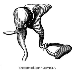
These ring-like structures on the left side of the image are partly responsible for our sense of equilibrium.
Semicircular canals.
300
moves the eye superiorly
superior rectus
300
3 main structures of the inner ear
semicircle canal, vestibule, and cochlea
300
The name of the "hairs" that bend to facilitate hearing and equilibrium.
Stereocilia.
300
The technical term for our "blind spot".
Optic disc.
300
name of the 3 ossicle bones
malleus, incus, stapes
400
This muscle contracts in response to bright light.
Radial muscle(s) of the iris.
400
the 3 ducts of the cochlea
Scala vestibuli, cochlear duct, scala tympani
400
connects stapes to cochlea
oval window
400
photoreceptors responsible for low light level vision
rods
400
structure that helps equalize the pressure between the outside of the ear and the middle ear
auditory tube
500
innervated by the trochlear nerve
superior oblique
500
smallest bone of the human body
stapes
500
membrane that the organs of corti sit on
basilar membrane
500
viscous fluid of the posterior cavity of eye
vitreous humor
500
boundary between outer and middle ear
tympanic membrane