eye muscles
middle/inner ear structures
receptor cells and sensory tissues
misc vision questions
misc hearing & equilibrium questions
100
main function of the fibrous tunic
give shape to the eye
100
The name of this group of bones.
ossicles
100
name of the center of the retina
macula lutea
100
two main structures of fibrous tunic
sclera + cornea
100
technical term for ear canal
external acoustic meatus
200
structure that contributes most to vascular tunic
choroid
200
technical name of visible ear
auricle
200
retina photoreceptors
rods + cones
200
focuses light on the retina
lens
200

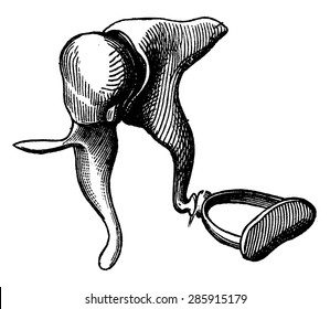
These ring-like structures on the left side of the image are partly responsible for our sense of equilibrium.
semicircular canals
300
structures connected by the fibers of the suspensory ligament
ciliary muscle + lens
300
boundaries of eardrum
outer ear + inner ear
300
photoreceptor responsible for dim light vision
rod
300
the technical term for our "blind spot"
optic disc
300
scala vestibuli fluid
perilymph
400
this muscle contracts in response to bright light
radial muscle(s) of the iris
400
the three ducts of the cochlea
1. cochlear
2. scala vestibuli
3. scala tempani
400
organ of corti membrane most associated with fluid waves
basilar
400
boundary of posterior chamber
iris + lens
400
organ of corti hair bending membrane
tectorial
500
the cranial nerve that controls the most extraocular muscles
oculomotor
500
the three main structures of the inner ear
1. cochlea
2. vestibule
3. semicircular canals
500
the name of the "hairs" that bend to facilitate hearing and equilibrium
stereocilia
500
orbital bone forming tear drainage system structure
lacrimal bone
500
order of ossicles from eardrum > inner ear
1. hammer (malleus)
2. anvil (incus)
3. stirrup (stapes)