Anatomy
Function
EKG
Circulation
Valves
100
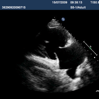
The valve in this image separates the left atrium and the left ventricle.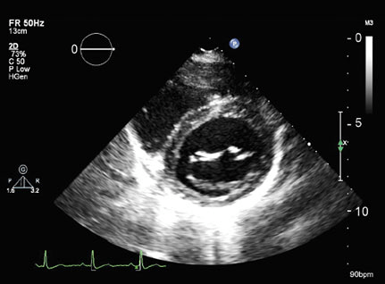
What is the mitral valve?
100
This is the major system that links body systems in order to fulfill many vital requirements including the transportation of blood.
What is the cardiovascular system?
100
This part of the conduction system receives electrical impulses from the atria and delays their transmission to the ventricles.
What is the AV node?
100
These are vessels that carry blood away from the heart.
What are arteries?
100
This valve can be visualized in the RVOT view.
What is the pulmonary valve (pulmonic valve)?
200
The name of this sonographic view.
What is the RVIT or Right Ventricular Inflow Tract?
200
This is the part of the cardiac cycle in which ventricular contraction occurs.
What is systole?
200
This is considered the pacemaker of the heart.
What is the SA node?
200
This chamber of the heart pumps deoxygenated blood to the lungs.
What is the right ventricle?
200
This valve has the largest area (7-9 cm2).
What is the tricuspid valve?
300
The anatomical structure that corresponds with letter H.
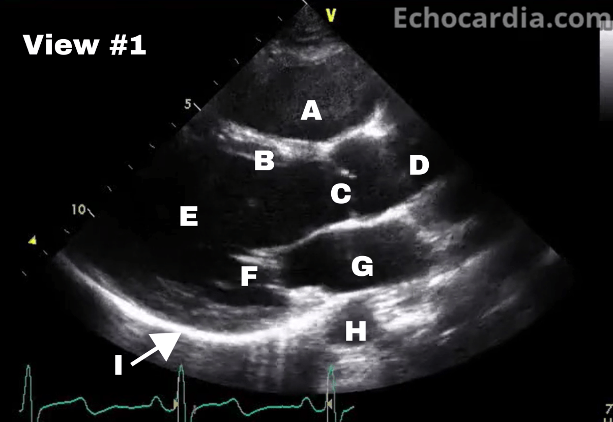
What is the descending aorta?
300
This is the pressure exerted by the blood on the walls of arteries within the systemic circulatory system.
What is blood pressure?
300
This "bundle" is located in the interventricular septum.
What is the Bundle of His?
300
This valve allows blood to exit the left side of the heart.
What is the aortic valve?
300
This valve has three cusps; the right coronary cusp, the left coronary cusp, and the non-coronary cusp
What is the aortic valve?
400
The anatomical structure identified in View #3, letter G.
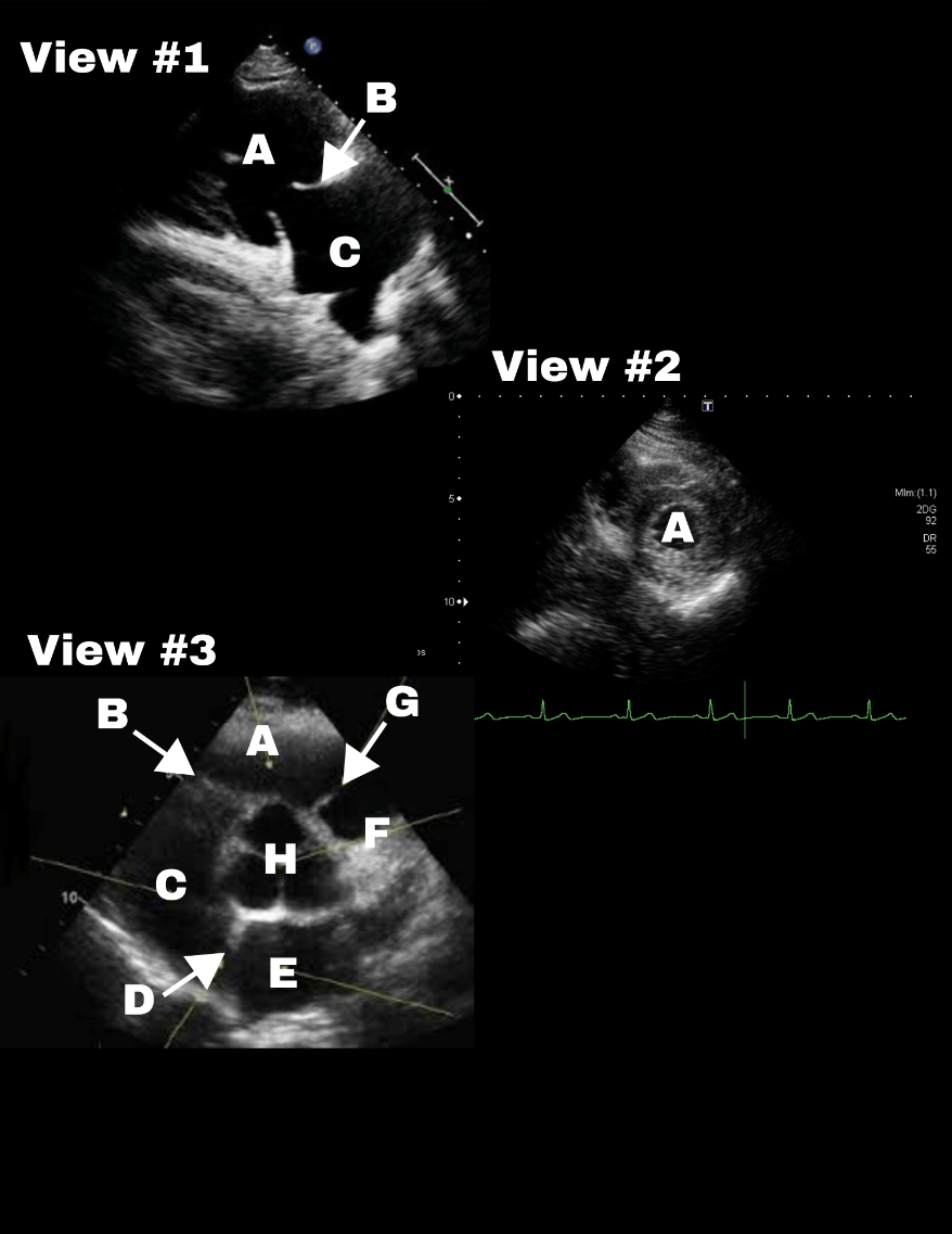
What is the pulmonary valve (or pulmonic valve)?
400
This would be considered a normal oxygen saturation level for the right side of the heart.
What is 75%
400
These fibers are located inside the ventricle walls and their primary function is to depolarize the ventricles.
What are the purkinje fibers?
400
While most veins transport deoxygenated blood, these veins are the only ones that carry oxygenated blood to the heart.
What are the pulmonary veins?
400
The only valve in the heart with two leaflets.
What is the mitral valve?
500
The anatomical structure identified in View #3, letter E.
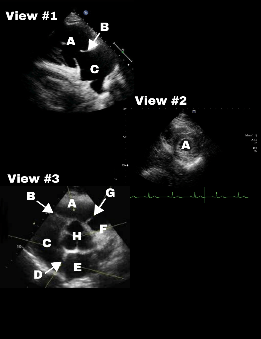
What is the left atrium?
500
Ventricular relaxation occurs during this phase of the cardiac cycle.
What is diastole?
500
This part of the conduction system is located at the bottom of the right atrium.
What is the AV node?
500
This is a fluid filled sac that protects the heart against infection and trauma.
What is the pericardium?
500
After the right ventricle contracts, blood passes through this valve to enter the lungs.
What is the pulmonary valve (or pulmonic valve)?