Head Bones
Head Muscles
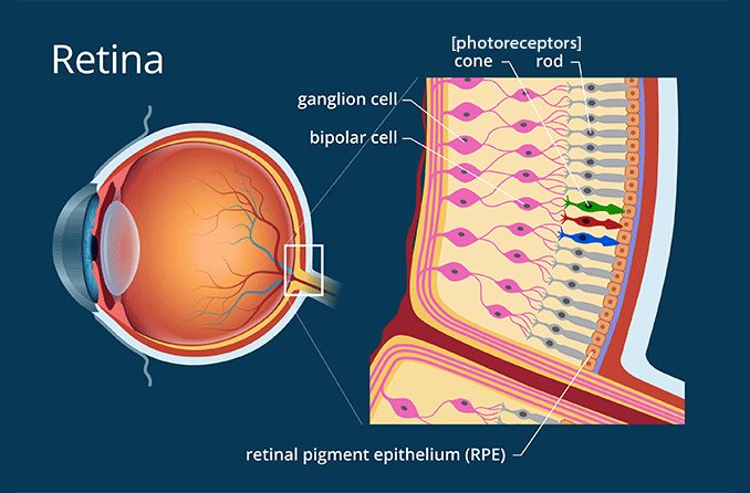
Special Senses
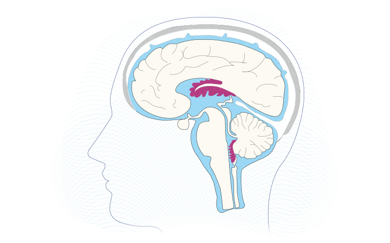
The Brain
Sensory and Motor
100
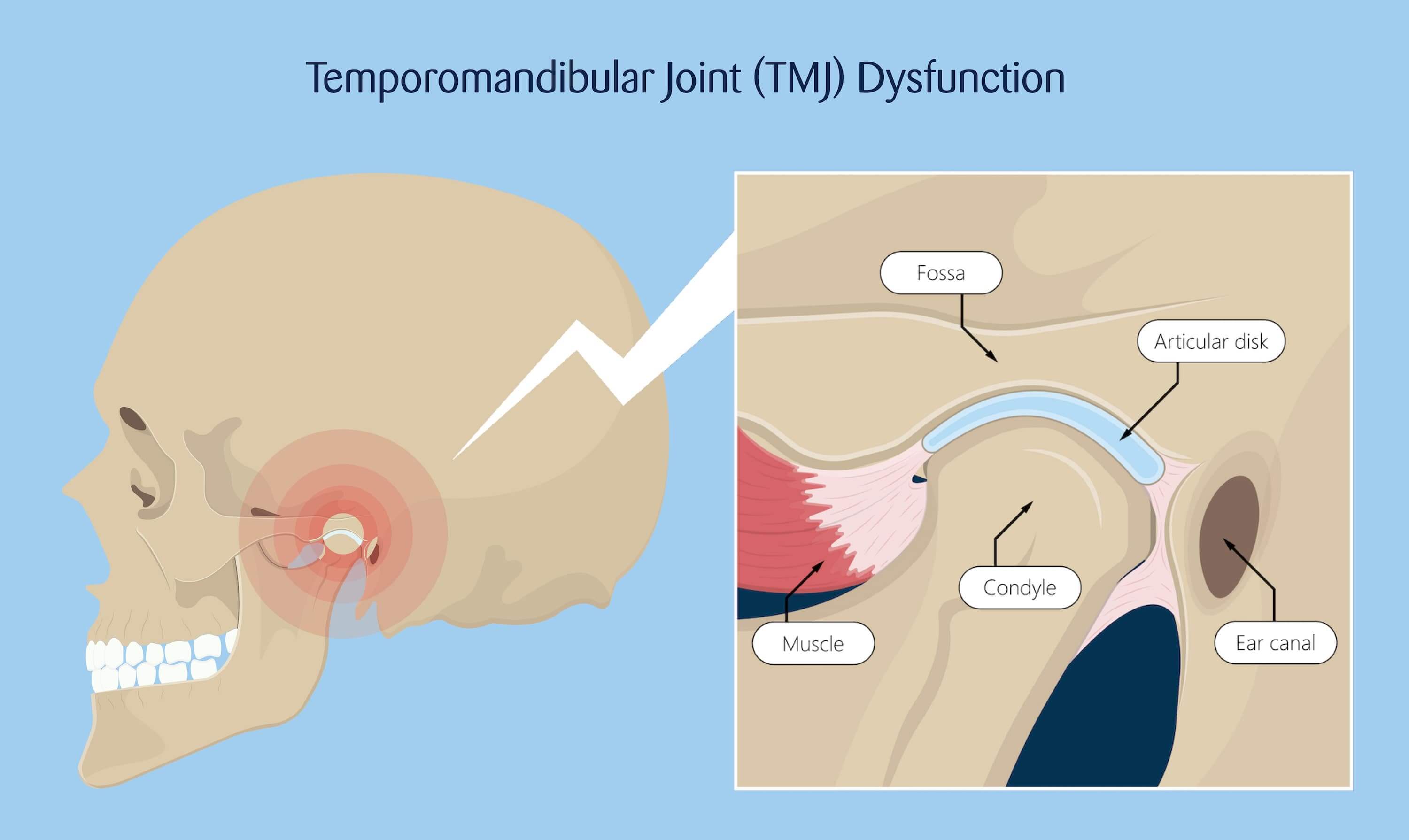
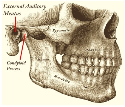
The mandibular condyle articulates with the temporal bone at this structure.
Mandibular fossa
100
The extrinsic eye muscles insert on this LAYER of the eye.
Fibrous Tunic

100
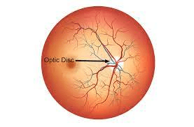
In this structure of the eye, there are no photoreceptors because the optic nerve must exit the eye.
Optic Disc or Blind Spot
100
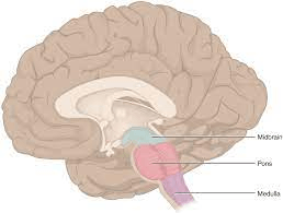
Autonomic centers that control blood pressure and heart rate are in this major brain region.
Medulla oblongata
100
Within the spinal cord, this tract of myelinated axons carries information about pain, temperature, and crude touch/pressure to the brain.
Spinothalamic Pathway
200
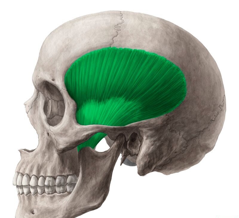
The temporalis muscle passes deep to this structure (made up of 2 bones).
Zygomatic arch
200
What raises her eyebrows?

Occipitofrontalis
200
These photoreceptors require abundant light to be stimulated, which allows them to be used only in light environments (daytime).
Cones
200
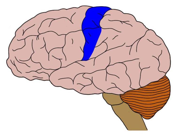
All sensory information from the body is routed to the Somatosensory Cortex, which is located on this.
(Structure AND Lobe)
Postcentral Gyrus of Parietal Lobe
200
The term for a neuron crossing the midline of the body (as it would have to do in order to inform the contralateral cerebral hemisphere of sensory stimuli).
Decussation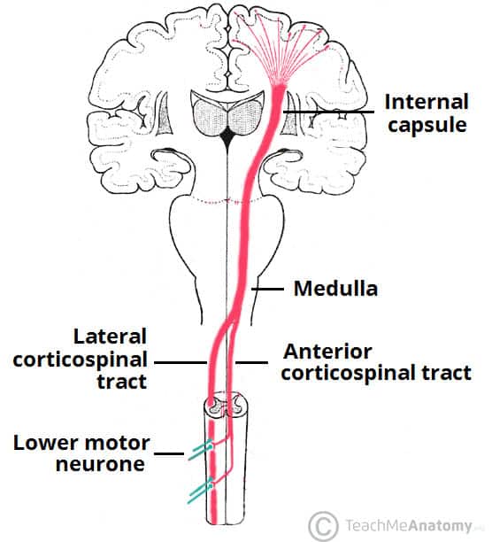
300
The hard palate (roof of the mouth) is formed by these boneS.
Palatine and Maxilla bones

300
Name an extrinsic eye muscle used to look at the bridge of the nose.
Medial Rectus
(Inferior Rectus looks at the tip of the nose)
300
When sound waves bend these, the hair cells depolarize, release a neurotransmitter, and trigger an action potential.
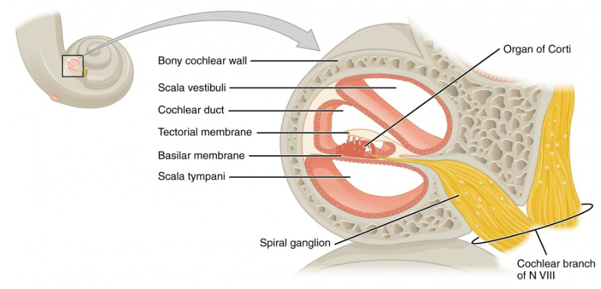 Stereocilia
300
Within each brain ventricle, this filters the blood to generate new Cerebrospinal Fluid.
Choroid Plexus
300
Receptors that monitor the position of joints are collectively called this.
Proprioceptors

400
The ear canal passes through this structure.
External Acoustic (Auditory) Meatus
400
These muscles originate on the sphenoid bone and can move the mandible left and right.
Pterygoids
![]()
400
When this muscle contracts, it makes the lens thicker, which refracts/bends the light from a nearby object more.
Ciliary muscle/body
400
This secondary brain vesicle is retained in the adult brain, and contains the thalamus.
Diencephalon
![]()
400
Within the skin, these detect very fine touch with fine spatial resolution.
Merkel Cell Fibers