I am so Humerus it hurts
Let's get Organized
IF I only hand a brain
I Abs-olutely know my anatomy
You got lot of nerve
OK, Twist my arm
Shoot from the hip
Slap my knee and stomp my foot
I'm so handy
100
This structure is the only bony attachment of the shoulder girdle to the trunk
Sternoclavicular joint
100
This bean-shaped organ filters blood and removes waste
Kidneys
100
This is the largest part of the brain and contains white and gray matter and gyri and sulci
Cerebrum
100
Name of all abdominals in the abdominal group
Rectus Abdominis, External Oblique, Internal Oblique, Transverse Abdominals
100
C-6 Dermatome
Thumb/Index

100
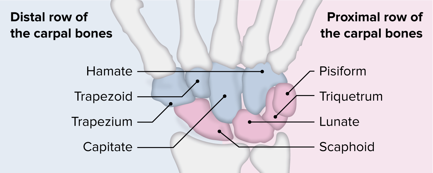
The wrist proximal carpal row
Scaphoid, lunate, triquetrum, pisiform
100
What two structures make up the Coxal joint?
Acetabulum, Femoral Head
100
This bony structure is considered a sesamoid bone
Patella
100
Name the small finger muscle Dr. Evil uses to plot world domination

Extensor digiti minimi
200
This ligamentous structure supports and reinforces the Glenohumeral joint and works in conjunction with the rotator cuff muscles to provide shoulder support and stability
Labrum
200
This organ produces and releases insulin through the endocrine gland
Pancreas
200
Name the 3 areas of the brainstem
midbrain, pons, and medulla
200
This intercostal assists with inhalation
External
200
Movement associated with brachial plexus nerve roots
Myotome
200
This muscle divides the flexors from the extensors
Brachioradialis
200
This is the longest muscle in the body and flexes, laterally rotates and abducts the hip. It also flexes and medially rotates the knee
Sartorius
200
Name the 3 Cuneiform bones
Medial, Middle (intermediate), and Lateral
200
What 2 tendons are in the 1st dorsal compartment?

Abductor Pollicis Longus and Extensor Pollicis Brevis
300
Pectoralis Major Innervation
Lateral and Medial Pectoral N.
300
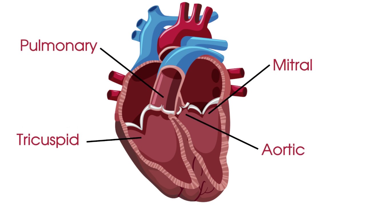
Name the 4 valves of the heart
Tricuspid, Pulmonary, Mitral, Aortic
300
this area of the brain is responsible for balance, movement, eye movement, and higher level thinking and action
cerebellum
300
This intercostal assists with exhalation
Internal
300
What 2 muscular structures are common for causing compression on the brachial plexus?
middle and anterior scalene, and under the pectoralis minor
300
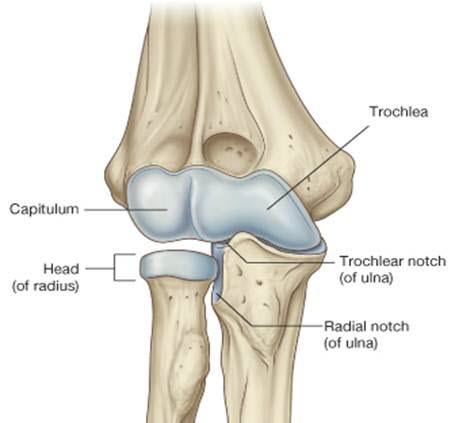
This structure articulates with the radial notch of the ulna
Radial Head
300
The hip adductor group (adductor magnus, adductor longus, adductor brevis, pectineus, and gracilis) are innervated by this nerve
Obturator
300
This muscle originates on the lateral condyle of the tibia and inserts on the middle and distal phalanges of the 2nd to 5th toes. It extends the toes and dorsiflexes the ankle
Extensor digitorum longus
300
Arches of the Hand
Distal transverse
Proximal Transverse
Longitudinal
400
The glenohumeral joint arthokinematics result in slide and glide in opposite directions
Convex on Concave
400
This organ system has a central and peripheral system. It communicates pain, temperature, and sensation
Nervous system
400
this cerebral lobe is responsible for vision
Occipital Lobe
400
Name the 3 distinct areas of the sternum
Manubrium, the body of the sternum, and Xiphoid Process
400
This cord gives rise to the upper and lower subscapular and thoracodorsal nerve
Posterior
400
What nerve innervates the Brachialis?
Musculocutaneous n.
400
This muscle that attaches at the top of your iliotibial (IT) band and is a vital muscle that helps stabilize the hip and knee. It assists with internal rotation, flexion, and abduction of the hip.
Tensor Fasciae Latae (TFL)
400
What 3 major structures travel in the inguinal triangle?
Femoral nerve, Femoral artery, Femoral vein
400
The abductor pollicis brevis, flexor pollicis brevis, and opponens pollicis, make up this muscle group
Thenar muscles
500
Ms. Smith arrived at the clinic with complaints of right shoulder pain. She followed up with the MD and she was diagnosed with right shoulder instability. You plan to begin strengthening to improve the dynamic stabilization of the Glenohumeral joint. What muscles do you plan to strengthen?
Supraspinatus, Infraspinatus, Teres Minor, Subscapularis (Rotator cuff), Deltoid, and Long head of the biceps
500
The heat and lungs work together to carry oxygen, nutrients, and hormones to the cells; and remove waste products (CO2). What is the name of this system?
Circulatory system
500
James was diagnosed with Parkinson's Disease and is currently experiencing poor movement and coordination making using utensils while eating difficult. What area of the brainstem is responsible for movement and coordination?
Midbrain
500
This muscular structure and its innervation that draws down the central tendon and increases the volume of the thoracic cavity during inhalation
Diaphragm/Phrenic N.
500
While leaning on his elbow, Mr. Jones discovered his small finger and 1/2 of his ring finger on the pinky side began going numb. What nerve did Mr. Jones just discover?
Ulnar N.
500
Mrs. Smith was referred for occupational therapy due to right hand numbness/tingling and decreased fine motor function. During your examination, you note loss of sensation in the thumb, index, and radial side of the ring finger. MMT noted weakness in the abductor pollicis brevis. What nerve is most likely involved and what structure is likely causing compression?
median and transverse carpal ligament
500
This muscle group extends the knee and flexes the hip. They are also innervated by the Femoral nerve. Name the group and all of the muscles associated with this group.
Quadriceps Femoris Group:
Rectus Femoris
Vastus Medialis
Vastus Lateralis
Vastus Intermedius
500
Johnny was playing football when he hurt his knee. The MD performed an anterior draw test and noted that Johnny had torn a ligament and would need surgery. What knee ligament did he most likely tear?
Anterior Cruciate Ligament (ACL)
500
Mr. James sustained a laceration over the dorsum of his index finger and he is now unable to extend the PIP joint. He followed up with the MD and had a surgical repair of the "Dorsal Apparatus." What muscles make up the dorsal apparatus?
The Extensor digitorum communis, lumbricals, and interossei