Vitiligo?
Buzz Words
Syndromes
Kodachromes
Random
100
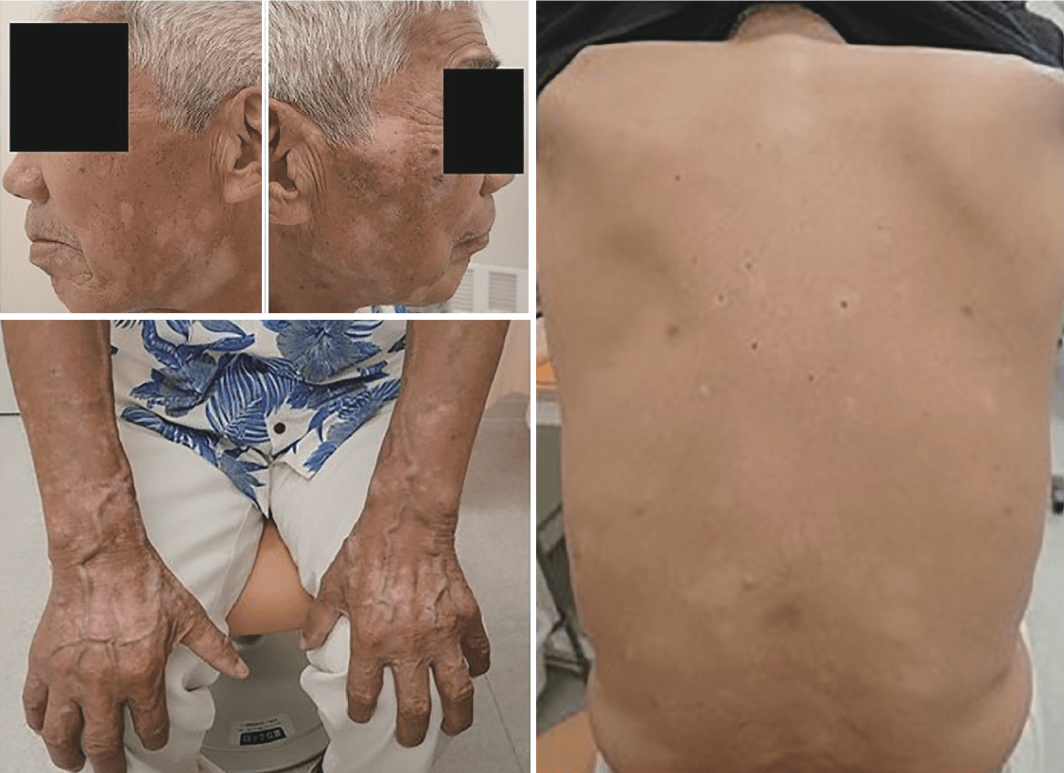
Hint:
Started on a medication for melanoma.
Medication-induced leukoderma
2/2 to Nivolumab (PD-1 inhibitor)
EGFR inhibitors (gefitinib), Bcr-abl inhibitors (imatinib and dasatinib), VEGFR inhibitors (pazopanib, sunitinib), BRAF inhibitors (vemurafenib), and interleukin-2 (IL-2) have all been reported to cause hypopigmentation
Tranexamic Acid - Decreases the production of prostaglandins, which are the precursor of tyrosine.
100
Vitiligo is associated most commonly with this autoimmune process...
Auto-immune thyroiditis (Hypothyroid)
15% of adults
5-10% of children
100
Most common inherited disorder that leads to hypomelanosis?
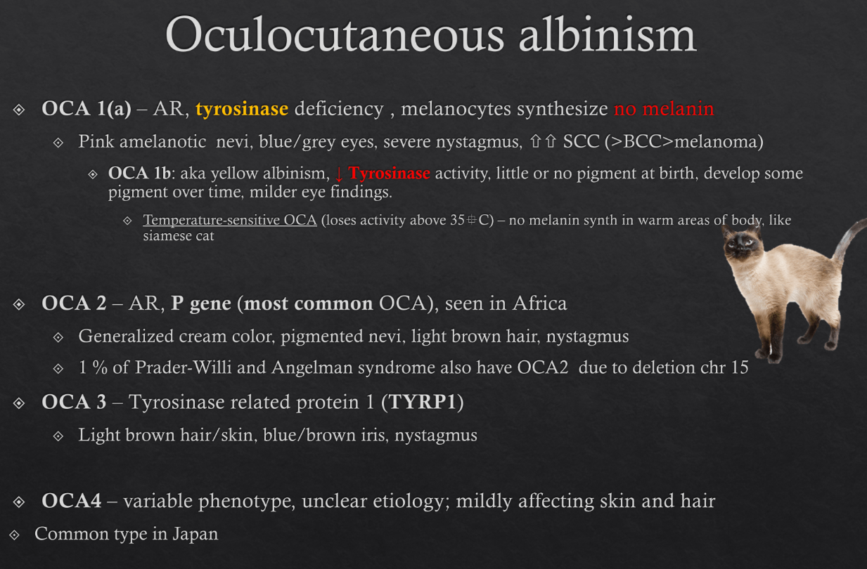
Oculocutaneous Albinism (OCA)
- group of genetic disorders
- diffuse pigmentary dilution from partial or total absence of melanin pigment in melanocytes of skin, hair follicles, eyes
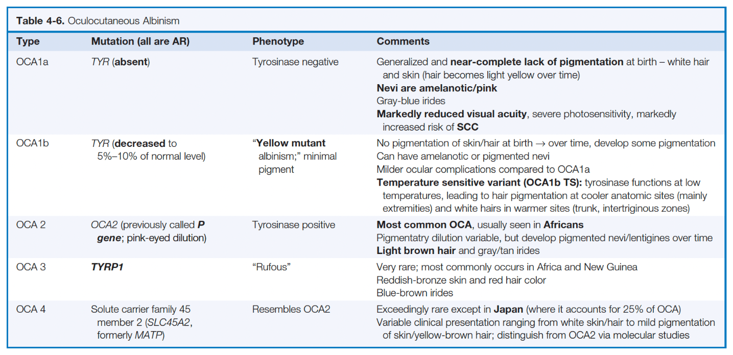
8 types identified based on molecular studies
- tyrosinase activity absent in OCA1A and reduced in OCA1B. 400 distinct mutations in tyrosinase gene (TYR) identified in OCA1
- OCA2 mutation (previously P) encodes the pink-eye dilution gene- influences tyrosinase activity
- OCA3 is tyrosinase-related protein 1 (TYRP1) gene mutation- stabilizes tyrosinase
- OCA4 is SLC45A2 mutation (encodes solute carrier family 45 member 2) which maintains deacidification in later stage melanosomes which is required for tyrosinase activity -- more common in patients from Japan, China, India
- OCA5-8 Pakistani family first, a few other groups since
Clinical:
OCA1A is classic "tyrosinase negative" with extreme UV sensitivity, white hair (can become yellow from UV) and skin, blue-gray eyes at birth
Both OCA1B and OCA2 have variable pigmentation "tyrosinase positive"
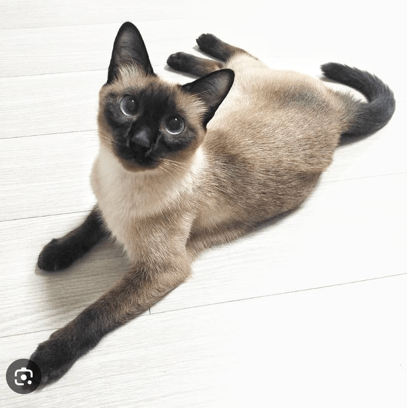
OCA1B can have "temperature sensitive" phenotype where tyrosinase loses activity above 35C-- melanin synthesis does not occur in warmer areas of the body-- Siamese cats have temp sensitive tyrosinase
Histopathology for OCA:
normal number of melanocytes, but reduced melanin content in the epidermis
Treatment - sun protection
100
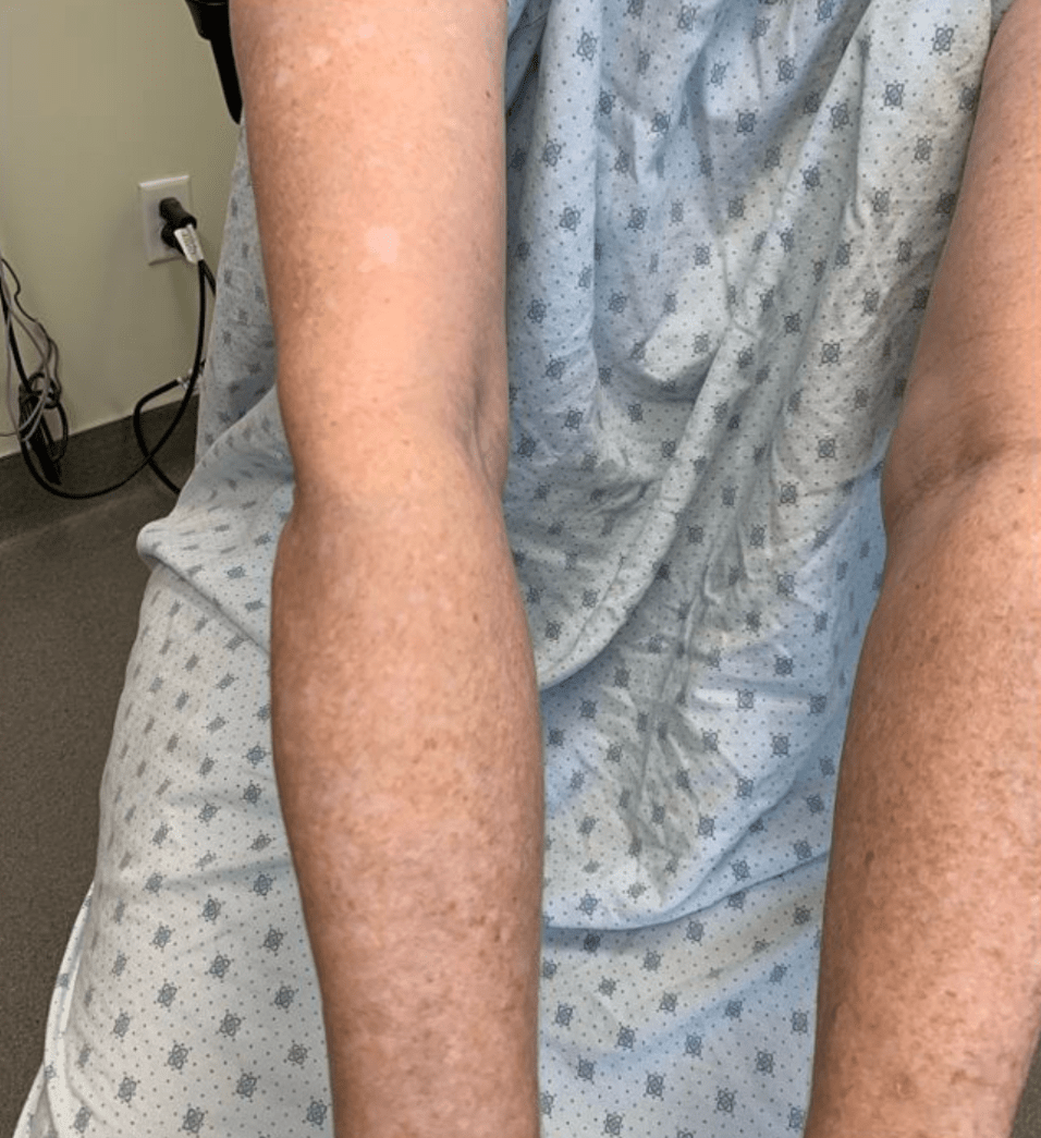
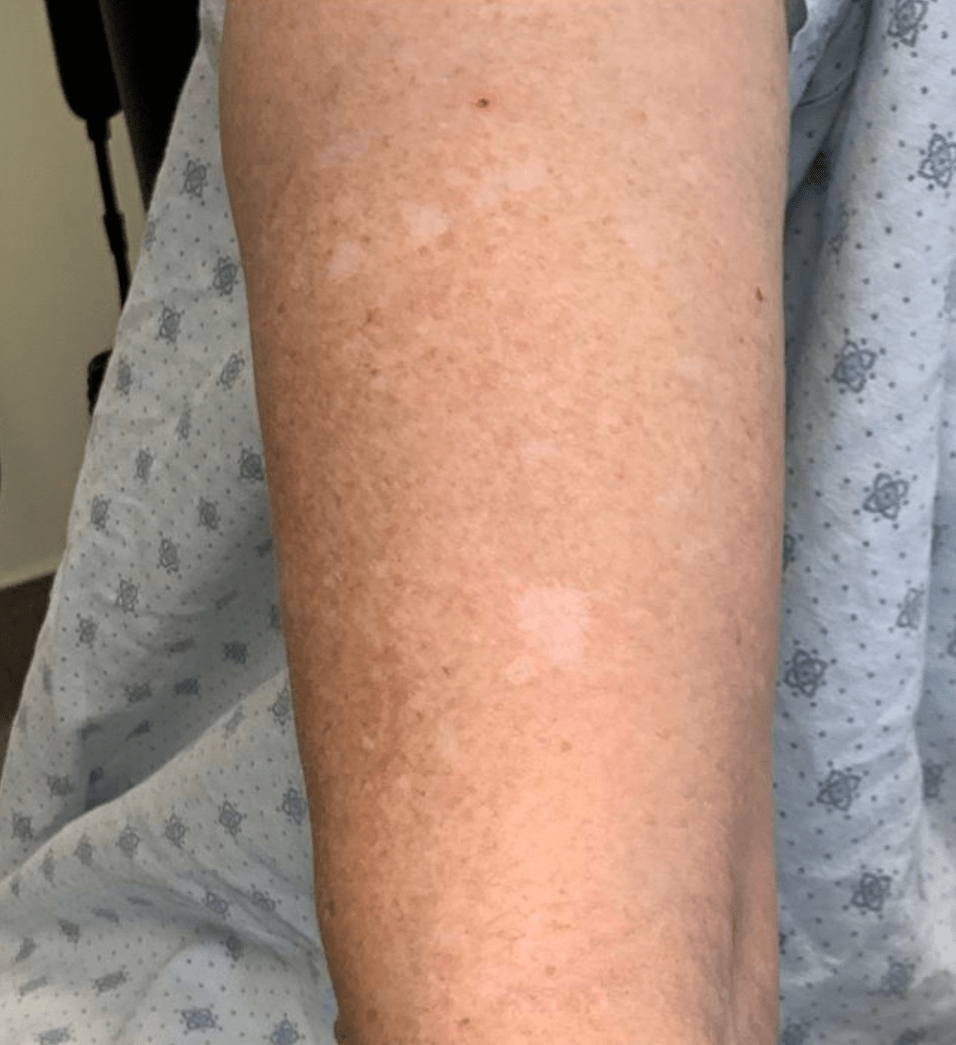
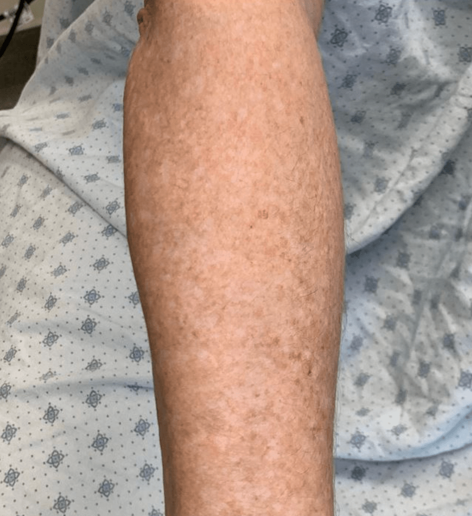

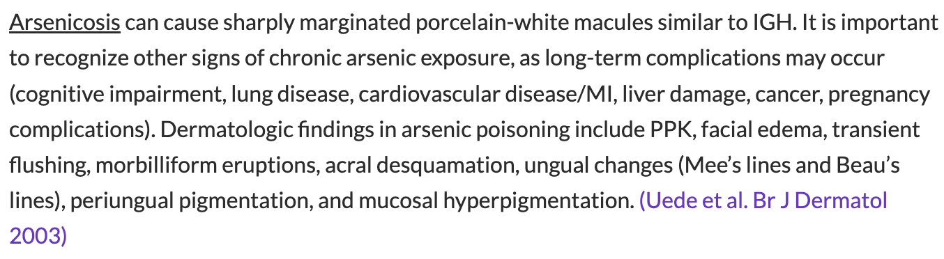
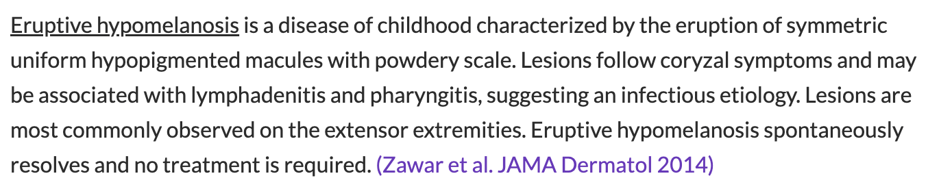
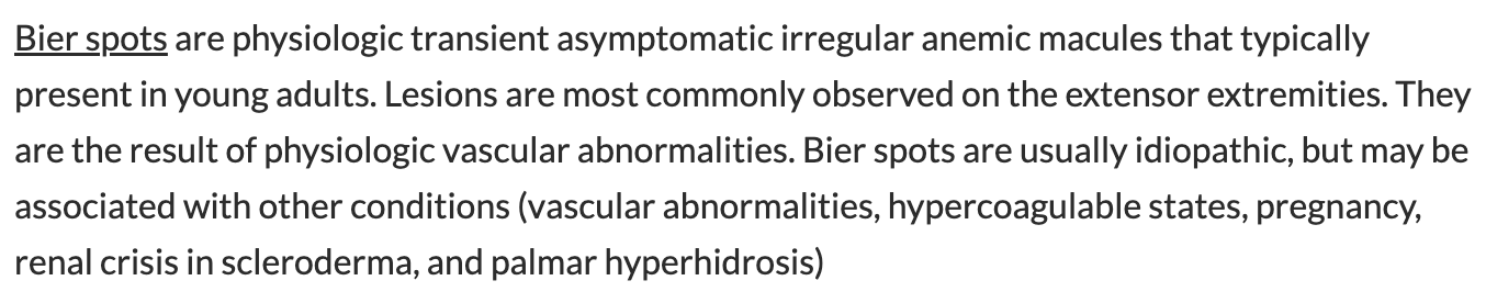
A. Arsenicosis
B. Eruptive hypomelanosis
C. Bier Spots
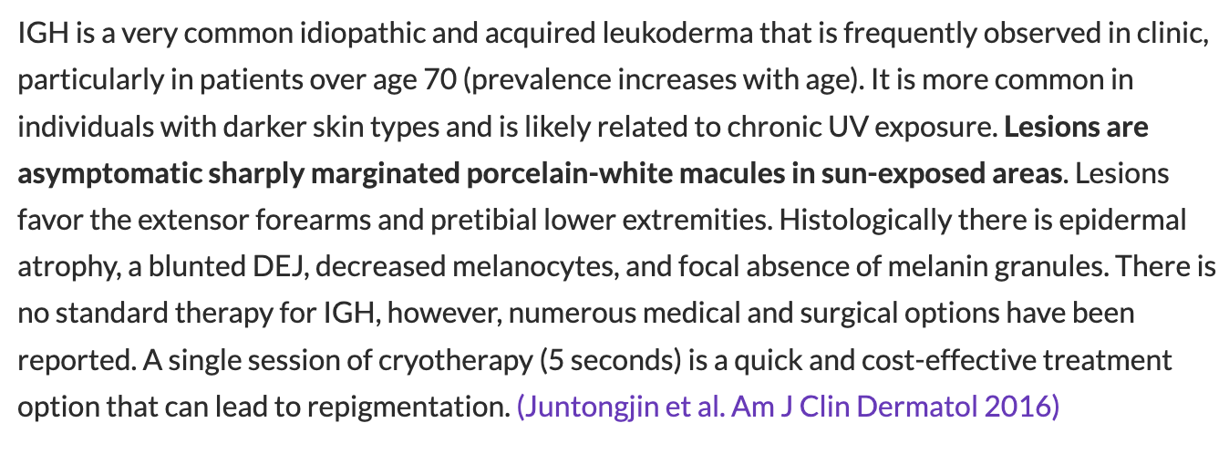
D. Idiopathic Guttate Hypomelanosis
D. Idiopathic Guttate Hypomelanosis
100
Name the below phenomenon.
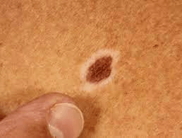
Halo Nevus
(1) appearance of the halo; (2) loss of pigment within the central nevus; (3) disappearance of the nevus; and (4) disappearance of the halo.
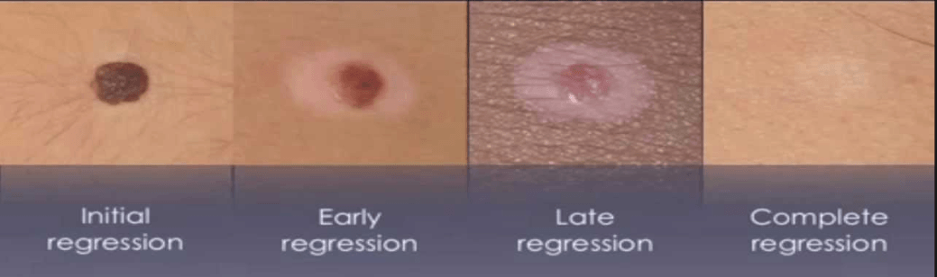
Individuals > 40 yo with new-onset halo nevi should be examined carefully for melanoma (ocular and cutaneous).
SIDE NOTE: At least three types of leukoderma are associated with melanoma:
(1) hypomelanosis within tumors (primary or metastatic) due to regression
(2) amelanosis around tumors (halo phenomenon)
(3) vitiligo-like depigmentation remote from the tumors.
200
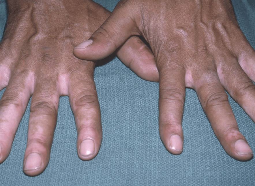
Chemical-induced leukoderma (contact vitiligo or chemical vitiligo)
Phenol-induced agent. Can be found in some insecticides, paint-strippers etc. Melanocytotoxic effect.
Other causative toxins include mercurials, arsenics, sulfhydryls, azo dyes, and paraphenylenediamine (PPD).
In some patients with chemical leukoderma, the depigmentation can spread from the site of contact with the offending chemical or even develop at distant sites, sometimes when patients are no longer exposed to the offending chemical It is difficult or impossible to determine whether these patients have a true diffuse chemical leukoderma or generalized vitiligo that was triggered by the contact with a toxic chemical
200
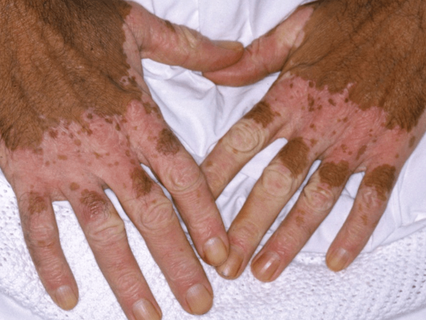
Salt and pepper dyspigmentation often relates to this condition
What is systemic sclerosus.
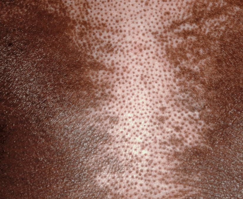
COMPARE TO:
leopard skin from onchocerciasis- over the shin classically 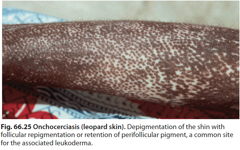
200
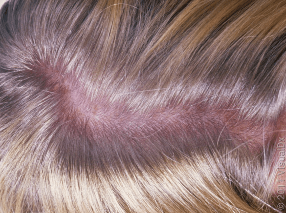
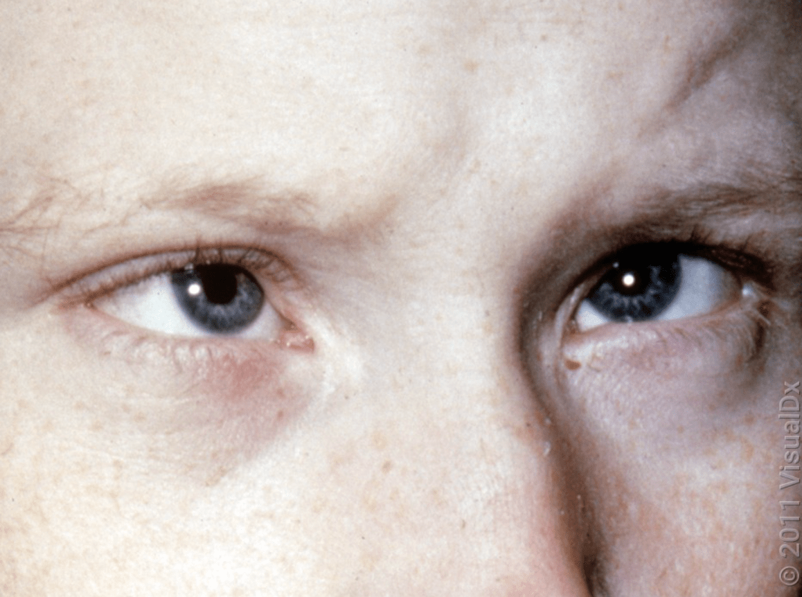
Hint:
Musty body odor
Phenylketonuria (PKU), disorder of phenylalanine metabolism.
The untreated state is characterized by a constellation of functional and physical manifestations including intellectual disability, a characteristic bodily appearance, and neurological impairments. Intellectual disability worsens from early childhood to adolescence during brain maturation. Patients develop the classic triad of blond hair, blue eyes, and light skin usually by the second year of life.
Primary treatment is dietary modification (low phenylalanine)
200
name the condition and three treatment options
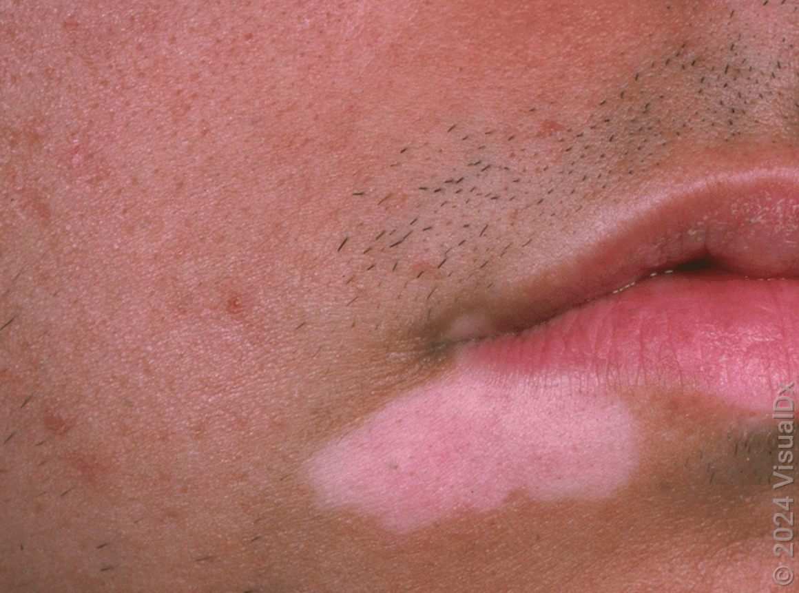
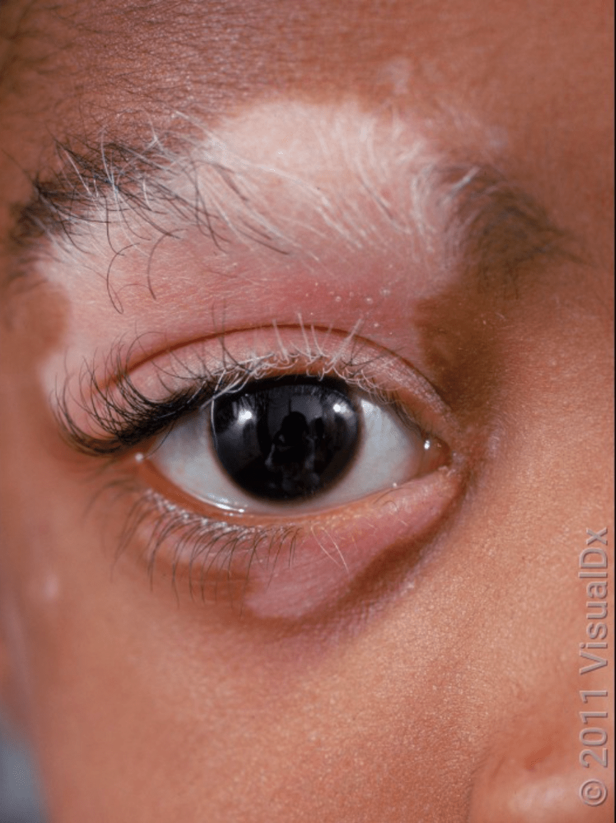
What is Vitiligo
- circumscribed depigmented macules and patches 2/2 loss of epidermal melanocytes
- 0.5%- 1% of population worldwide
- Can appear any time from shortly after birth to late adulthood (average onset ~30 years old)
- pathogenesis: genetic (multiple susceptibility loci and candidate genes- involved in melanogenesis, immune regulation, apoptosis), intrinsic abnormalities of melanocytes making them more susceptible to oxidative stress, destruction of melanocytes from immune activation
- Clinical: totally amelanotic patches (most common) with discrete borders, can be hypomelanotic at their onset or if actively spreading, can be blue color from developing in areas of postinflammatory dermal pigmentation. Enlarge centrifugally. Usually asymptomatic, can be pruritic especially if active. Common areas of involvement (places that are normally more hyperpigmented): face (around eyes, mouth), dorsal hands, elbows, knees, digits, flexor wrists, dorsal ankles, shins, nipples, axillae, umbilicus, sacral, inguinal, anogenital. Koebnerization. Leukotrichia (10-60%) from follicular melanocytes being affected.
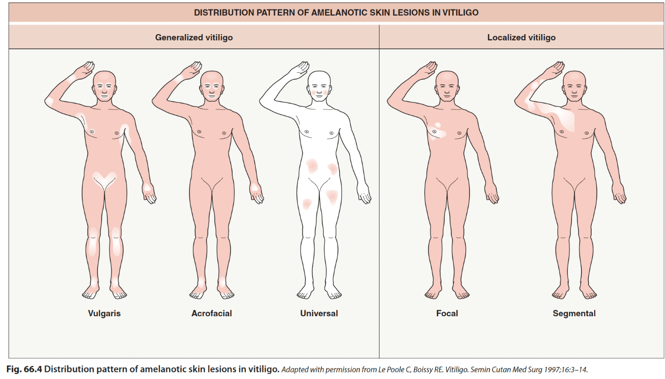
>90% are vulgaris or acrofacial variants. Segmental more common in children (15-30% pediatric vitiligo)
- Course: hypomelanotic lesions with poorly defined borders and confetti-like lesions are a/w progression. Generalized vitiligo usually slow spread, segmental vitiligo usually reaches full extent in 1-2 years and restricted to initial segmental area. Halo nevi or leukotrichia increases likelihood of progressing from segmental to mixed.
- Can have ocular involvement: Vogt-Koyanagi-Harada syndrome (uveitis, aseptic meningitis, otic involvement, vitiligo) and Alezzandrini syndrome (unilateral whitening of sclap hair, eyebrows, eyelashes with ipsilateral facial skin depigmentation and visual changes)
- most patients otherwise healthy, but generalized vitiligo has some associated autoimmune diseases (up to 20%)- autoimmune thyroid (15%), alopecia areata (2-4%), pernicious anemia, T1DM, myansthenia gravis, connective tissue disease (eg lupus, RA, Sjogren)
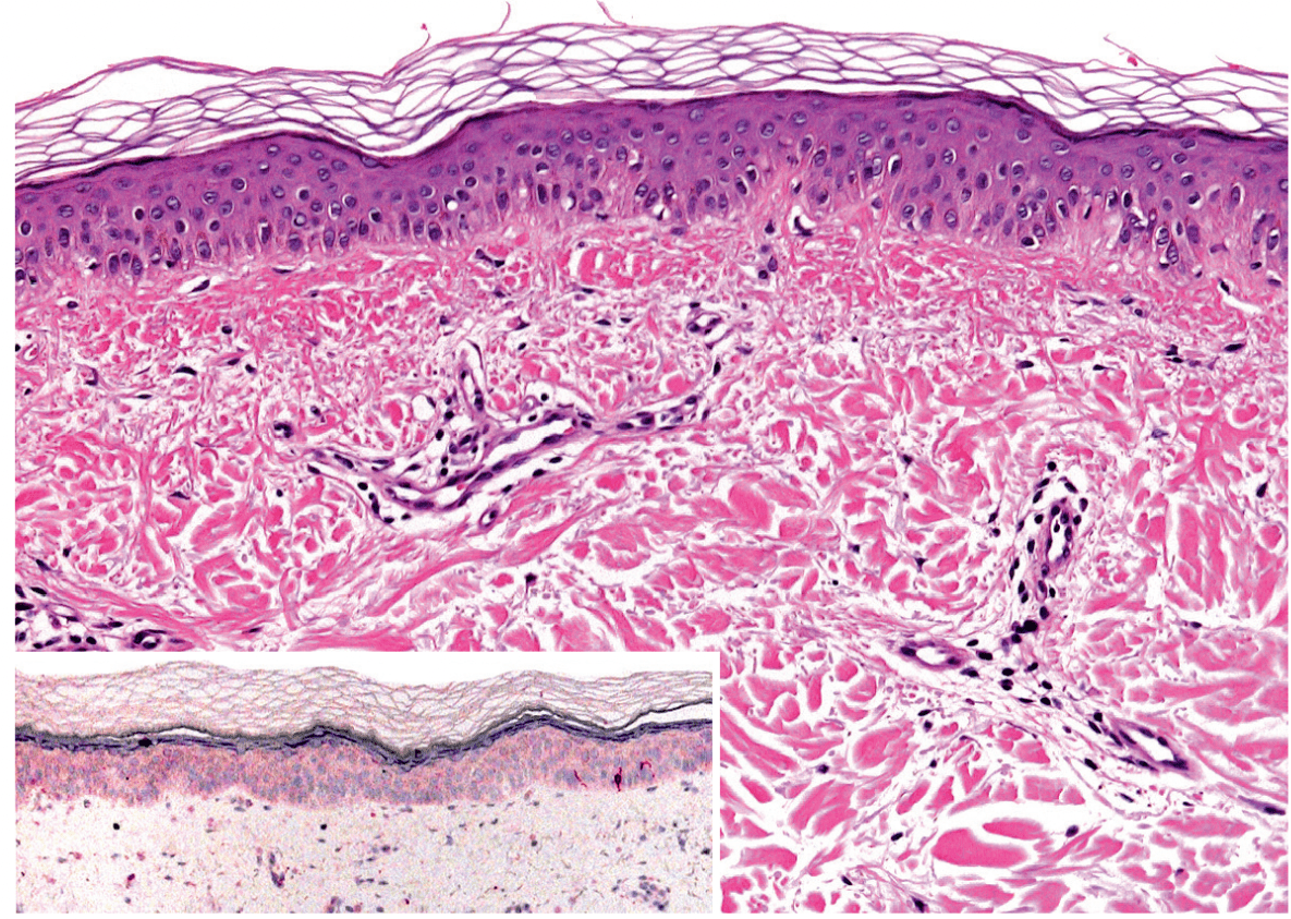
complete absence of melanocytes in basal layer highlighed with Melan-A (MART1) stain
Treatment;
- repigmentation requires repopulation of epidermal melanocyts from neighboring pigmented hair follicles, surrounding uninvolved epidermis, or lesional interfollicular melanocyte precursors.
- generally 6 months needed to determine if treatment is effective
- face, neck, mid-extremities, and trunk have best response. Distal extremities and lips most resistant.
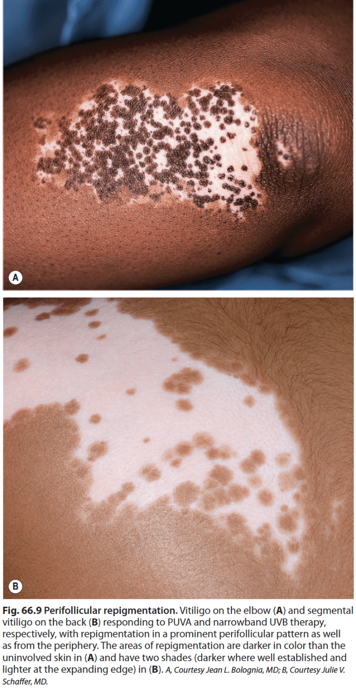
- 40% chance of recurrent depigmentation
- Topical steroids- superpotent or high potentency in 6-8 week cycles or twice weekly, alternating with tacrolimus, ruxolitinib, or weaker steroid)
- Topical calcineurin inhibitor- facial sites, can be synergistic with UVB or excimer, can use for maintenance
- Topical JAKi- ruxolitinib 1.5% cream, FDA approved for non-segmental vitiligo ≤10% BSA in ≥12 yo, acne at site of application most common AE
- nbUVB 2-3x/week (311 nm)- first line for adults and children ≥6 yo with generalized vitiligo (esp ≥15% BSA or cosmetically sensitive area)
- PUVA- oral PUVA no longer recommended (greater efficacy and lack of SE with nbUVB), topical PUVA for localized lesions
- Excimer laser and lamp (308 nm) for segmental vitiligo or <10% BSA, compared to nbUVB efficacy the same or greater, shorter duration of treatment, avoids darkening of unaffected skin. Can enhance with TCI, TCS, topical JAK, or PO steroid pulse.
- systemic immunomodulators- MTX or JAKi (need more studies)
- surgical grafting
- depigmentation if widespread- lifelong strict photoprotection, 20% monobenzyl ether of hydroquinone (MBEH) 1-3 mo for response to start, apply for 9-12 mo or longer
Steroids for acute vitiligo:
- mini-pulses (RCT for efficacy and safety are lacking)
Oral dexamethasone 2.5 mg (16.7 mg pred equiv) on two consecutive days weekly for 3-6 months. halts disease expansion in 90%, does not induce significant repigmentation usually, relapse often occurs
Alternatives, 40 mg IM Kenalog or Oral Pred ~10-20 mg daily for 2 weeks. Repeat monthly PRN.
200
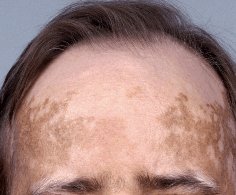
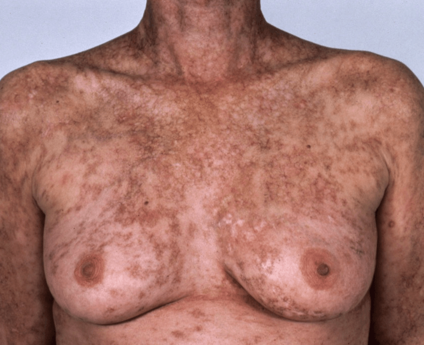
Hint: recent worsening of skin findings with prior rash, associated with diarrhea and elevated bilirubin.
Graft versus host disease
Depigmented variant:
May be punctate or confetti-like; generally considered to be a postinflammatory phenomenon; may be associated with dermal fibrosis of varying depths and appear “leopard-like”; spontaneous depigmentation suggestive of vitiligo may be prominent
Grading of acute GVHD:
- SKIN (rash BSA)
- LIVER (Bilirubin)
- GUT (Diarrhea)
300
these spots are a major criteria for which condition?
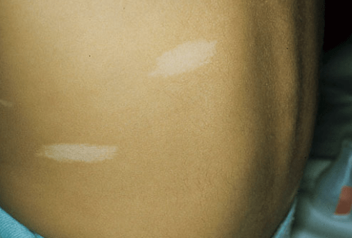
Tuberous Sclerosus
AD disorder of hamartomas in multiple organs (skin, brain, eye, heart, kidney)
Ash leaf spots (hypomelanotic macules- decreased size and poor melanization of melanosomes) are first presenting finding.
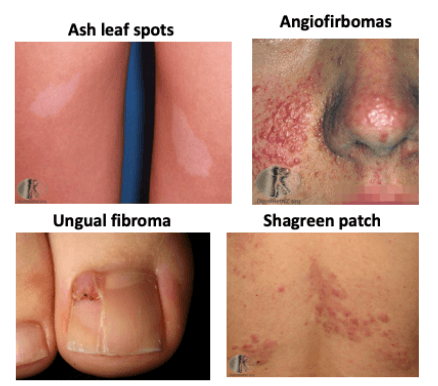 Major skin findings (2M = definite TS):
>3 hypopigmented macules (earliest finding), facial angiofibromas, Fibrous cephalic plaque, Shagreen patch, Periungal fibromas
DDX:
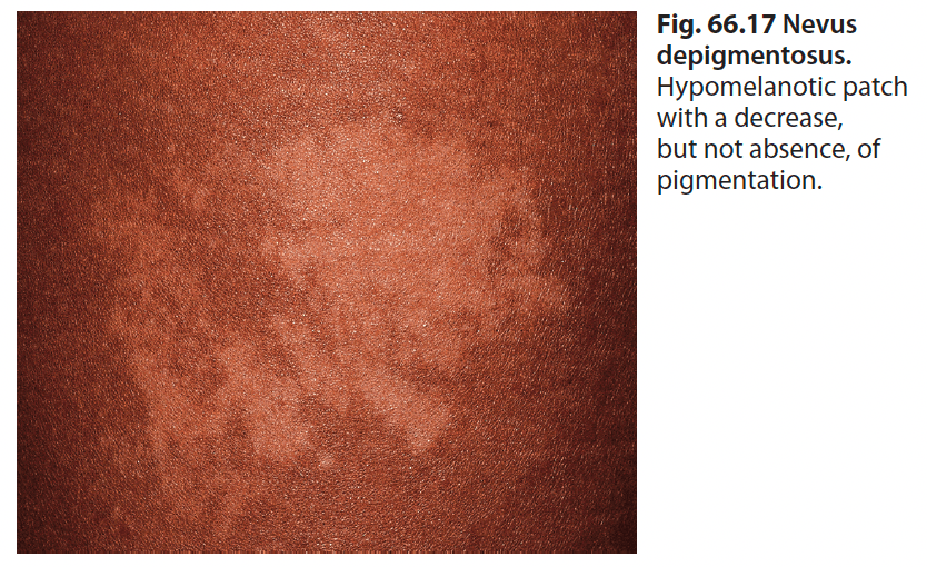
Nevus depigmentosus
300
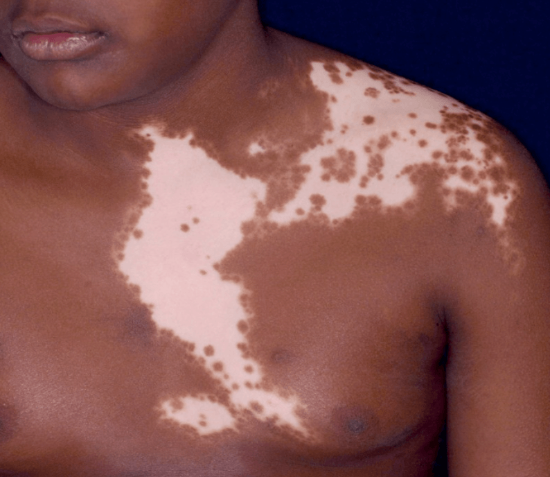
This variant of vitiligo is much more common in children compared to adults
What is segmental vitiligo
(~15-30%)
Often progresses for 1-2 years then remains stable.
300
Which syndrome is shown below?
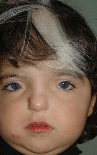
Waardenburg Syndrome (Four types)
Deafness
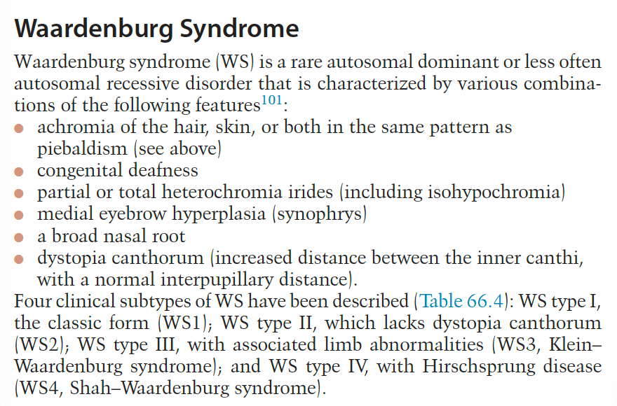
Genes: PAX3, MITF, SOX10
- WS type 1: Characteristically presents with dystopia canthorum (lateral displacement of the inner canthi) and frequently associated with poliosis (white forelock) and vitiligo on face or upper extremities. Complete or segmental heterochromia iridis and sensorineural hearing loss may also be seen. It is caused by a mutation of the PAX3 gene, and in most cases inherited in an autosomal dominant fashion.
- WS type 2: Characterized by the absence of dystopia canthorum and more frequently associated with heterochromia iridis and congenital sensorineural hearing loss. In most cases, it is due to a mutation of the MITF gene and is inherited in an autosomal dominant fashion.
- WS type 3 (Klein-Waardenburg syndrome): Present with severe features of WS type 1 in association with upper limb abnormalities. It typically results from a mutation of the PAX3 gene that may be inherited in an autosomal dominant fashion. Severe cases may be related to the mutation homozygosity.
- WS type 4 (Waardenburg-Shah syndrome): Manifested by typical features of WS in association with Hirschsprung disease (aganglionic megacolon). It is caused by a homozygous mutation of the EDNRB or EDN3 gene or by a heterozygous mutation in the SOX10 gene.
histopathology: absent or minimal number of melanocytes seen (as in piebaldism)
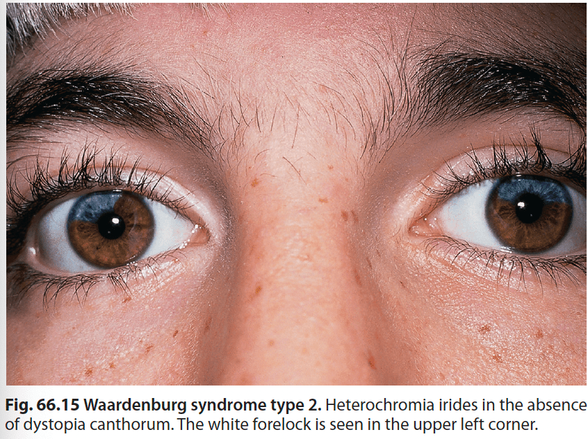
"Jail Warden seeing an AD for a trip to Alaska (has white snow on his head) so he is "PAX your MITtens, and PAX your SOX" (Type 1, 2, 3, 4 Waardenburg genes) while wearing earmuffs. 2nd suitcase with mittens has extra pair of earmuffs (↑ rates of deafness), 3rd suitcase has shirts (upper limb abnormalities), 4th suitcase is a colon bag that is paralyzed and full (Hirschsprung)."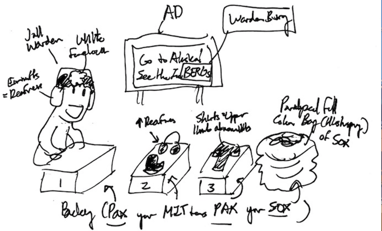
300
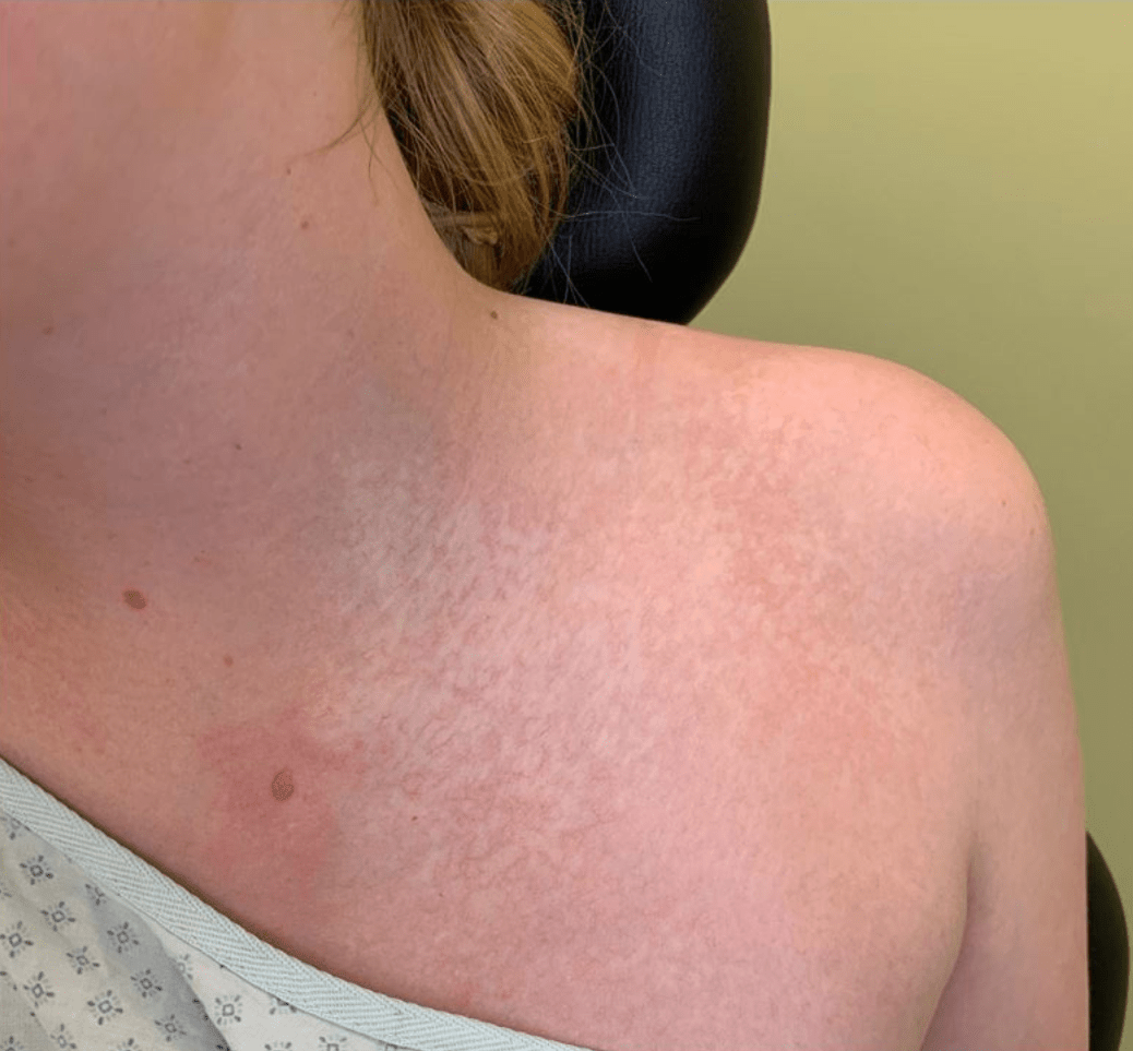
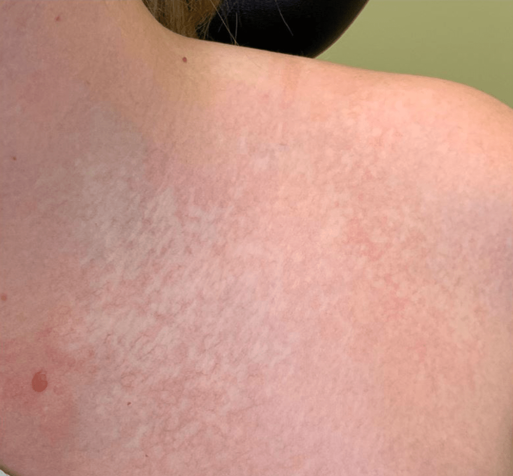
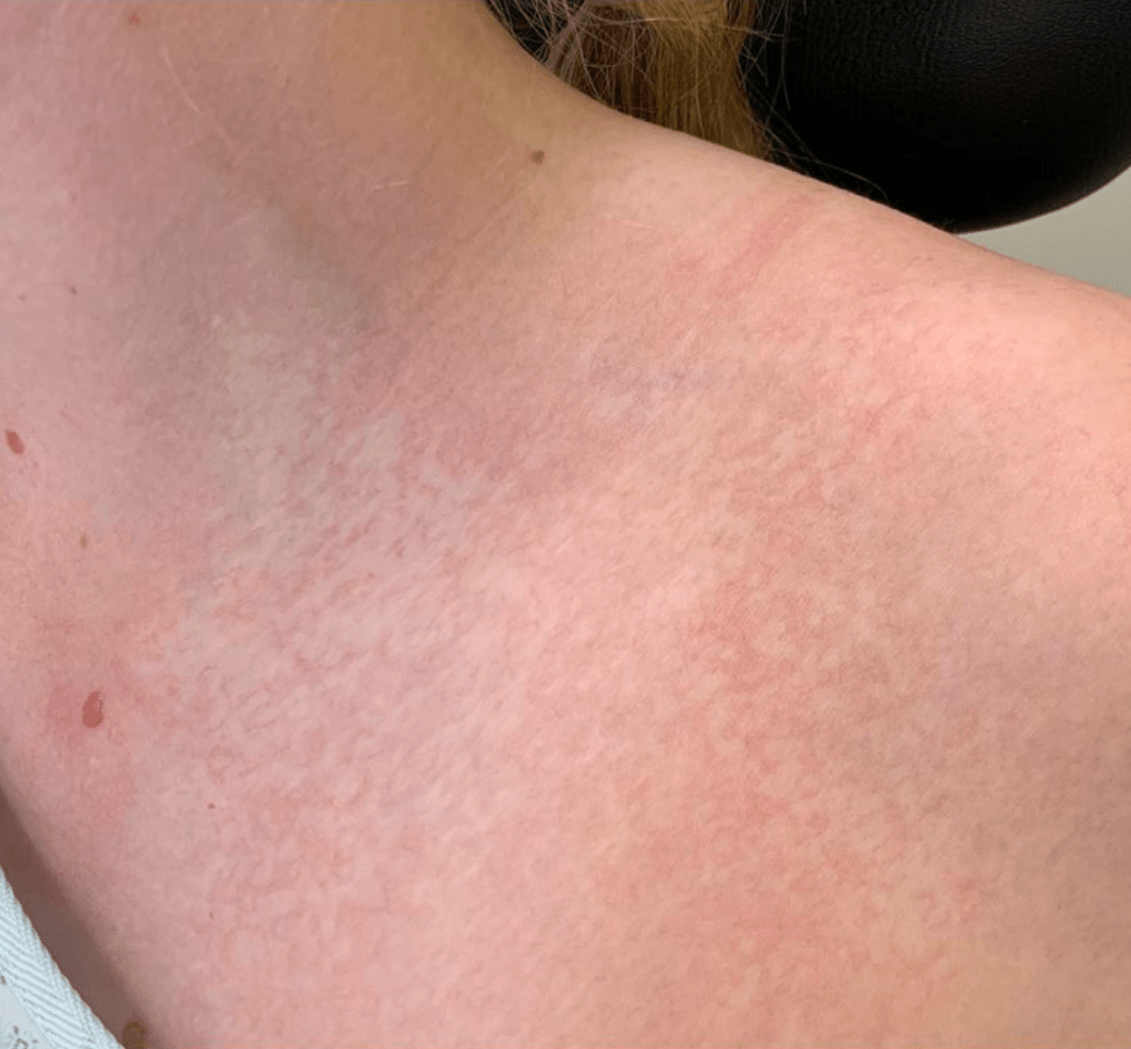
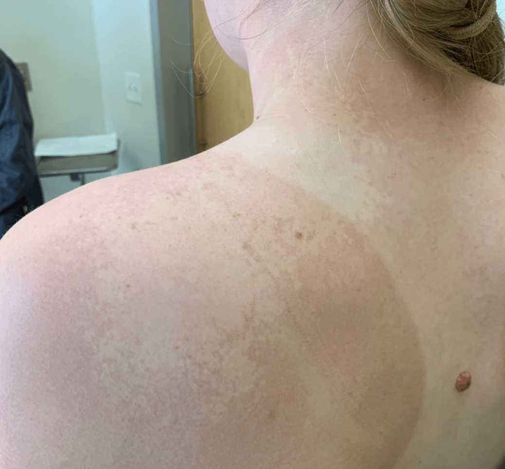
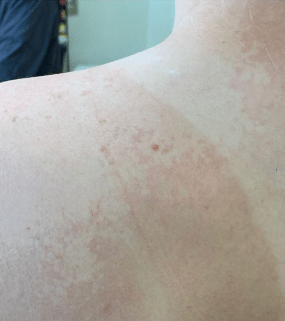
This pictured disorder is related to which of the below?
A. Autoimmune Destruction of Melanocytes
B. Increased Sensitivity to Catecholamines
C. Defective melanogenesis and abnormal transport of melanosomes
D. Inactivating c-Kit Mutation
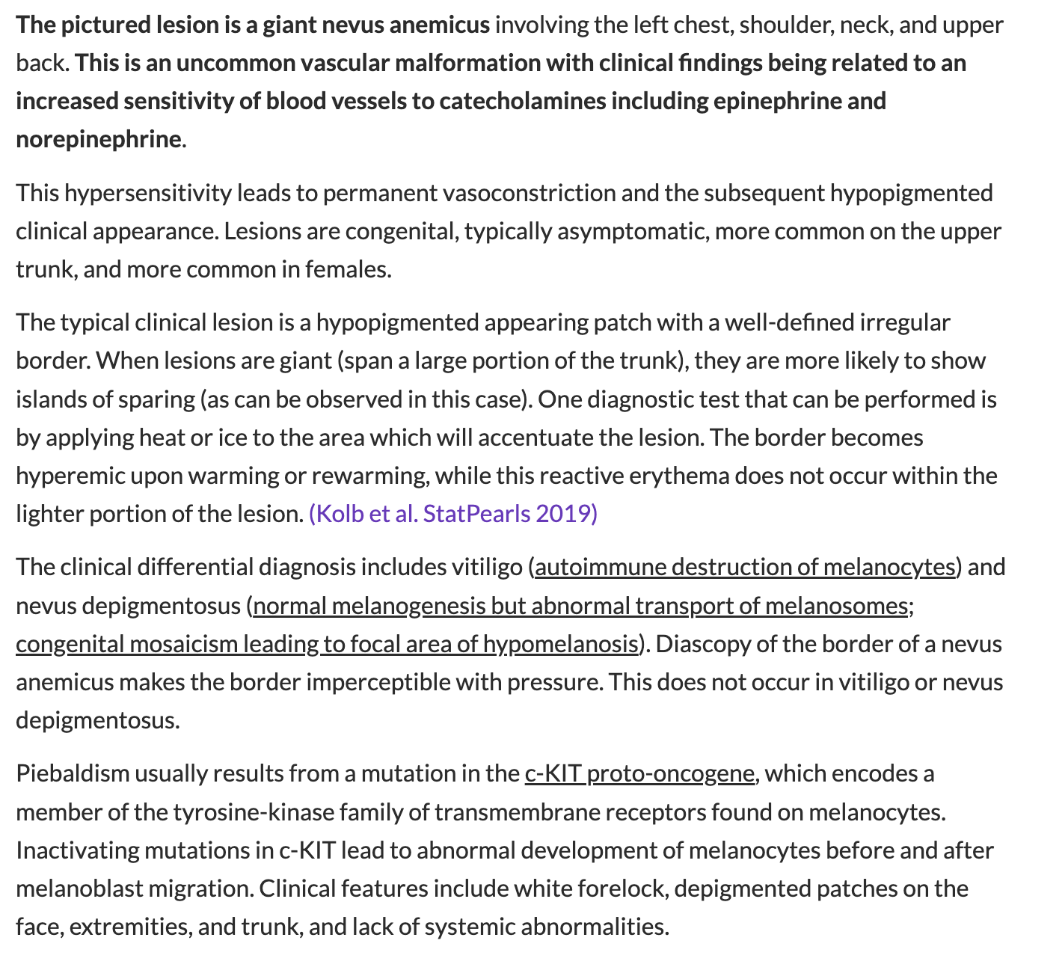
B. Increased Sensitivity to Catecholamines
- Most commonly detected later in life
- Most noticeable when there is surrounding vasodilation due to heat or emotional stress
300
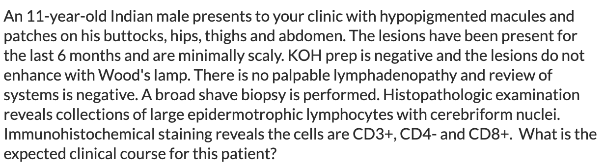
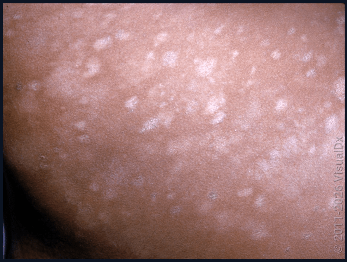
A. Waxing and waning course with disease limited to skin
B. Progressive worsening to systemic disease
C. Complete and permanent resolution without treatment
D. Permanent hypopigmentation despite appropriate treatment
A. Waxing and waning course with disease limited to skin - Hypopigmented MF
400
4 stages of this condition
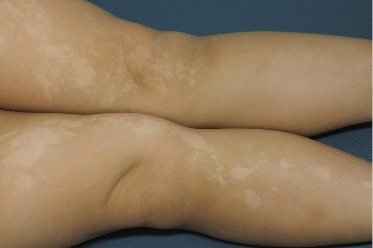
Icontinentia Pigmenti
Vesicular, Verrucous, Hyperpigmented, Hypopigmented
XLD -- males typically do not survive.
Mutation in NF-kappa B essential modulator (NEMO)
400
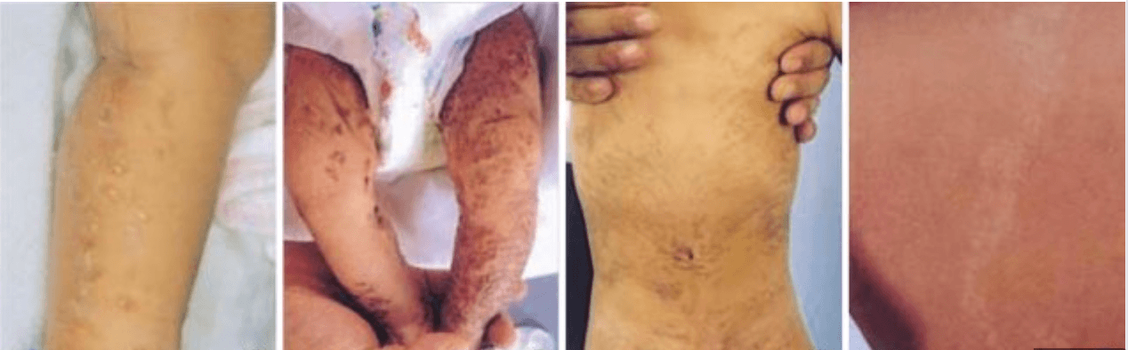
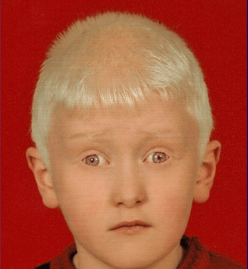
Classic 'Tyrosinase Positive" Oculocutaneous Albinism.
OCA2 (P gene)-- broad clinical spectrum
OCA1B also "tyrosinase-positive"
400

A. Granulomatous colitis
B. Non-melanoma skin cancer
C. Pancytopenia
D. Pigmented nevi
E. Pulmonary fibrosis
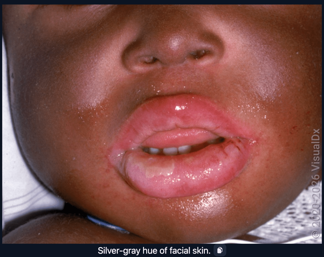
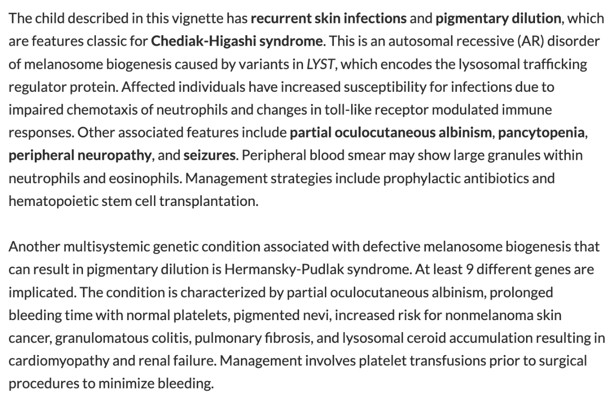
C. Pancytopenia - Chédiak-Higashi syndrome
400
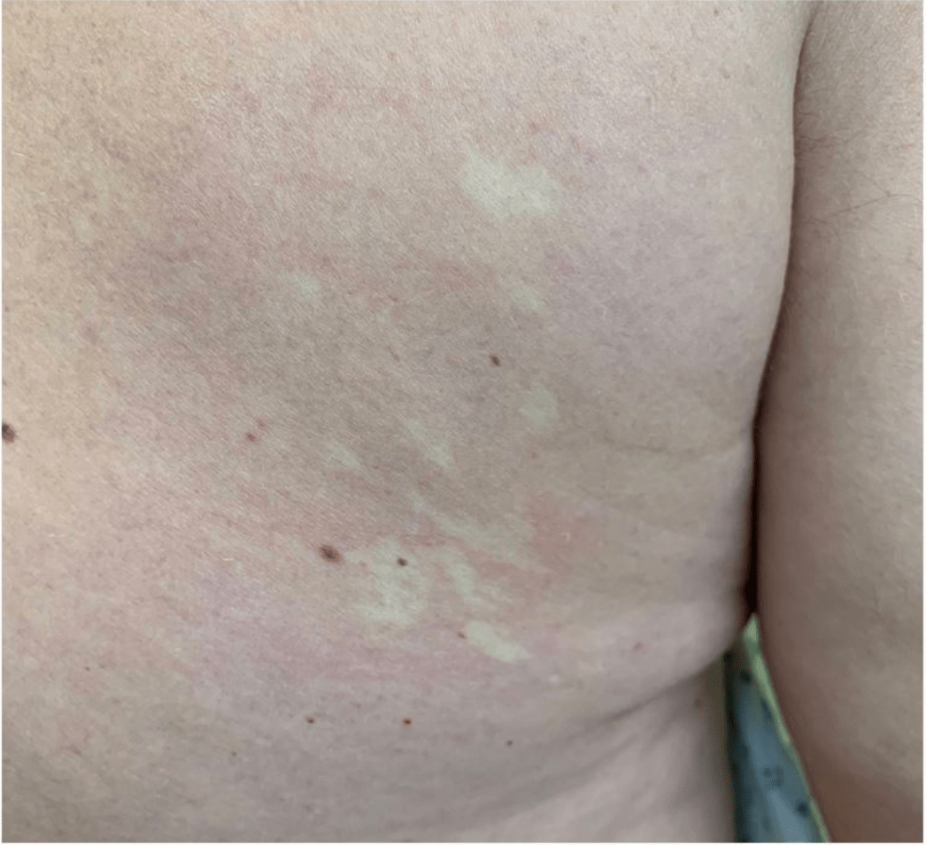
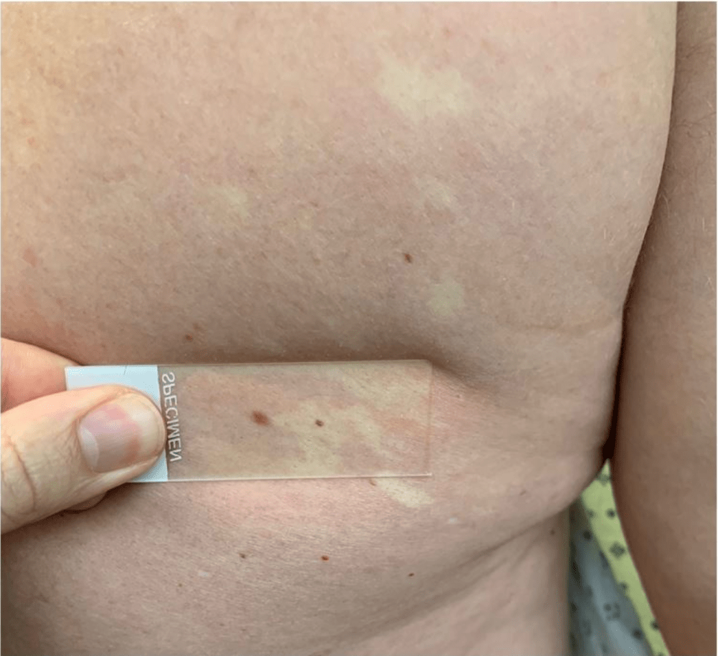

A. Segmental vitiligo
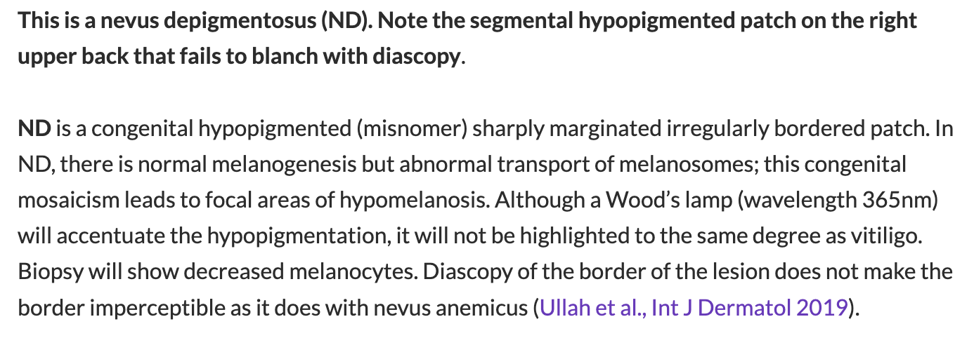
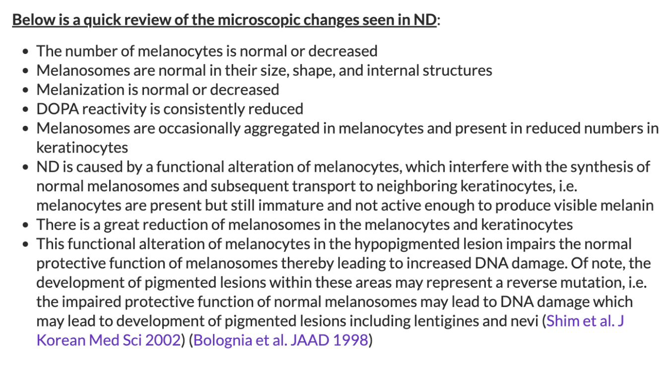
B. Nevus depigmentosus
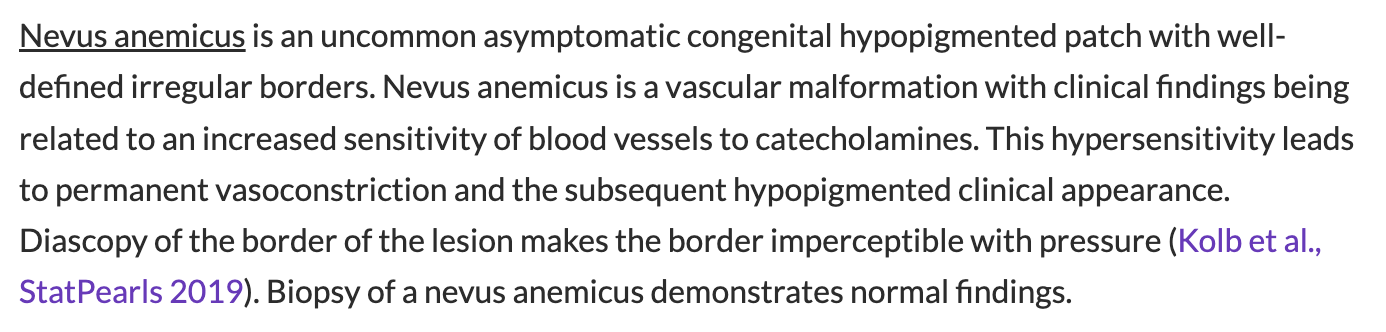
C. Nevus anemicus
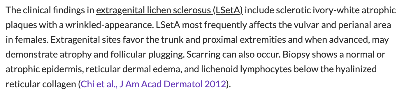
D. Extragenital lichen sclerosus
B. Nevus depigmentosus

400
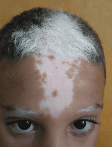
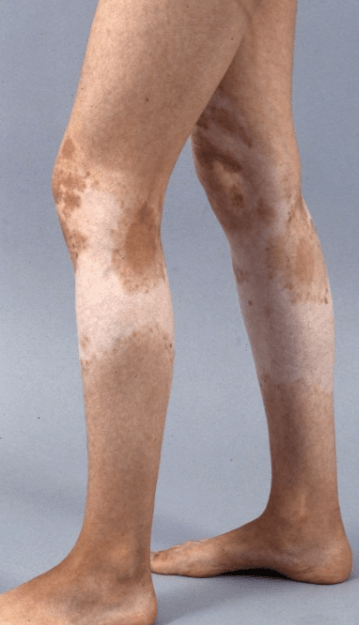
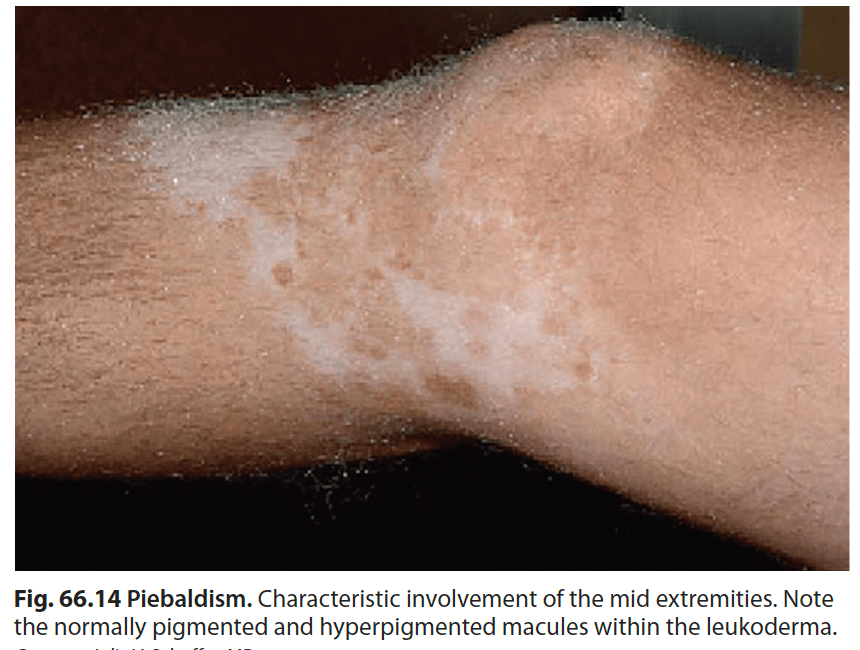
Name the condition
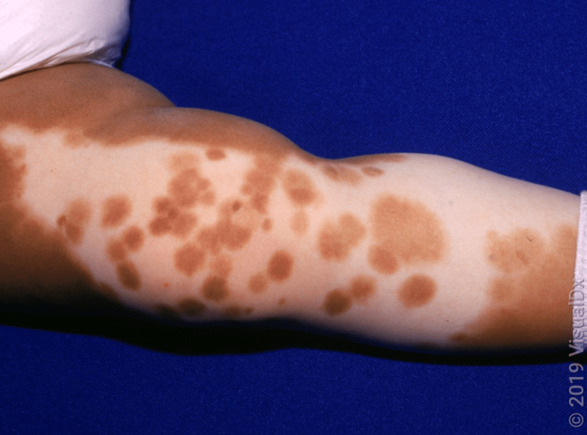
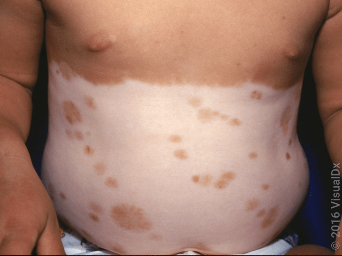
Piebaldism
Autosomal dominant, uncommon
KIT proto-oncogene encoding a member of the tyrosine kinase family of transmembrane receptors on the surface of melanocytes (functioning KIT receptor is required for the normal development of melanocytes)
Clinical
- Poliosis (AKA white forelock, 80-90%)
- congenital, stable, circumscribed areas of leukoderma due to an absence of melanocytes within involved sites- characteristic distribution favors the central anterior trunk, mid extremities, central forehead, midfrontal scalp. Distinctly irregular shape, well circumscribed, milk-white color with normally pigmented macules/patches within.
Stable since birth
Exclude Waardenberg syndrome with an ocular and hearing exam
Tx: autografting normal skin (multiple procedures required), color matching products, protect from sunburn
500
Hint:
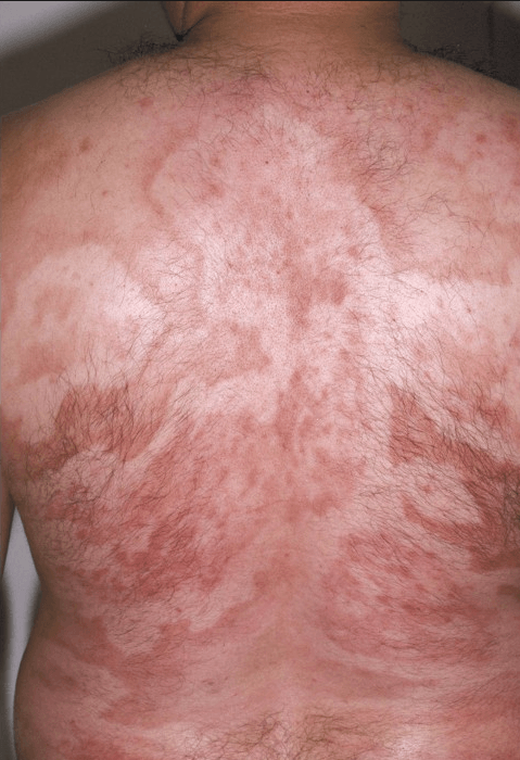
Patient also with new and worsening neuropathy.
Hansen Disease (Leprosy)
Chronic granulomatous infection caused by Mycobacterium leprae (Hansen's bacillus)
Dapsone, rifampin, and clofazimine (for LL)
Lepromatous Leprosy
- Hypomelanotic macules may be the earliest expression of lepromatous leprosy
- Lesions are usually small, multiple, subtle, and ill-defined
- Normal sensation within
- Face, extremities, and buttocks are favored, and the warmer parts of the body are usually spared
- Later, plaques, nodules, or diffuse infiltration of the skin appear
Tuberculoid leprosy
- hypopigmented patches have discrete edges and can be quite large, up to 30 cm in diameter
- A raised border, uniform infiltration, or a characteristic pebbled appearance may be observed
- surface can be slightly scaly, dry, or minimally atrophic
- asymmetrically distributed on the posterolateral aspects of the extremities, back, buttocks, and face
- associated alopecia, anhidrosis, and loss of tactile and heat sensations
Borderline leprosy
- plaques and annular lesions - "swiss cheese appearance"
Indeterminate leprosy
- hypomelanotic macules, can be erythematous, hypoesthesia, asymmetric distribution
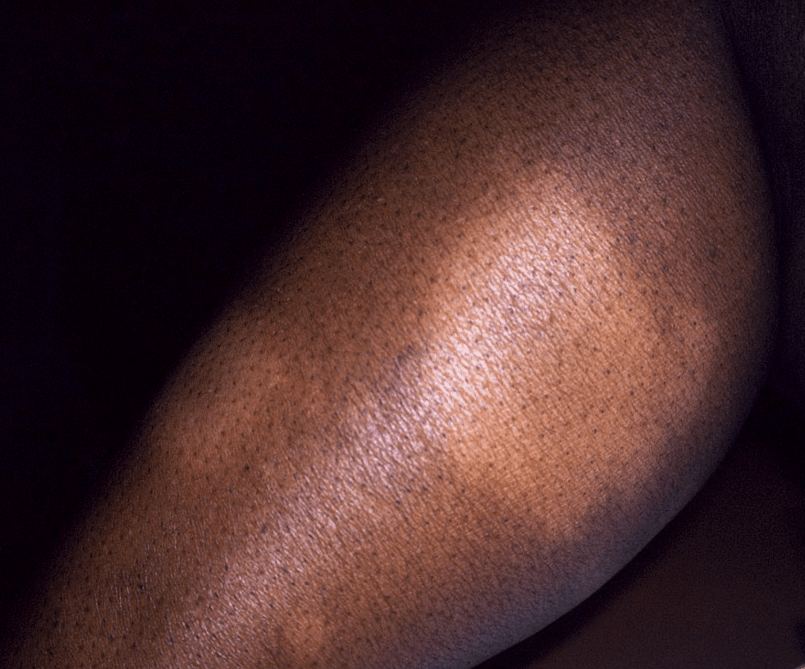
500
Hint: condition related to Treponema infection
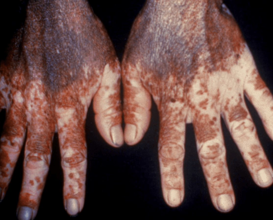
Pinta
Nonvenereal endemic treponemal infection caused by the spirochete Treponema pallidum subsp carateum. The disease is confined to rural areas of northern South America and Mexico.
Irregular, vitiligo-like areas of depigmentation favor skin overlying bony prominences and are often surrounded by brown to blue–gray hyperpigmentation
500

A. Ophthalmology
B. Pulmonology
C. Cardiology
D. Hematology/Oncology
A. Ophthalmology
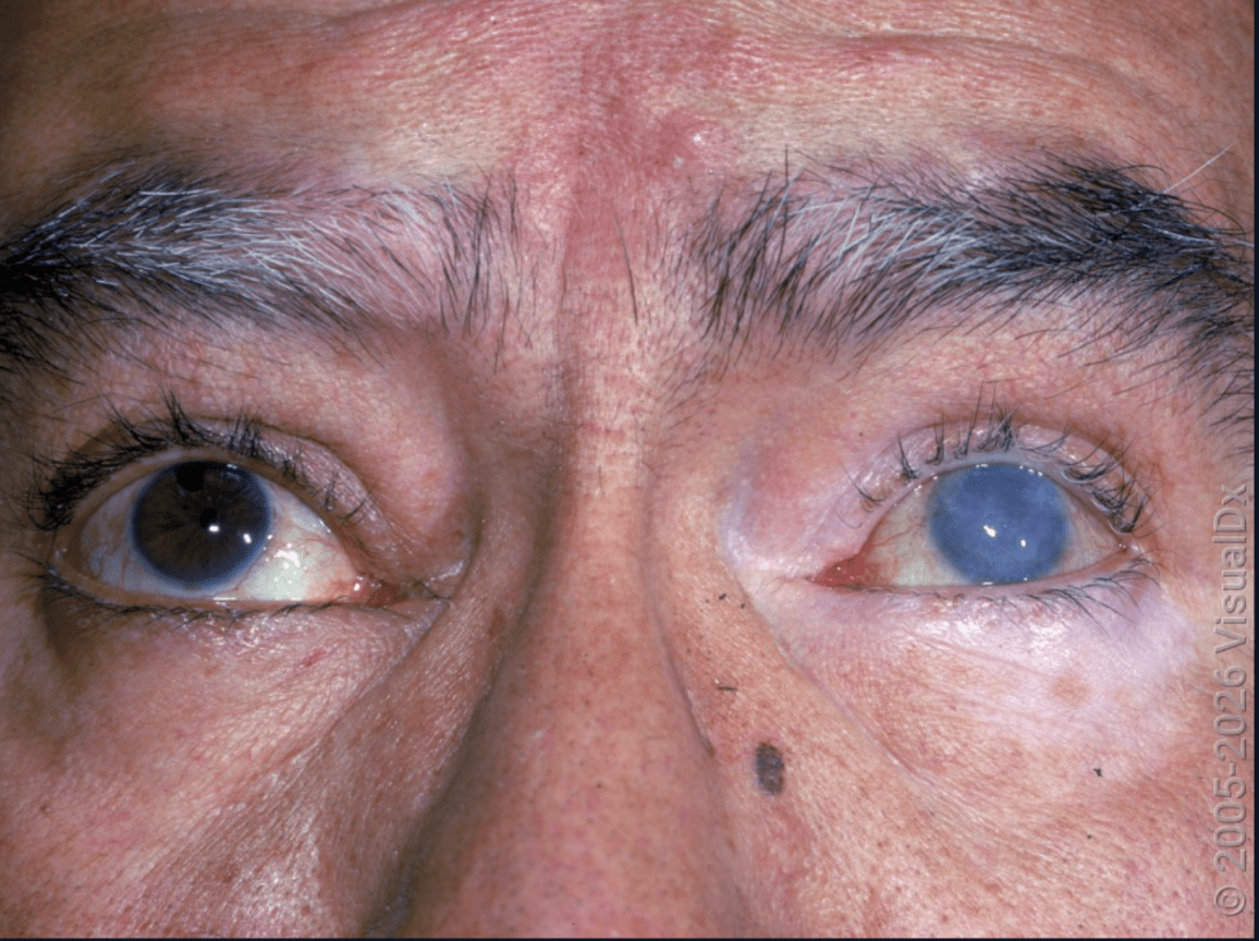
T-cell-mediated autoimmune response to melanocytes of central nervous system and skin in genetically susceptible individuals
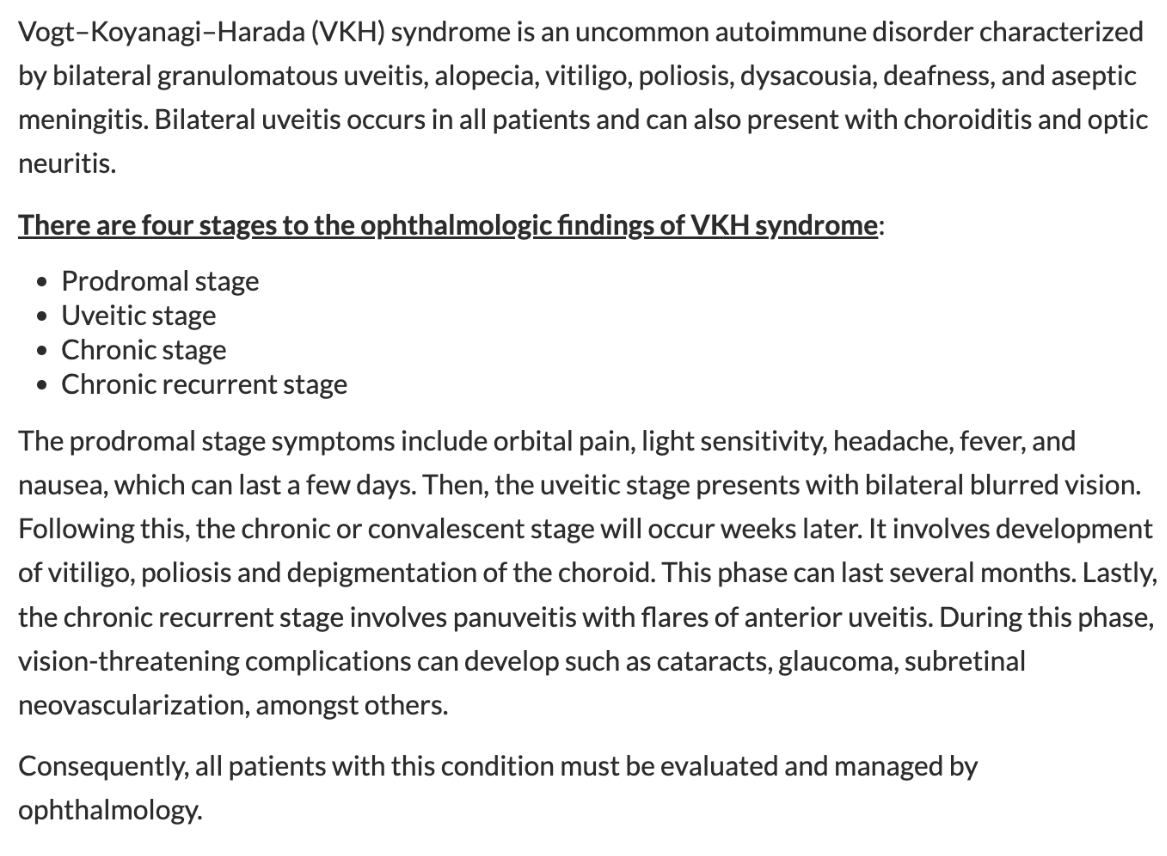
Skin features of the syndrome never precede the neurological, auditory, or ocular manifestations
500
name the condition
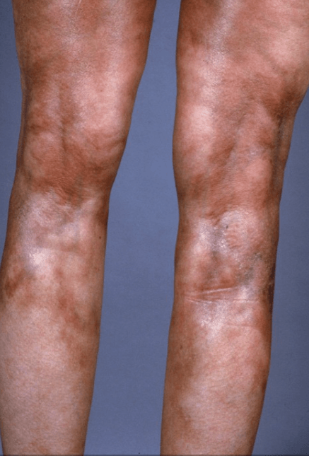
Hint:
Indurated hypo-de pigmented plaques
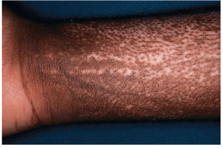
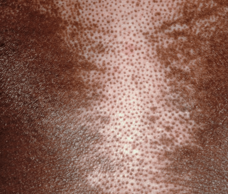
What is scleroderma (systemic sclerosis)
Anti-topoisomerase I
Characteristic leukoderma in both sclerotic and non-sclerotic skin (characteristically circumscribed areas of complete pigment loss except for perifollicular and supravenous retention of pigment)
500

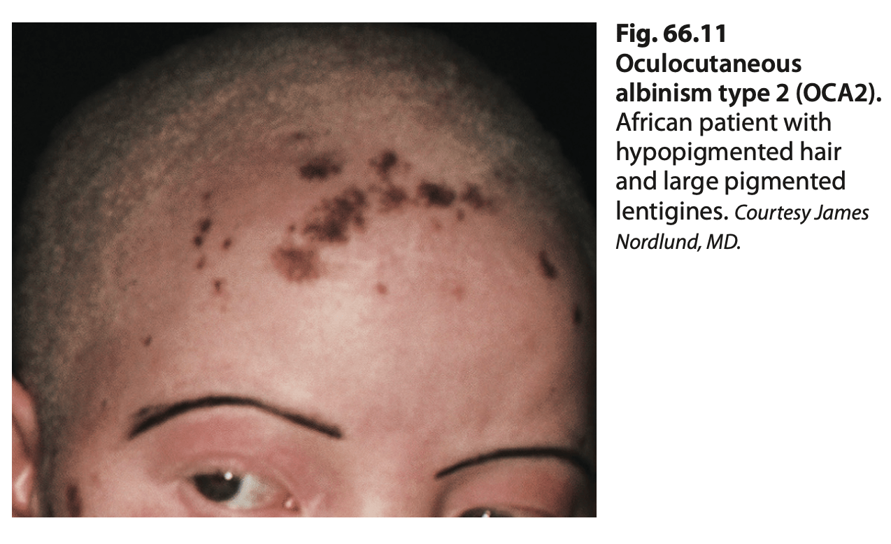
A. OCA1A
B. OCA1B
C. OCA2
D. OCA3
E. OCA4
C. OCA2
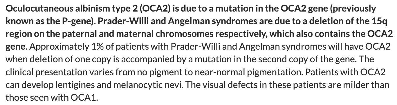
Prader-Willi Syndrome:
- Hyperphagia
- Intellectual disability
- Short stature
Angelman Syndrome:
- Happy personality
- Intellectual disability
- Hand flapping movements
- Speech impairment

OCA1B^

"Rufous:" reddish-brown or rust-colored

