Photon counting, by the numbers
Right heart, Right On!
Calls at night
Surprising slices
100
This technical limitation of PCCT's occurs when multiple X-ray photons arrive at the detector within a short time
What is pile up effect ?
Pile up effect occurs when multiple photons arrive at the same time causing overlapping/additive signals and compromise accurate energy measures. Can be reduced with lower detector dead time.

100
Name the pulmonary artery branch abnormality

What is Criss cross branch pulmonary artery ?
100
Patient referred for burning chest pain to the back

What is Anomalous left main coronary artery yfrom the pulmonary artery ALCAPA.
100
A patient with a murmur has this abnormality
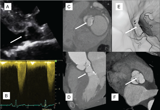
What is subaortic membrane ?
200
this paper in Jack and also feature to NJ C CT, showed significant improvements and CAD diagnostic accuracy measures for PCCT versus EID. Name one measure
which is diagnostic accuracy, specificity, or positive predictive value
Sakai K, et al JACC 2024
COMPARED TO EID-CT VERSUS PCCT FOR CAD ACCURACY VERSUS INVASIVE ANGIOGRAPHY. ALSO SHOWED DECREASED INVASIVE ANGIOGRAPHY AND INCREASED CORONARY REVASCULARIZATION WITH PCCT
200
in a patient with L loop or congenitally corrected transposition of the greater arteries, this is the name of this valve

TRICUSPID VALVE OR SYSTEMIC AV VALVE
THE VALVE FOLLOWS THE VENTRICLE. SYSTEMIC AV VALVE ( TRICUSPID ) ABNORMALITIES AND REGURGITATION IN L TGA ARE COMMON AND SHOULD BE ROUTINELY ASSESSED
200
ED patient referred for chest pain

What is anomalous LCX from RCA ?
200
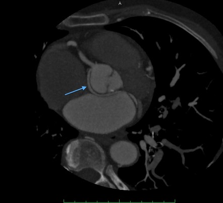
NAMED THE NATIVE CORONARY ANOMALY DESIGNATED BY ARROWS

WHAT IS CORONARY ARTERY ANEURYSM ?

300
when imaging coronary artery stents, PCCT has the potential to decrease this kind of artifact compared to EID scan was
What is blooming artifact ?

300
Four CTA measurments of the MPA are recommended pre--transcatheter pulmonary valve implantation. Name 2 MPA CTA measurements.
What is MPA perimeter, diameter, area, landing zone length.
300
Patient referred for chest pain and weight loss

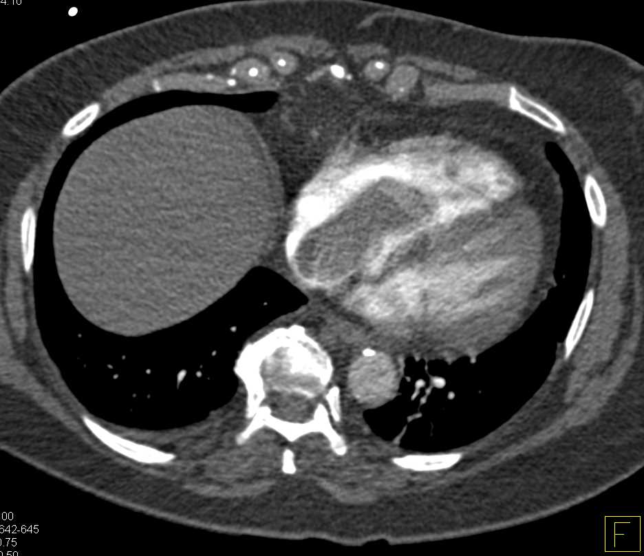
What is IVC mass(renal cell cancer) with extension to right atrium
300
This TAVI valve abnormality is less common with antithrombotic use.

What is hypoattenuation leaflet thickening HALT ?
400
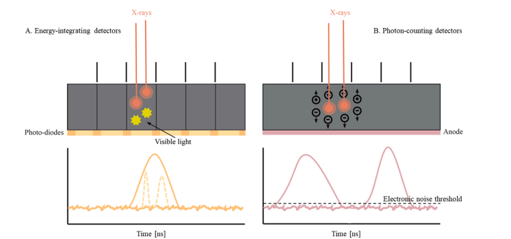
THIS CHANGE IN DETECTOR MATERIAL FROM ENERGY INTEGRATING/SCINTILLATION DETECTORS ALLOWS FOR CURRENT PHOTON COUNTING CT SCANNERS. NAMED DETECTOR TYPE OR DETECTOR MATERIAL
WHICH IS SEMICONDUCTOR, CADMIUM TELLURIDE, OR SILICONE ?
PCCT use semiconducting material such as cadmium telluride/silicone to detect individual signals that counts and measures voltage peak of photons

400
BASED ON THE 2018 ACHD GUIDELINES IN 2024 AND SCCT RVOT WHITE PAPER, CORONARY ARTERY SHOULD BE ASSESSED FOR THIS ABNORMALITY PRIOR TO PERCUTANEOUS PULMONARY VALVE IMPLANTATION
WHAT IS CORONARY ARTERY ACROSS BETWEEN PULMONARY ARTERY AND AORTA ?
RISK OF CORONARY COMPRESSION 5-6% WITH ANOMALOUS CORONARY COURSE WITHIN LANDING ZONE
400
Patient with Heartmate III LVAD with "Alarms". Echo is unchanged.

What is LVAD outflow cannula stenosis/thrombus ?
400
LIKELY CAUSE FOR THE PATIENT PRESENTING WITH DIZZINESS AND ELEVATED TROPONIN

WHAT IS LEFT ATRIAL APPENDAGE THROMBI ?
![]()
500
changes in his detector structure allow higher spatial resolution for photon counting CT compared to energy integrating CT detectors. Name 1 change
what is 1. Smaller detector elements size, 2. Removal of septal collimators
scintillation detectors require larger size for light detection and require septa to limit light diffusion between detectors. Fulton counting CT allowed to take 2 elements size is small as 0.1 x 0.1 mm
500
IN INFECTIVE ENDOCARDITIS, CTA IMAGING FOR THIS ABNORMALITY IS REASONABLE BY AHA/ ACC VALVE GUIDELINES (CLASS 2A ) AND FEATURED IN A CONSENSUS STATEMENT IN THE SCCT PROSTHETIC VALVE EXPERT CONSENSUS DOCUMENT
WHAT IS PERIANNULAR ABSCESS OR MYCOTIC ANEURYSM ?
CT HAS REPORTED SENSITIVITY AND SPECIFICITY TO DETECT PERIANNULAR COMPLICATION OF 80% AND 82%, SIMILAR TO TEE ( SENSITIVITY 77%, SPECIFICITY OF 86%) IN 1 SYSTEMIC REVIEW
500
Changes in cardiac CT contrast protocol are suggested in this patient with a prior cardiac fontan per medical history presenting with a right leg swelling and new dyspnea. Name one contrast protocol change.
High contrast load, dual venous phase
500
Pre-TAVI with this CT scan and has cold extremities. Name the primary cause.

What is occluded descending aorta ?
