MCQ 1
MCQ 2
MCQ 3
OSPE 1
OSPE 2
100
Which component gives elastic cartilage its flexibility and recoil?
A. Thick type I collagen bundles
B. A dense network of elastic fibres
C. Calcium hydroxyapatite crystals
D. Network of reticular fibres
B. A dense network of elastic fibres
100
Why does the matrix of hyaline cartilage appear glassy and homogeneous in H&E sections?
A. It contains no collagen
B. It is filled with elastic fibres
C. It is heavily calcified
D. It contains type II collagen fibrils
D. It contains type II collagen fibrils
100
•Which of the following statements is true concerning Hyaline cartilage?
a. Is a highly vascular tissue.
b. Capable of extensive repair.
c. Matrix contains type I collagen
d. Surrounded by a perichondrium.
d. Surrounded by a perichondrium.
100
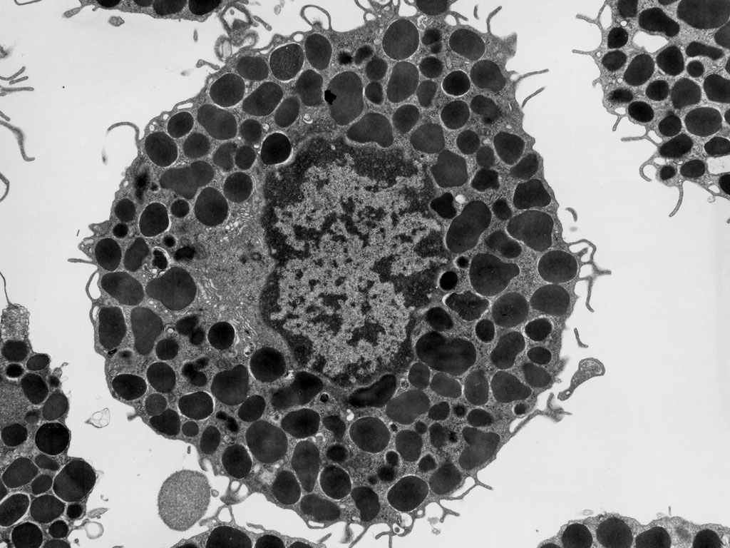
Identify the CT cell.
Mention one character.
Mast cell
Large dense granules
100
 Identify the CT cell
Mention the stain
Fat cell
Sudan III
200
How do isogenous groups form within cartilage?
A. Chondroblasts migrate from the bone marrow
B. Perichondrial cells invade the matrix
C. Chondroblasts divide in perichondrium
D. Chondrocytes divide within their lacunae
D. Chondrocytes divide within their lacunae
200
Which feature of cartilage best explains its slow growth and limited capacity for repair?
A. The presence of a dense calcified matrix
B. The presence of an abundance of type I collagen
C. The absence of blood vessels and nerves
D. The absence of living cells in the matrix
C. The absence of blood vessels and nerves
200
Which statement best describes the role of the perichondrium?
A. It supplies nutrition and growth for the cartilage
B. It secretes elastic fibers and reticular fibers
C. It calcifies the cartilage matrix
D. It forms isogenous groups by division
A. It supplies nutrition and growth for the cartilage
200
 Identify the type of epithelium.
Mention the site
Simple cubical epith.
Thyroid follicle
200
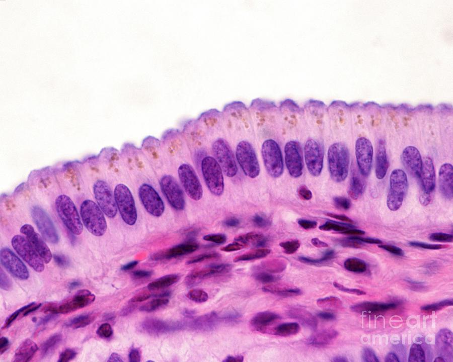
Identify the type of epithelium.
Mention one site
Simple columnar epith
Stomach
300
A section shows rows of chondrocytes between thick collagen bundles and no perichondrium. Which tissue and site is most likely?
A. Hyaline cartilage of a costal cartilage
B. Elastic cartilage of the epiglottis
C. Fibrocartilage of an intervertebral disc
D. Hyaline cartilage of the trachea
C. Fibrocartilage of an intervertebral disc
300
A pathologist examines a biopsy of the temporomandibular joint articular disc. Which structure will be absent?
A. Perichondrium
B. Chondrocytes
C. Collagen bundles
D. Lacunae
A. Perichondrium
300
. Which tissue is damaged in TMJ osteoarthritis?
A. Hyaline cartilage
B. Elastic cartilage
C. Dense connective tissue
D. Fibrocartilage
D. Fibrocartilage
300

Identify the type of epithelium
Mention one site
Stratified squamous epith non keratinized
Oral cavity
300
Identify the type of epithelium.
Mention one site
Stratified squamous keratinized
Skin
400
A hematoma forms between the perichondrium and the auricular cartilage. Which mechanism best explains the cartilage necrosis that follows?
A. Excess collagen synthesis by chondroblasts
B. Separation from the perichondrium blocks nutrition to chondrocytes
C. Digestion of the elastic fibres by the blood
D. Direct viral infection of chondrocytes
B. Separation from the perichondrium blocks nutrition to chondrocytes
400
Why does articular hyaline cartilage repair more poorly than costal cartilage?
A. It contains elastic fibres
B. It is richly vascular
C. It is covered by a thick perichondrium
D. It lacks a perichondrium, so it has no source of new chondroblasts
D. It lacks a perichondrium, so it has no source of new chondroblasts
400
Which feature best explains why fibrocartilage resists both compression and tension better than hyaline cartilage?
A. A dense network of elastic fibres
B. Thick type I collagen bundles
C. A thick perichondrium
D. Abundant ground substance with no fibres
B. Thick type I collagen bundles
400
 Identify the type of cartilage
Mention one character
Mention one location
Hyaline cartilage
Homogenous matrix, cell nest, covered by perichondrium
Codstal cartilage
400
 Identify the type of cartilage
Mention one character
Mention one site
Elastic cartilage
Elastic fibers in matrix
Ear pinna
500
A researcher plans to inject chondrocytes into a damaged articular surface and expects rapid regeneration. Which assessment best reflects the histological outcome?
A. Rapid success is likely because the tissue is richly vascular
B. Success is likely to be slow and limited, because the tissue is avascular
C. Success is guaranteed because chondrocytes divide quickly
D. Treatment is unnecessary because the perichondrium regenerates articular cartilage
B. Success is likely to be slow and limited, because the tissue is avascular
500
You are designing a replacement for a damaged TMJ disc. Which design feature best mimics the native tissue?
A. A rigid, calcified plate
B. A thin, highly elastic sheet like the epiglottis
C. A smooth, glassy gel with no fibres
D. A flexible pad with oriented collagen bundles
D. A flexible pad with oriented collagen bundles
500
•58-year-old man with a history of ischemic heart disease is scheduled for open heart surgery. Before cutting the costal cartilage to open the chest cavity, the surgeon is mindful that healing of cartilage is problematic. He understands that hyaline cartilage has a limited capacity to undergo repair and regeneration due to which of the following intrinsic attributes?
a. Avascularity of the cartilage matrix.
b. Cell cycle arrest of differentiated chondrocytes.
c. Deficiency of chondroblasts in the perichondrium.
d. Overgrowth of fibroblasts at the wound site.
a. Avascularity of the cartilage matrix.
500
 Identify the type of cartilage
Mention one character
Mention one site
Fibrocartilage
Collagen fibers type I, parallel plates of hyaline cartilage
TMJ
500
 Mention type of epithelium.
Mention one site
Mention one character
Transiltional epith
Urinary bladder
Dome shaped cells