Anatomy/Pathology
Positioning
I can't think critically
Methods annoy me
Rotations confuse me
100
What is the name of the term associated with the medial end of the clavicle?
What is the sternal extremity?
100
How much CR angulation is required for a PA oblique (scapular Y) projection?
What is no CR angle is required?
100
A radiograph of an AP axial projection of the clavicle demonstrates that the clavicle is within the mid aspect of the lung apices. What should you do to correct the error? Should you increase or decrease the cephalic angle during repeat exposure?
What is increase the cephalic CR angle during repeat exposure?
100
How much posterior CR angulation is required for the supine version of the tangential projection (Fisk modification) for the intertubercular (bicipital) sulcus?
What is 10 to 15 degrees?
100
Which rotation of the humerus will result in a lateral position of the proximal humerus?
What is internal rotation?
200
What is the name of the anterior surface of the scapula?
What is the costal surface?
200
What is the name of the shoulder projection that best demonstrates the glenoid cavity in profile?
What is the Grashey method?
200
A radiograph of the AP oblique (Grashey method) projection for the glenoid cavity reveals that the anterior and posterior rims of the glenoid process are not superimposed. Should you increase the rotation of the body by turning the patient toward the IR or away from it?
What is increase rotation of the body toward the IR?
200
How much is the affected limb raised superiorly for the PA axial transaxillary (Bernageau method) projection?
What is 160 to 180 degrees?
200
Which AP projection of the shoulder and proximal humerus is created by placing the affected palm of the hand facing inward toward the thigh?
What is the neutral rotation?
300
What type of joint movement does the scapulohumeral joint have?
What is a ball and socket (spheroidal) type?
300
Where is the CR centered for the AP oblique (Grashey method) position?
What is 2 inches medial and 2 inches inferior to the superolateral border of the shoulder?
300
A radiograph of a PA oblique (scapular Y) lateral reveals that the scapula is slightly rotated (the vertebral and axillary borders are not superimposed). The axillary (medial) border of the scapula is determined to be more lateral compared with the vertebral (lateral) border. Should you increase or decrease the rotation of the thorax (toward or away from the IR)?
What is increase rotation of the thorax (toward the IR)?
300
What is the name of the method that best demonstrates the coracoacromial arch and the supraspinatus outlet?
What is the Neer method?
300
Which AP shoulder projection will demonstrate the lesser tubercle in profile medially?
What is internal rotation?
400
What is the name of the structure marked number 3?
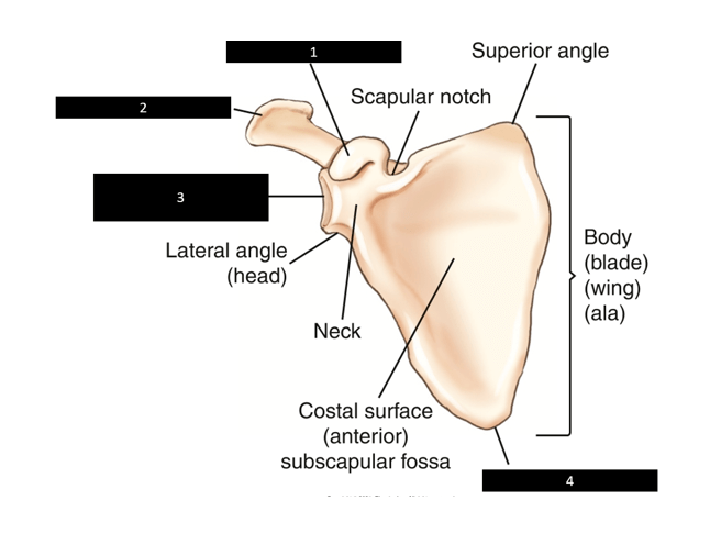
What is the glenoid cavity (fossa)?
400
How many degrees is the central ray (CR) directed medially from horizontally to the axial and humeral head for the inferosuperior (transaxillary) shoulder (Lawrence method)?
What is 25 to 30 degrees?
400
A patient enters the ED with a dislocated shoulder. The technologist attempts to position the patient into the transthoracic lateral projection, but the patient cannot completely raise the unaffected arm over his head. How do you compensate for the patient's inability to raise his arm completely?
What is angle the CR 10 to 15 degrees cephalad?
400
The inferosuperior axial projection (Clements modification) requires a CR angle of ___ toward the axilla if a patient cannot fully abduct the extremity 90 degrees.
What is 5 to 15 degres?
400
Which projection will demonstrate the greater tubercle in profile medially?
External, internal, neutral, or none of the options.
What is none of the options?
500
What is the common term for idiopathic chronic adhesive capsulitis?
What is a frozen shoulder?
500
A radiograph of a transthoracic lateral projection reveals that it is difficult to visualize the proximal humerus due to the ribs and lung markings. The exam was performed on suspended respiration. What change will improve the visibility of the proximal humerus by blurring the ribs and lung markings out of the way?
What is use of an orthostatic (breathing) technique?
500
A patient comes to the ED with a possible right AC joint separation. Right clavicle and AC joints are ordered. The clavicle is taken first and there is a fracture to the clavicle. What should you do in this situation?
What is consult the ED physician before continuing with the AC joint study?
500
A patient is scheduled for an arthrogram. During the study, the radiologist requires a projection to demonstrate the intertubercular sulcus. What shoulder method or modification will best demonstrate this structure?
What is the Fisk modification?
500
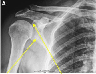
The image below indicates that the patient is in neutral, external, or internal rotation of the proximal humerus.
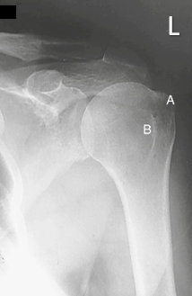
What is the external rotation?