MCQ 1
MCQ 2
MCQ 3
OSPE 1
OSPE 2
100
Which stain is used for the demonstration of fat cells?
A. Sudan III.
B. Silver.
C. PAS.
D. Orcein
A. Sudan III.
100
Which of the following cells secrete histamine?
A. Macrophage
B. Mast cell
C. Neutrophil
D. Plasma
B. Mast cell
100
Macrophage is characterized by having which of the following?
a. Signet ring appearance.
b. Abundant rER.
c. Highly granular cytoplasm masking the nucleus
d. Plenty of lysosomes.
d. Plenty of lysosomes.
100
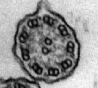 Identify the structure in the EM photo.
Cilia
100
 Identify the organelle in the EM Photo
Centriole
200
Which of the following applies to the mast cells?
a. Contain metachromatic granules.
b. Are derived from B-lymphocytes.
c. Contain bilobed nuclei.
d. Are derived from T-lymphocytes
a. Contain metachromatic granules.
200
Which of the following is the character of the nucleus of the cell secreting antibodies?
a. Bilobed.
b.Cartwheel shaped.
c.Kidney-shaped.
d.Multinucleated.
D.Adipocyte
b.Cartwheel shaped.
200
Which of the following represents the Plasma cell?
A.Activated T lymphocyte.
B.Antibodies producing cell.
C.Active phagocytic cell.
D.Antigen presenting cells.
B.Antibodies producing cell.
200

Identify the structure in the EM photo
Primary lysosomes
200
A B
Identify the EM photos.
A. Euchromatic nucleus
B. Heterochromatic nucleus
300
Which one of the connective tissue cells can differentiate into an endothelial cell and a fibroblast?
A.Macrophage.
B.Plasma cell.
C.Pericyte.
D.Mast cell.
C.Pericyte.
300
Which of the following connective tissue cells plays a major role in wound healing?
a.Fibroblast.
b.Mast cell.
c.Plasma cell.
d.Fat cell.
a.Fibroblast.
300
A new skin cream is supposed to stimulate collagen production, which of the following is the responsible cell?
a.Mast cell
b.Fibroblast
c.Macrophage
d. Fat cell
b.Fibroblast
300
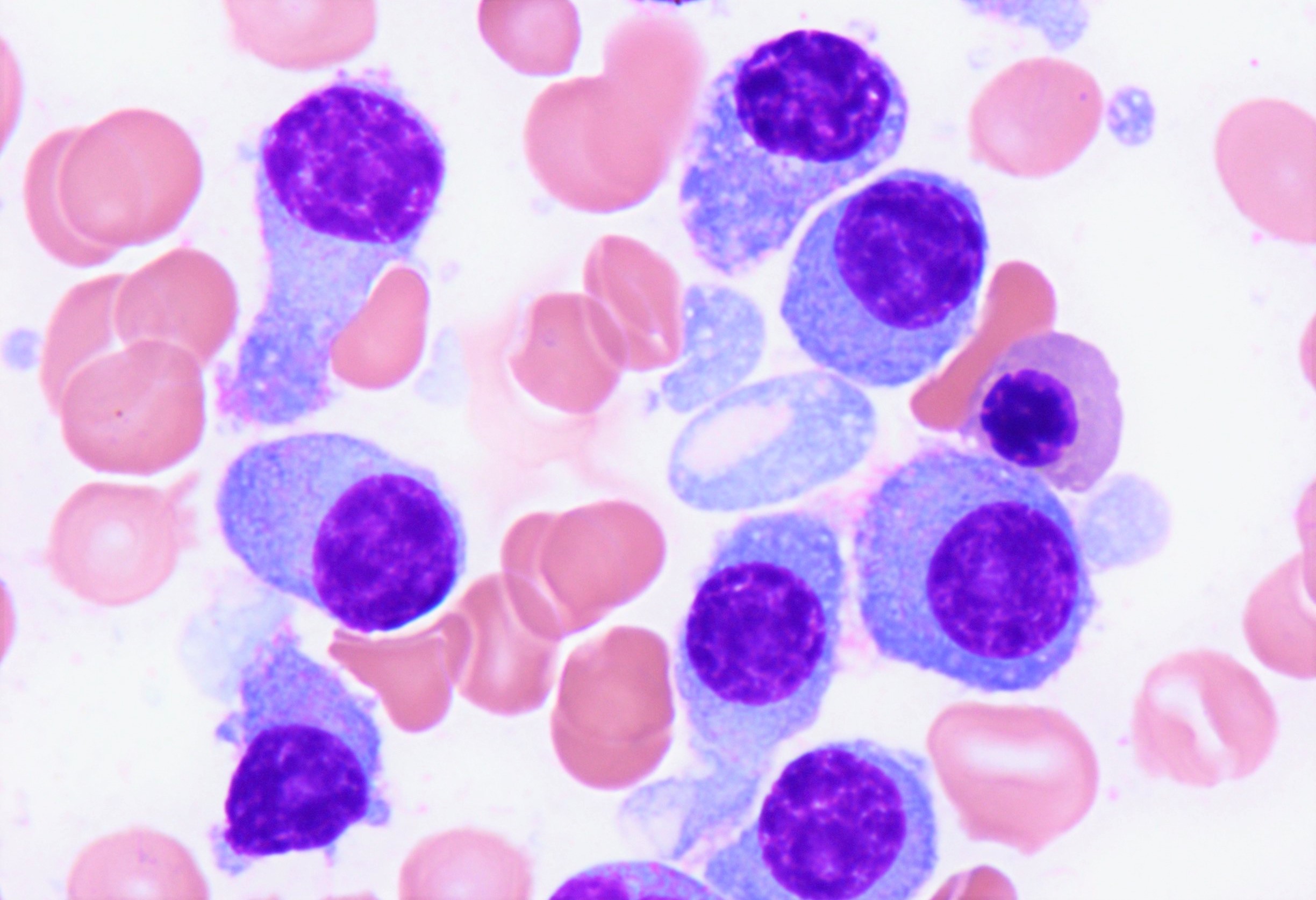 Identify type of connective tissue cell
Plasma cells
300
 Identify the type of CT cells, mention the stain
Fat cells, Sudan III
400
A section of the periodontal ligament shows parallel acidophilic fiber bundles with rows of flat nuclei between them. Which tissue type is this?
A. Loose areolar connective tissue
B. Dense regular connective tissue
C. Dense irregular connective tissue
D. Reticular tissue
B. Dense regular connective tissue
400
A lymph node section treated with silver impregnation shows a fine black network. Which fiber type is demonstrated?
A. Type I collagen
B. Elastin fibers
C. Reticular fibers
D. Type IV collagen
C. Reticular fibers
400
A gingival biopsy shows cells with eccentric cartwheel nuclei and a pale perinuclear zone. Which cell is this, and what is its function?
A. Mast cell: releases histamine
B. Plasma cell: secretes antibodies
C. Macrophage: phagocytoses bacteria
D. Eosinophil: kills parasites
B. Plasma cell: secretes antibodies
400
Identify the CT cell in the EM photo
Mast cell
400
 Identify CT fiber, Mention the stain
Reticular fibers, silver stain
500
Which feature best distinguishes a macrophage from a plasma cell on electron microscopy?
A. An eccentric nucleus
B. Numerous lysosomes with little rER
C. A basophilic cytoplasm
D. Derivation from mesoderm
B. Numerous lysosomes with little rER
500
You are designing a scaffold to repair a torn tendon. Which design best mimics the native tissue?
A. A randomly arranged elastin network seeded with adipocytes
B. Type II collagen seeded with chondrocytes
C. A fine type III reticular network seeded with plasma cells
D. Parallel-aligned type I collagen seeded with fibroblast-like cells
D. Parallel-aligned type I collagen seeded with fibroblast-like cells
500
Why are elastic fibers poorly visible in H&E sections, whereas collagen fibers are prominent?
A. Elastic fibers are destroyed during tissue processing
B. Collagen is selectively stained by silver impregnation
C. Elastic fibers are thin, whereas collagen bundles are thick
D. Elastic fibers are present only in embryonic tissue
C. Elastic fibers are thin, whereas collagen bundles are thick
500
 Identify CT fibers, mention the stain
Elastic fibers, orcein
500
 Identify the CT fibers, mention the stain
Collagen fibers, H&E