Mixed Epidermal Growths
Benign Epidermal Growths
Cancers
Dermal & Subcutaneous Growths
Random
100
Medium to dark brown macule, subtly accentuated skin lines
junctional melanocytic nevi
100
Risk factors for Verruca
!INOCULATION of SKIN by 1 or MORE HPV! atopic dermatitis, immunosuppression (HIV, meds, malignancies), sexual activity, nail biting, occupations (e.g. Wet Work or Butcher)
100
main etiology for melanoma
UVB and/or UVA exposure (more UVA)
100
have characteristic "dimple sign" due to connection with underlying tissue
Dermatofibroma
100
characteristic appearance of "warty/waxy and stuck-on" appearance
Seborrheic Keratosis
200
Intradermal melanocytic nevi
200
HPV types associated with increased risk of malignant transformation
16, 18, 31, 33, 35
200
Why Acral Melanoma is concerned "scary", especially that under finger nail?
usually discovered late, nail unit worse because there is no subcutaneous layer so it metastasizes faster
200
etiology of dermatofibroma
REACTIVE response to minor trauma (insect bite, impacted hair, shaving injury)
200
pathogenesis of freckles
hypertrophic, hyperactive melanocytes in response to UV radiation (more melanosome deposition in keratinocytes
300
the definition of A B C D E of atypical (dysplastic) nevi
A - asymmetrical B - border (uneven) C - color (variety, not the same)
D - diameter (over 6 mm) E - Evolving (change in size, shape, color, elevation)
300
transmission for verruca (warts)
direct contact, autoinoculation, fomite
300
Major origin of squamous cell carcinoma (SCC is most likely to develop from this)
actinic keratosis
300
Firm, mobile, well-defined subcutaneous nodule with a central puncture, is spherical to oblong in shape
Epidermal inclusion (sebaceous) cyst
300
Erythematous scaling papules or patches, usually rough surface, adherent yellow crust may develop
Actinic keratosis
400
Verry common in adults (50-60% occurrences over 60 years old), but higher incidence in those with type II DM
Acrochordons (skin tags)
400
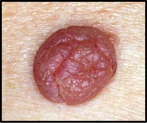
Contain threaded capillaries
Verruca Vulgaris & Verruca Plantaris
400
How are Lentigo Maligna and Lentigo Maligna Melanoma different?
Lentigo Maligna is non-invasive (flat and grows outwards)
Lentigo Maligna Melanoma is invasive (spreads lower and into dermis)
400
Difference between keloid & hypertrophic scar
hypertrophic scar stays within original wound borders, keloids spread beyond boundary of original wound
400
Difference between freckles and lentigines
lentigines are larger than freckles and are not transient (always present), also have small risk of transformation into malignant melanoma
500
Difference between acrochrodon & neurofibroma
neurofibroma has "buttonhole" invagination on compression during palpitation
500
Difference between Clavus & Verruca Plantaris
clavus is more symmetrical & has a plug (invagination) rather than seeds [presentation of thrombosed capillaries]
500
type of basal cell carcinoma that resembles scar tissue
Morpheaform
500
What should you do if you find melanoma at depth over 0.75 mm?
send to surgical oncology & consider lymph node biopsy or gene profile assay
500
A 45-year-old woman presents with multiple new, asymptomatic, pigmented papules on her upper back. On exam, the lesions are round, sharply demarcated, have a "stuck-on" appearance, and vary in color from tan to dark brown. Dermoscopy reveals comedo-like openings and milia-like cysts. She is worried they may be cancerous. Which of the following best supports your diagnosis over malignant melanoma?
A. Irregular borders with surrounding erythema
B. Positive dimple sign with central pallor
C. Warty, waxy surface with lack of ulceration or bleeding
D. Rapid change in lesion size and color with spontaneous regression
warty, waxy surface with lack of ulceration or bleeding supports a diagnosis of seborrheic keratosis, not malignant melanoma. Seborrheic keratoses are benign epidermal neoplasms with characteristic "stuck-on" appearance, often featuring comedo-like openings and milia-like cysts under dermoscopy. They generally lack the asymmetry, irregular borders, color variation, and evolving nature seen in melanomas