Red Flags
Single Ventricles
Syndromes
I think it's normal
Misc
100
Left-sided obstructive lesions have decreased flow to
BODY
100
Name a common type of single ventricle.
HLHS
Tricuspid Atresia
Ebstein Anomaly
DORV
DILV
AVC
100
What is demonstrated here that is a common finding in Connective Tissue Disorders?
Dilated aortic root
100
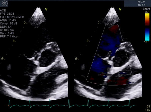
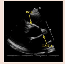 Do these coronaries have normal or anomalous origin?
Normal
100
Is this function normal or abnormal?
Abnormal
200
What lesion is this demonstrating?
L-TGA
200
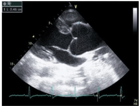
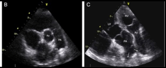
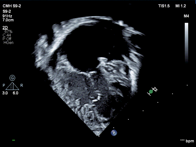
What type of single ventricle is this?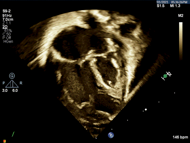
HLHS
200
What syndro?me is associated with COA
Turner Syndrome
200
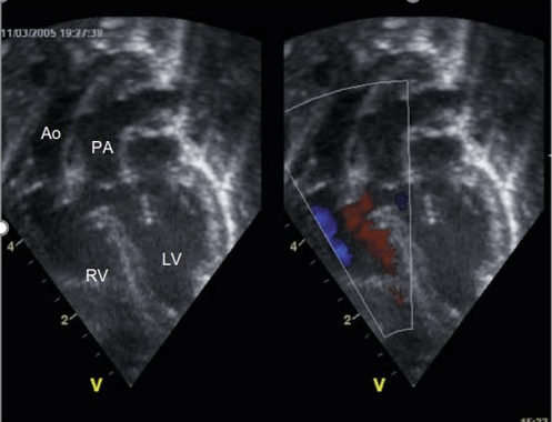
This PLAX view demonstrates what about the relation?
The are in continuity.
200
What is the likely mechanism of the MR?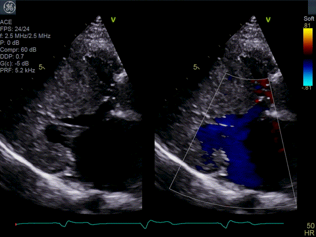
SAM from HOCM
300
Which image is a red flag?
The one on the right
300
Name the 3 stages of repair for a single ventricle.
Norwood
Sano
Fontan
300
What syndrome is associated with this defect and what defect is it?
Trisomy 21
AVC
300
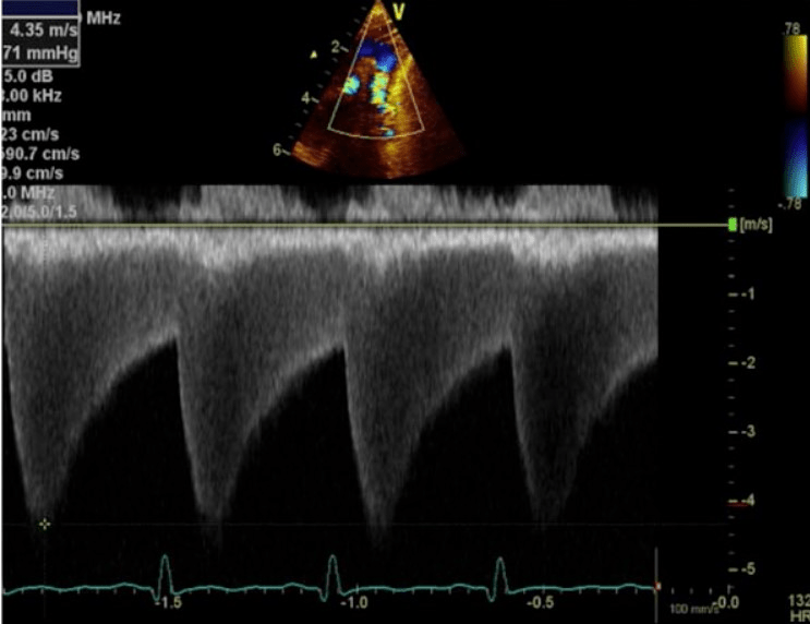
 The Doppler waveform of this Glenn looks normal. Why or why not?
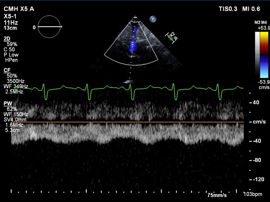
Normal, low velocity, passive flow
300
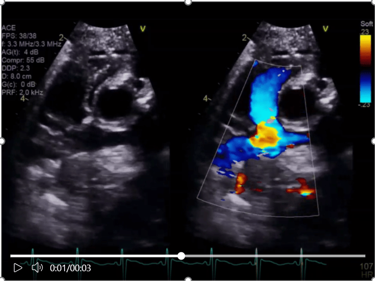
What does this demonstrate about the PV and TV?
They are not in continuity.
400
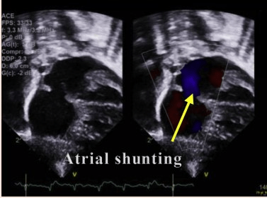
What is the red flag seen here?
Right to left atrial shunting
400
What are normal oxygen saturations for a repaired single ventricle?
78%
400
Williams syndrome is associated with what echo finding?
Supravalvar aortic stenosis
400
What is the laminar, blue flow in the image below?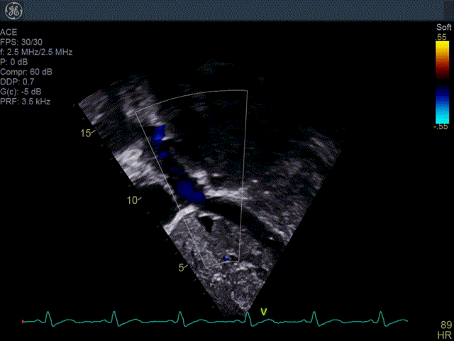
Fontan flow
400
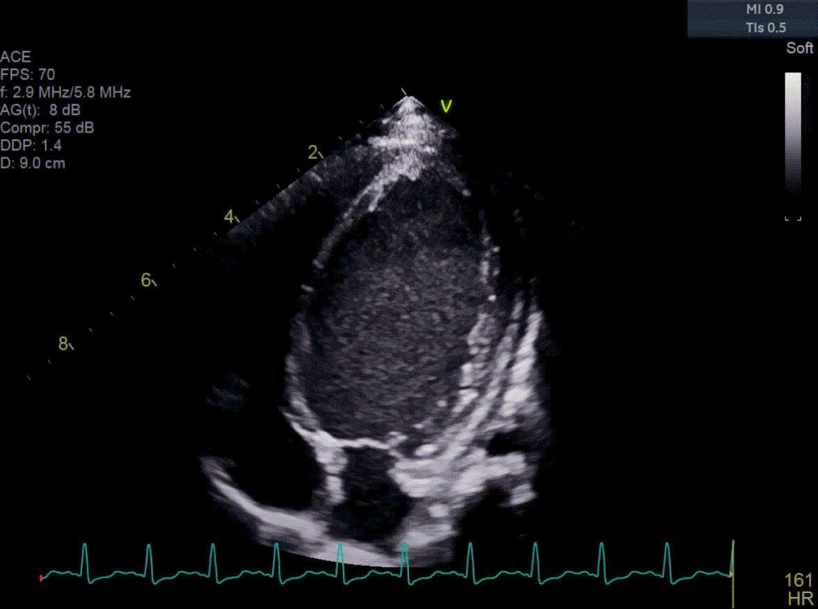
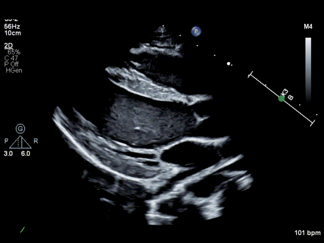
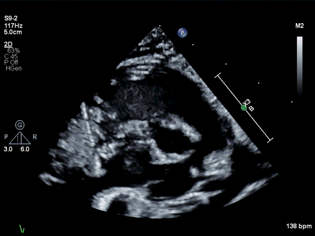
What type of Single Ventricle?
HLHS
500
Name the 5 T's of Ductal Dependent lesions.
1. Truncus
2. TGA
3. TA
4. TOF
5. TAPVR
500
Name the 3 parts of the Norwood operation.
Atrial septectomy
BTT shunt or Sano
DKS
500
Name 3 types of Connective Tissue Disorders?
Marfan
Loey Dietz
Ehler Danlos
500
What repair was performed?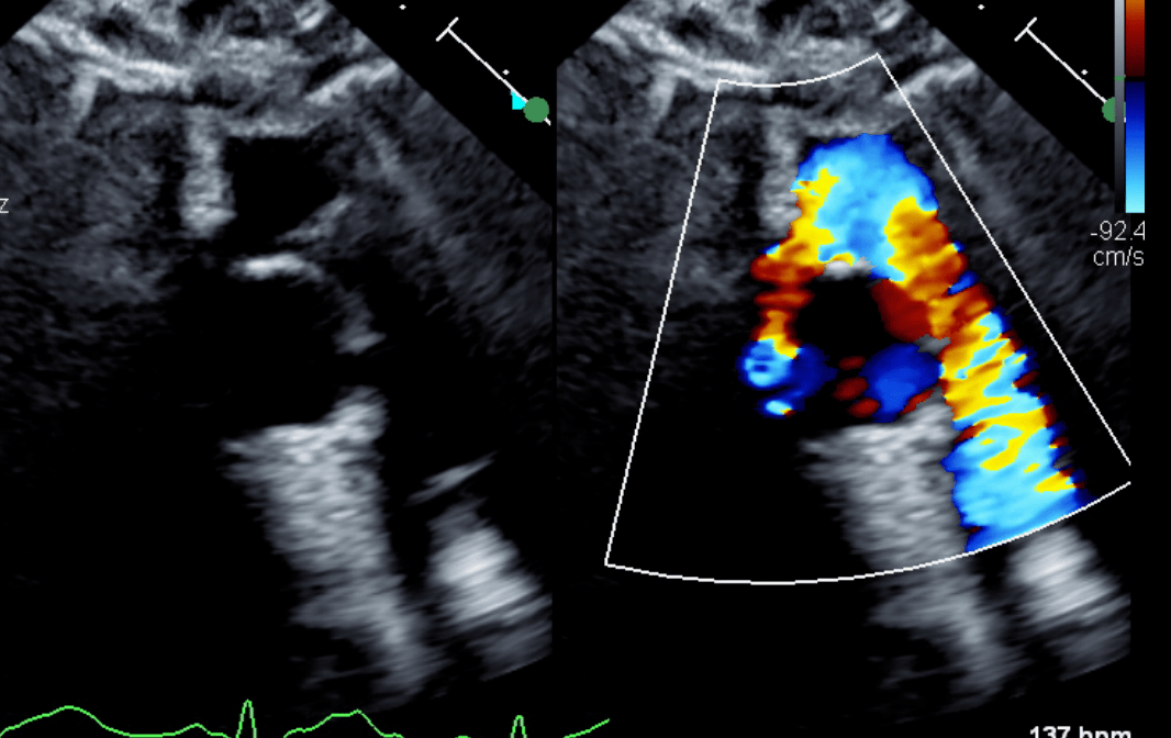
ASO, Le Compte
500
What is this demonstrating post repair?
VSD patch leak