A
B
C
D
E
100
Pulmonary circuit meaning
to and from the lungs
100
How many sulci does the heart have?
The heart has 3 sulci.
100
what are the 2 primary layers of pericardium?
1. Fibrous pericardium
2. Serous pericardium
100
Where will you find the epicardium?
the epicardium covers the outer surface of the heart
100
what divides the left and right ventricles (internally)?
the interventricular septum
200
what artery carries deoxygenated blood?
pulmonary artery (slide 7/47)
200
describe the location of the coronary sulcus.
The coronary sulcus runs between the atria and the ventricles
200
Which layer of the pericardium is dense, irregular tissue?
The fibrous pericardium is dense, irregular tissue.
200
what is the myocardium?
the myocardium is the muscular layer of the heart.
200
True or False:
the right ventricle is thicker than the left ventricle.
False, the left ventricle is thicker.
200
systemic circuit meaning
to and from body tissues
200
describe the location of the anterior interventricular sulcus
the anterior interventricular sulcus runs between the left and right ventricles
200
True or False:
The heart is encased within a membranous fluid-filled sac, the pericardium.
True
200
What is the endocardium?
The endocardium makes up the inner surface of the heart.
200
What is the name of the ridges inside the anterior right atrium?
pectinate muscles
400
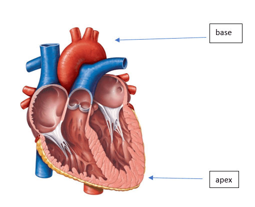
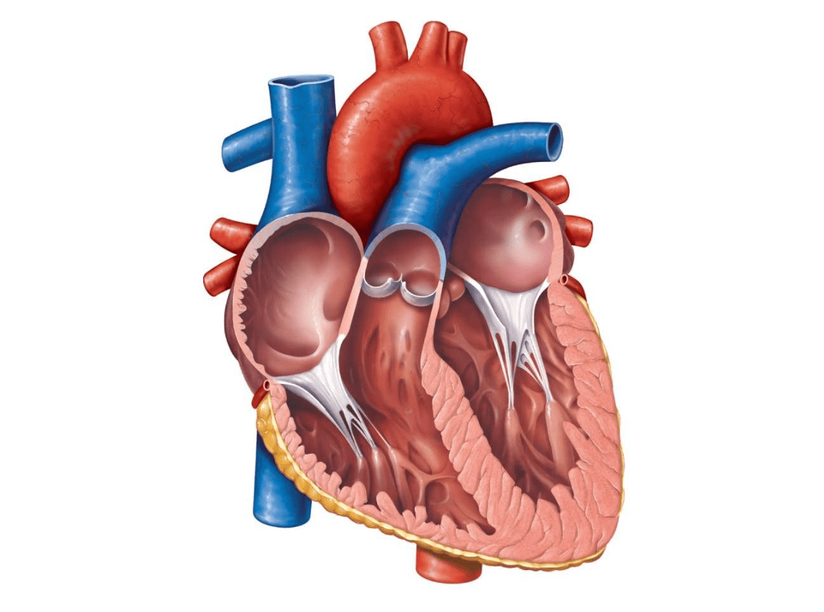 Where is the apex of the heart? Where is the base?
400
describe the location of the posterior interventricular sulcus
the posterior interventricular sulcus lies between the ventricles.
400
What is the purpose of pericardial fluid?
Lubrication
400
How is the myocardium arranged?
The myocardium (muscle) is arranged in spiral and circular patterns.
400
what is the fossa ovalis?
a depression in interatrial septum
[remnant of foramen ovale]
500
True or False:
blood vessels anchor the apex of the heart.
False: Blood vessels anchor the base of the heart.
500
The heart is enclosed and held in place by what?
Pericardium
500
What are the three layers of the heart wall?
1. Epicardium
2. Myocardium
3. Endocardium
500
how many chambers are there in the human heart?
4 chambers: 2 atria, 2 ventricles.
500
These three things make of the internal walls of the right ventricle
Trabeculae carneae
papillary muscles
chordae tendineae