Diastolic dysfunction is a problem with what portion of the cardiac cycle?
Ventricular Relaxation
What does the left ventricle’s inability to relax affect
LV filling pressures, affecting inflow from the left atrium
What grade of diastolic dysfunction is also referred to as pseudo normal?
Grade II
What grade of diastolic dysfunction is also referred to as restrictive?
Grade III
What does the “E wave” represent in regard to Mitral Inflow with PW Doppler?
Passive filling phase
or
Initial phase of rapid LV filling
What effect does increased pressure in the left atrium have on the left atrial size
Causes it to be enlarged
With grade I diastolic dysfunction what is the expected mitral inflow pattern
E to A reversal
Decrease in E wave, increase in A
What does the “A wave” represent in regard to Mitral Inflow with PW Doppler?
Atrial contraction
What can we assume about LA pressures if left atrial volume is enlarged?
Increased left atrial pressures
What value is considered normal for the Septal e’
8 cm/s
When determining left atrial size what is the ideal measurement used?
Left atrial volume
What type of Doppler do we use to obtain tissue Doppler?
PW, CW, or Color
PW
What is the normal E/A ratio parameter?
E/A 1-1.5
What is the normal E-wave max velocity?
<1.3 m/s
What is the normal E-wave Deceleration Time?
150 - 240 m/s
What is the normal A-wave max velocity?
0.2 – 0.35 m/s
What value is considered normal for the Lateral e’
10 cm/s
When grading diastolic function what left atrial volume index value is considered normal?
< 34 mL/m2
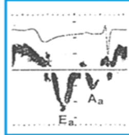
What grade of diastolic function would be expected with the above tissue Doppler pattern?
Normal diastolic function
What is the parameter of the E to A ratio with Grade II diastolic dysfunction
E/A 0.8 - 2.0
What is the parameter of the E to A ratio with Grade I diastolic dysfunction
E/A < 0.9
What average E/e’ ration is considered normal in our scan lab?
< 15
When grading diastolic function what tricuspid regurgitation velocity is considered normal?
< 2.8 m/s
When grading diastolic function what IVRT duration is considered normal
60 – 100 ms
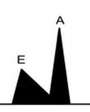
What grade of diastolic dysfunction would be expected with the above mitral inflow pattern, with normal LV systolic function?
Grade I
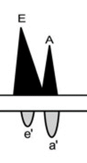
What grade of diastolic dysfunction would be expected with the above mitral inflow pattern (flow noted on top), and the bottom flow pattern obtained from tissue Doppler?
Grade II
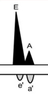
What grade of diastolic dysfunction would be expected with the above mitral inflow pattern (flow noted on top), and the bottom flow pattern obtained from tissue Doppler?
Grade III
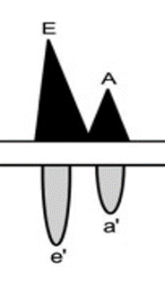
What grade of diastolic dysfunction would be expected with the above mitral inflow pattern, with normal LV systolic function, and the bottom flow pattern obtained from tissue Doppler?
Normal diastolic function
With grade II diastolic dysfunction what is the expected mitral inflow pattern, and the expected tissue doppler pattern.
Pseudo-normal
Appears at first to be a normal inflow pattern, with E larger than A. Tissue doppler will demonstrate E to A reversal with E smaller than the A wave.
With Grade III diastolic dysfunction will the deceleration time increase or decrease?
Decrease