Pediatrics
Quick Facts
Adults
Special
Considerations
Considerations
100
6-month-old female infant presents to clinic with parents complaining of congestion and decreased energy. She has decreased po intake and urine output. There has been no vomiting. Baby is afebrile, RR 50, O2 is 94 On physical exam, baby is lightly fussy, and you notice nasal flaring with mild subcostal retractions. You auscultate and hear diffuse rhonchi. What is the most likely diagnosis?
RSV
Learning point: Bronchiolitis is the most common respiratory illness affecting infants and young children worldwide. It most often occurs between 2 and 6 months of age. Can present with mild fever, respiratory distress, and rhonchi.
100
The most common bacterial cause of pneumonia across all age groups
Streptoccocus pneumonia.
Empiric antibiotics are normally tailored to the following most common organisms:
Streptococcus pneumoniae, Haemophilus influenzae, Mycoplasma pneumoniae, Staphylococcus aureus, Legionella species, Chlamydia pneumoniae, and Moraxella catarrhalis.
100
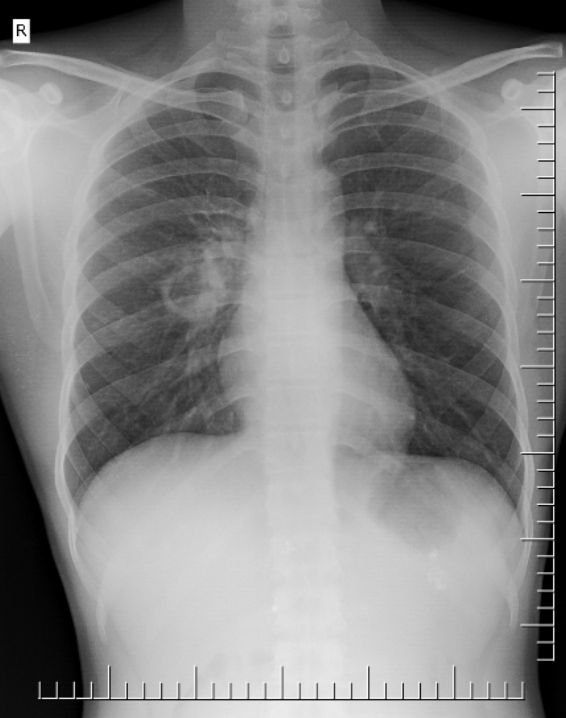
64 yo male with hx of COPD presenting for 2 day hx of cough, shortness of breath, increased sputum production which has started to turn yellow. Physical exam notable for diffuse expiratory wheezing. CXR is below. List top 3 ddx.
COPD exacerbation, viral respiratory infection, bronchitis, pneumonia.
100
A 38-year-old man recently moved from the Philippines presents with 3 weeks of productive cough, intermittent low-grade fever, drenching night sweats, and a 5-kg weight loss.
Vitals: 38.2°C, Pulse: 98 bpm, BP 116/70, RR 20, O2 96% on RA.
He appears thin and mildly diaphoretic. Pulmonary auscultation reveals crackles and bronchial breath sounds over the right upper lung field.
Laboratory studies including CBC, CMP, and respiratory viral panel are unremarkable except for a mild leukocytosis. Sputum Gram stain is negative for bacteria.
Chest radiograph is pictured below:
Guess the organism.
Tuberculosis.
Other differentials to consider include fungal infections (histoplasmosis, invasive aspergillosis, blastomycosis), septic emboli, malignancy, sarcoidosis
200
An 8-year-old male presents to ED with parents complaining of shortness of breath and decreased energy for 1 day. He has decreased appetite and urination. He is febrile to 38.5C, RR 30 (normal <20), O2 is 96. On physical exam, his eyes are listless, and you notice nasal flaring. You auscultate and hear crackles on the right anterior chest. Chest X-Ray reveals a focal RML consolidation. What is the most likely diagnosis?
Bacterial Community Acquired Pneumonia
Learning objective:
Bacterial pneumonia tends to have abrupt onset of symptoms which include fever, cough, and focal chest pain. Physical exam can be notable for focal lung findings of crackles or decreased lung sounds.
200
Top 3 common causes of CAP in children less than 2
RSV (42%), Rhinovirus (29%), Adenovirus (18%).
Bacterira: Strep pneumo (3%), S. aureus (1%), mycoplasma 2%
200
35 yo female presented to ED complaining of congestion, cough, chest tightness, 2 days. She has a couple of bouts of nausea and vomiting. Vitals a notable for low grade fever but are otherwise normal. On physical exam is notable for diffuse wheezing. CXR demonstrates multifocal consolidation in b/l lungs with peribronchial thickening.
What is the most likely diagnosis?
Bonus: Do you start antimicrobials?
Answer: Influenza Pneumonia/viral pneumonia
Bonus: Depends
Influenza is the most common viral cause of pneumonia in adults Tamiflu within 2 days of illness (5 days for paxlovid for COVID infections). Per 2019 American thoracic society guidelines, it is recommended to start empiric abx for influenza pneumonia due to higher risk of coinfection. Influenza tends to predispose to s. aureus infections.
200
A 15-year-old boy with known sickle cell disease (HbSS genotype) presents with 2 days of fever, pleuritic chest pain, increasing cough, and shortness of breath. He reports new onset of hypoxia and worsening back pain.
Vitals: T: 38.8°C, P: 120 bpm, BP: 102/64, RR 28/min, and O2: 91% on RA.
PE: He appears ill and tachypneic. Lung sounds reveal decreased breath sounds and crackles at the right lower lung field. There is mild scleral icterus and evidence of digital clubbing. No focal neurologic deficits are present.
Laboratory studies show: hemoglobin 7.2 g/dL (baseline 8.5), WBC 16,000/μL (neutrophil predominant), reticulocyte count 8%, platelets 320,000/μL, and total bilirubin 3.2 mg/dL. Blood cultures are pending. Chest radiograph demonstrates a new right lower lobe infiltrate with mild volume loss and no pleural effusion.
List 3 differential diagnoses
Acute chest syndrome, community acquired pneumonia, vasoocclusive pain, severe anemia, Pulmonary thrombosis, cardiac ischemia
300
24-month-old male presented to ED with parents complaining of progressively worsening congestion and decreased energy for 4 days. He has decreased appetite and urination. He is afebrile, RR 55, O2 is 93. He placed on HFNC with improved RR to 30 (normal) and O2 now is at 97. On physical exam, he is sleeping, and you notice mild nasal flaring with intercostal retractions. You auscultate and hear diffuse wheezing. CXR demonstrates multifocal consolidation in b/l lungs with peribronchial thickening. RVP is positive for human metapneumovirus. What is the most likely diagnosis? Bonus: Do you start antibiotics?
Answer: Viral Pneumonia
Bonus: No
Viral Pneumonia is usually distinguished from bacterial pneumonia in that fevers tend to be more mild if present, onset of symptoms are gradual, and symptoms are often diffuse and bilateral. Antibiotic initiation is typically avoided, but in severe illness, or atypical features, antibiotics could be considered.
300
Top 3 causes of CAP in children 10-17 years old
Answer: Mycoplamsa pneumoniae (23%), Rhinovirus (19%), influenza (11%).
Virus: RSV (7%), Coronavirus (4%), human Metapneumovirus (4%).
Bacteria: Strep Pneumo (3%), S. aureus (1%), Strep pyogenes (<1%)
300
A 34-year-old male with a hx of iv drug use presents with 7 days of fever, productive cough with foul-smelling sputum, and progressive dyspnea. He reports mild hemoptysis and malaise.
Chest radiograph demonstrates a thick-walled cavitary lesion with an air-fluid level in the right upper lobe, surrounded by consolidation.
What is the most likely causative organism?
Staph Aureus.
Other organisms to consider: oral anaerobes, endemic fungal pneumonia, M. tuberculosis, atypical mycobacteria
Risk factors for S. aureus infection include injection drug use, influenza, and structural lung disease.
300
A 58-year-old female with rheumatoid arthritis, currently taking prednisone 30 mg daily for the past 6 weeks, presents with 10 days of progressive exertional dyspnea, nonproductive cough, and low-grade fever. She is febrile to 38.0°C, pulse 102 bpm, she is normotensive, respiratory rate 24/min, and oxygen saturation 87% on room air. Lung exam reveals diffuse fine crackles bilaterally. CT demonstrates extensive ground-glass infiltrates.
What is the most likely causative organism?
Answer: Pneumocystis jirovecii pneumonia
Corticosteroid therapy is a well-established risk factor for PJP, with increased risk at doses ≥20 mg prednisone daily for ≥4 weeks, as described by the American Thoracic Society
400
A 15 yo male presents to clinic with gradual onset of malaise, sore throat, low grade fever, and persistent dry cough over 5 days. He has never had these symptoms before. Vitals are notable for fever of 100.8F and are otherwise unremarkable. You hear mild diffuse wheezing on physical exam.
What is the best next step in his diagnostic workup?
What is the most likely diagnosis?
Answer: CXR- looking for
Bonus: Atypical pneumonia
Learning Point: Atypical Pneumonia typically presents with a nonproductive cough and low-grade fever with a milder disease progression than typical bacterial pneumonia
Radiographic findings may be greater than expected from the clinical features and commonly include uni- or bilateral lower lobe or perihilar bronchopneumonia with reticulonodular opacity, bronchial cuffing and linear atelectasis.
400
What autoimmune condition is the most notorious mimicker in pulmonary radiography?
Sarcoidosis.
Learning point: Like lupus for everything else medicine, sarcoidosis can mimic many other pathologies including CAP in the right clinical context. Classic presentation of Sarcoidosis occurs in a young or middle-aged adult—often female—presenting with bilateral hilar lymphadenopathy on chest imaging (bat wing sign), with or without pulmonary symptoms such as dry cough, dyspnea, and chest discomfort.
400
A 45-year-old male with a hx of alcohol use disorder presents with confusion, decreased appetite, and mild cough after a night of drinking a 5th of vodka. He is has a mild fever and is tachypneic with crackles in the right lower lung field. Chest radiograph shows a new consolidation in the RLL.
What organisms if any would you be concerned about in this case?
Oral anaerobes/gut enteric bacteria
Learning point: Aspiration pneumonia/ pneumonitis often will cause pneumonia in the RLL due to the steeper angle of the right mainstem bronchus. Alcohol use is a risk factor for aspiration due to suppression of the gag reflexes and relaxation of the lower esophageal sphincter.
400
A 22-year-old woman with cystic fibrosis presents with 5 days of increased cough, thicker and more purulent sputum, and worsening dyspnea. She reports mild pleuritic chest pain and has noticed a 2-kg weight loss over the past week. She denies fever or hemoptysis.
Vitals: T: 37.6°C, P: 104 bpm, BP: 110/68, RR 22/min, and O2 93% on RA.
PE: She appears thin. Lungs notable for diffuse coarse crackles and digital clubbing. No cyanosis is present.
Chest radiograph demonstrates increased peribronchial markings, patchy infiltrates, and upper lobe bronchiectasis without new consolidation or effusion.
What are the 2 most likely organisms
1. Pseudomonas aeruginosa
2. Staph aureus
3. Burkholderia cepacia
500
A 2 week old AGA female born Term via uncomplicated SVD presents to the ED with parental concerns for tachypnea and increased work of breathing. Vitals are notable for fever of 101F. Sepsis workup obtained and notable for CXR with Left Lower lobe infiltrate with air bronchograms. RVP is pending. List 3 most likely causative infectious organisms that will guide your antimicrobial management.
Bacterial pneumonia (GBS is most common, E. Coli, C. trachomatis, Listeria monoctyogenes, S. aureus)
Viral pneumonia (HSV, Flu, possibly covid)
500
What fungal causes of community acquired pneumonia are native to Texas?
Coccidioides, Aspergillus, Histoplasmosis, Cryptococcus +/-Blastomyces
Learning point:
Lots of sources of possible fungal infections. Some endemic areas can have 30% of CAP cases come from Coccidiodes.
"In patients with suspected CAP from the endemic area for coccidioidomycosis, we suggest initial serological testing with close clinical follow-up and serial testing (conditional recommendation, moderate-quality evidence)"
500
A 56-year-old man with a history of COPD presents to the emergency department with 3 days of high fever, malaise, headache, and watery diarrhea. He reports a nonproductive cough and mild shortness of breath. He recently returned from a business trip where he stayed at a hotel.
Patient is febrile to 39.7°C, heart rate 92 bpm, blood pressure 110/68 mmHg, respiratory rate 24/min, and oxygen saturation 92% on room air. He has crackles at the left lung base.
Labs demonstrate: WBC 8,500/μL, sodium 128 mmol/L, AST 68 U/L, ALT 54 U/L, and C-reactive protein markedly elevated. Sputum Gram stain shows many neutrophils but no organisms.
Chest radiograph demonstrates a patchy left lower lobe infiltrate with early consolidation and no pleural effusion.
Which of the following is the most likely diagnosis?
Classic features of Legionella infection: high fever, relative bradycardia, gastrointestinal symptoms (notably diarrhea), confusion, hyponatremia, mild transaminitis, microscopic hematuria, and a nonproductive cough with patchy or rapidly progressive infiltrates on imaging.
Risk factors include COPD, Hotel/cruise ship stay
500
A 36-year-old man with HIV on ART presents with 3 days of fever, productive cough with yellow sputum, pleuritic chest pain, and dyspnea. He denies hemoptysis or night sweats.
Vitals: T 38.4°C, Pulse 102, BP 120/76, RR: 22, O2: 95% on RA.
He appears mildly ill but is alert and oriented. Pulmonary auscultation reveals decreased breath sounds and crackles over the right lower lung field; no egophony or wheezing is present.
Laboratory studies show: Leukocytosis, elevated CRP, and normal renal and hepatic function. His viral load and CD4 count are pending but his last one from 3 months ago showed a CD4 count of 300 (AIDS defined as <200). Chest radiograph demonstrates a right lower lobe segmental consolidation without effusion or cavitation.
What is the most likely diagnosis?
Bonus- How much is the mortality risk increased for this patient because of his diagnosis of HIV?
Answer: Streptococcus pneumoniae
Bonus: None*
Learning point: Common things remain common in HIV, the most number one cause for viral pneumonia in HIV is influenza and the most common bacterial cause of CAP is strep pneumoniae. *2025 Retrospective cohort study found no in-hospital mortality difference in patients with HIV without AIDs compared to those without HIV