LEAD the Way
Mind Your Ps & Qs
Achy Breaky Heart
What's taking so long?
PotPourri
100
Triaxial diagram
Leads I, II, and III joined at the middle
100
A normal PR Interval
0.12 - 0.20 seconds
100

Asystole
100
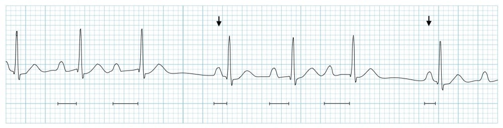
Wenckebach
100
Flight or Fight
Sympathetic nervous system
200
The six leads placed on the patients chest
Precordial Leads
200
What you count to determine Heart Rate
R to R Interval
200

Ventricular Tachycardia
200

SR with 1st AV block
200
S2, the second heart sound, reflects the closure of these
Aortic & pulmonic valve
300
V1 is placed here
Fourth Intercostal space, right sternal boarder
300
Represents ventricular repolarization on the ECG
T wave
300

Trigeminy
300

2:1 Heart Block
300
Returns deoxygenated blood to the right atrium from the lower chest and abdomen
Inferior Vena Cava
400
Used for accessing rhythms with wide QRS complexes, and can pinpoint abnormalities in ventricular conduction
MCL
400
The flat line between the T wave of one beat and the P wave of the next beat
Basline / isoelectric line
400

Agonal
400

3rd Degree Heart Block
400
The outermost layer of the heart
Epicardium
500
Einthoven's Law
Lead I + Lead III + Lead II
500
In this period, a strong stimulus will result in depolarization
Relative refractory period
500

Ventricular Fibrillation
500

2nd degree type II, Mobitz II
500
This separates the right and left ventricle
Interventricular septum