IT ALL STARTS SOMEWHERE
TWO LAYERS BECOME THREE
NEURULATION + NEURAL CREST
MESODERM, SOMITES, AND ADULT DERIVATIVES
FOLDING, GUT, AND BODY ORGANIZATION
100
This single diploid cell is formed when the male and female haploid gametes fuse.
What is the zygote?
100
This process converts the bilaminar embryonic disc into a trilaminar embryonic disc.
What is gastrulation?
100
Signals from the underlying notochord cause this midline region of ectoderm to thicken.
What is the neural plate?
100
Mesoderm becomes organized into these three major populations after gastrulation.
What are paraxial, intermediate, and lateral plate mesoderm?
100
During Week 4, folding incorporates the upper portion of the yolk sac into the embryo to form this structure.
What is the primitive gut?
200
During this process, the zygote undergoes rapid mitotic divisions without increasing in overall size.
What is cleavage?
200
this elongated structure develops from proliferating epiblast and establishes the cranio-caudal and left-right axes.
What is the primitive streak?
200
As the neural folds rise, the depression between them is called this.
What is the neural groove?
200
Paraxial mesoderm becomes segmented into these structures.
What are somites?
200
The region of the primitive gut receiving the vitelline duct is called this.
What is the midgut?
300
At approximately this cell number, the developing conceptus becomes known as a morula.
What is 16?
300
The primitive streak is located at this end of the embryonic disc.
What is the caudal end?
300
Fusion of the neural folds converts the neural groove into the lumen of this structure.
What is the neural tube?
300
Name the three major regions of a somite and one adult derivative of each.
Sclerotome → axial skeleton
dermatome → dermis
myotome → skeletal muscle.
300
The coelom divides lateral plate mesoderm into these two layers.
What are parietal and visceral mesoderm?
400
Fertilization normally occurs in this region of the uterine tube.
What is the ampullary region?
400
Mesoderm normally occupies the space between ectoderm and endoderm except at these two membranes.
What are the oropharyngeal/buccopharyngeal membrane and the cloacal membrane?
400
This migratory cell population forms structures including nerve ganglia, Schwann cells, melanocytes, adrenal medulla, and many cranial connective tissues.
What is the neural crest?
400
This mesodermal region lies between paraxial and lateral plate mesoderm and develops into the urogenital system.
What is intermediate mesoderm?
400
Cephalocaudal folding moves the developing heart from a position cranial to the oropharyngeal membrane to this new relationship.
What is underneath and caudal to the oropharyngeal membrane?
500
Trace the developmental sequence from fertilization through the first named multicellular stages, ending with formation of the blastocyst.
Zygote → blastomeres through cleavage → morula → blastocyst.
500
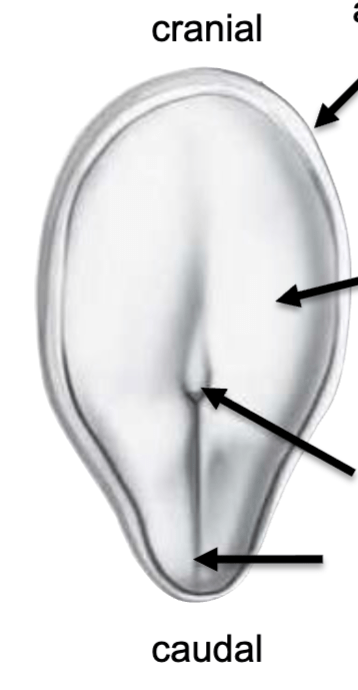
Name the structures cranial to caudal that have arrows pointing to them.
What is:
Amnion
Non-Proliferating Epiblast
Primitive Node
Primitive Groove
500
Trace the developmental sequence from the notochord to formation of the neural tube.
Notochord signaling → neural ectoderm → neural plate → neural folds and neural groove → fusion of neural folds → neural tube.
500
Adult vertebrae, ribs, spinal nerves, and intercostal vasculature all show a repeated segmental organization. What embryonic developmental process helps explain this shared pattern?
Somitogenesis, or segmentation of paraxial mesoderm into somites.
500
Trace two major consequences of Week 4 folding: one involving the digestive system and one involving the cardiovascular system.
Folding incorporates part of the yolk sac into the embryo to form the primitive gut, while also repositioning the cardiogenic mesoderm and developing heart from cranial to the oropharyngeal membrane to underneath and caudal to it.