Ch. 9.1 - Skeletal Muscle Overview
Ch. 10 - Muscle Tissue
Ch. 10 - Muscle Tissue II
Ch. 11 - Nervous Tissue
Ch. 11 - Nervous Tissue II
100
What is a muscle insertion?
The portion of a muscle that attaches to bone & moves.
100
What is distensibility and elasticity?
Distensibility - The ability for muscle cells to stretch up to 3 times their resting length without damage.
Elasticity - The ability for muscle cells to return to their original shape after stretching.
100
What is muscle tone?
A small amount of tension generated by a muscle at rest due to involuntary activation of motor units by the brain & spinal cord.
100
What type of neural circuit would be useful for sending information to a singular part of the brain?
Converging circuit
100
What divisions and subdivisions of the nervous system would be responsible for stomach pain?
Peripheral nervous system, Sensory (afferent) division, Visceral sensory division
200
What is a pennate muscle?
A muscle that attaches to a tendon at an angle in a way that resembles a feather.
Unipennate - Single tendon, "feather" sticks out on one side
Bipennate - Single tendon, "feathers" stick out on both sides
Multipennate - Multiple tendons; multiple "feathers" attached to each other
200
What are the main types of energy sources for skeletal muscle?
Immediate energy - Creatine phosphate can readily donate its phosphate an ADP to produce ATP.
Glycolytic - ATP acquired from glycolysis.
Oxidative - ATP acquired from oxidative phosphorylation.
200
How is isotonic concentric contractions different from isotonic eccentric contractions?
Isotonic concentric contractions shorten muscle by increasing force. (i.e. Lifting an object)
Isotonic eccentric contractions lengthen muscle by decreasing force. (i.e. Lowering the object)
200
How do electrical synapses differ from chemical synapses?
Electrical synapses are bidirectional, have nearly instantaneous transmission, & use gap junctions
200
List the catecholamines
Norepinephrine (noradrenaline), Epinephrine (adrenaline), Dopamine
300
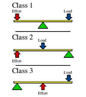
How are the parts in a secondary lever arranged?
The fulcrum is on one end of the lever, the load is in the middle, and the force/effort is on the other end of the lever.
300
What proteins are thin filaments made of and what do they do?
Actin - Have active site to bind to myosin
Tropomysoin - Wraps around actin to block its active site
Troponin - Binds to calcium to shift the tropomyosin to reveal the actin active sites
300
What is wave summation and what can occur as a result?
Repeated stimulation of a muscle fiber by a motor neuron to generate muscle contractions with progressively greater tension. Can result in unfused or fused tetanus.
Unfused Tetanus - Partial relaxation between contractions
Fused Tetanus - No relaxation between contractions
300
What are the two types of summation?
Temporal summation - A single presynaptic neuron repeatedly releases neurotransmitters to cause an action potential
Spatial summation - Multiple presynaptic neurons simultaneously release neurotransmitters to cause an action potential300
What could happen to neurotransmitters after a synaptic transmission ends?
Diffusion & Absorption - Some neurotransmitters diffuse through away from the synaptic cleft
Degradation - Enzymes break down certain neurotransmitters
Reuptake - Some neurotransmitters return to the presynaptic neuron they were released from
400
Describe how are skeletal muscle fibers organized into muscles (organ level).
Skeletal Muscle Fibers wrapped by Endomysium -> Fascicles wrapped by Perimysium -> Multiple fascicles wrapped by Epimysium -> Entire muscle wrapped by Muscle fascia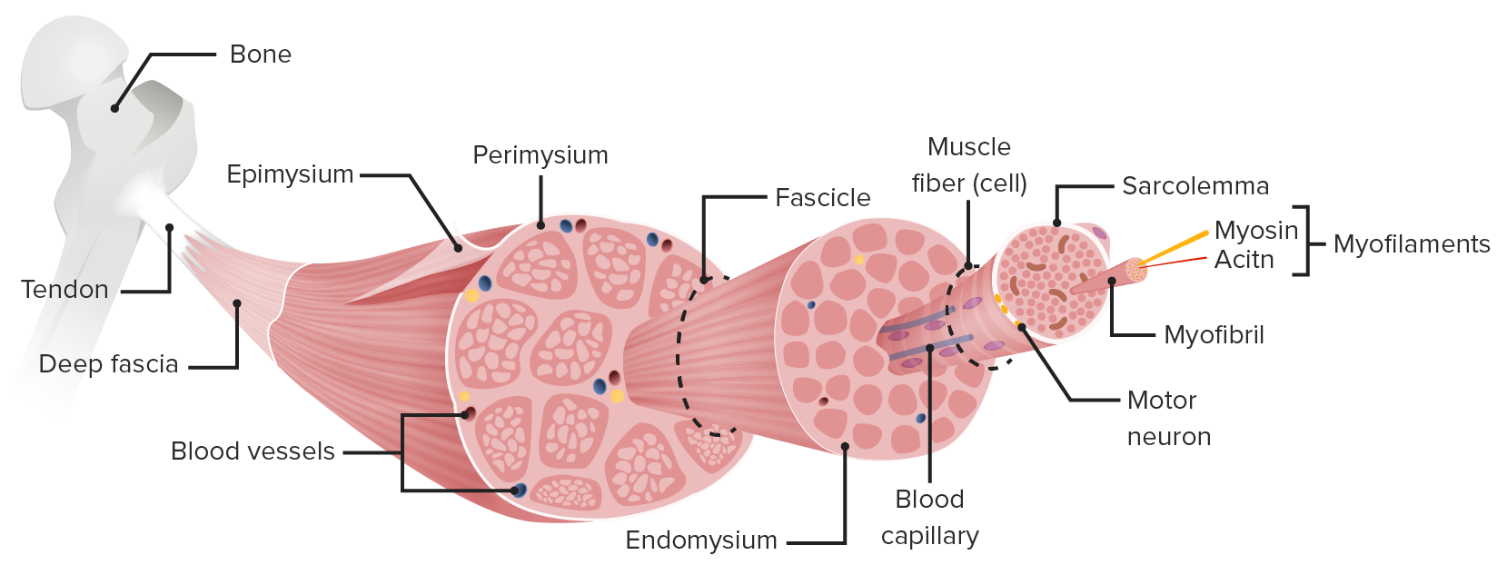
400
What factors contribute to the generation of the resting membrane potential?
Sodium ion leak channels allow sodium ions in to the cell, potassium ion leak channels allow potassium ions out of the cell, & potassium ions diffuse more easily than sodium ions through their respective leak channels; more cations leak out of the cell than leak in, which causes the inside of the cell to become less positive/more negative.
400
Describe the different classes of muscle fibers.
Slow Twitch (Type I) - Small/intermediate diameter, slow & less forceful contraction, maintain extended contraction, slow oxidative
Fast Twitch (Type II) - Large diameter, faster contractions, tire more quickly
Type IIa - Fast oxidative glycolytic
Type IIx - Fast glycolytic, extremely fast & powerful contractions
400
What are the neuroglia in the brain and what do they do?
Astrocytes - Maintain the blood-brain barrier
Oligodendrocytes - Myelinate certain axons
Microglial cells - Immune function/Act as phagocytes
Ependymal cells - Produce & spread cerebrospinal fluid (CSF)
400
Why is continuous conduction slower than saltatory conduction?
In continuous conduction, each section of the axolemma must be depolarized to threshold.
In saltatory conduction, the myelin sheaths help insulate electrical charge, so current flow is more efficient.
500
What functional muscle groups are there other than the prime mover and what do they do?
Antagonist - Muscle that opposes and slows the prime mover (agonist)
Synergist - Muscle that works with the agonist by guiding movement and ensuring it's smooth; some help with joint stability
Fixator - Muscle that holds bone in place
500
How does acetylcholine (ACh) cause calcium to be released into the muscle cell's cytosol?
ACh binds to ligand-gated channels, which allows cations into the muscle cell & causes local depolarization. Local depolarizations open voltage-gated channels, allowing an action potential to form & then propagate down the T-tubules. The depolarization of T-tubules causes calcium ions to be released from the SR.
500
In smooth muscle contraction, how does calcium activate the crossbridge cycle?
1. Calcium ions will bind to a protein called calmodulin (Cam).
2. Calcium ion-Cam complex will activate the myosin-associated enzyme myosin light-chain kinase (MLCK).
3. MLCK causes activation of myosin ATPase.
4. Crossbridge cycle begins
500
What occurs in a chemical synapse after calcium ions flow in to the presynaptic neuron?
Calcium ions cause synaptic vesicles carrying neurotransmitters to fuse with the presynaptic membrane to release their content.
The released neurotransmitters will bind to ligand-gated ion channels on the postsynaptic neuron to allow cations in to cause an action potential.
500
How does an action potential travel down an axon?
Via propagation; a section of the axolemma depolarizes to create an action potential to open nearby voltage-gated sodium ion channels, allowing another action potential to form and the previously depolarized part to repolarize. This process repeats until the action potential reaches the axon terminal.