A picture is worth a thousand words
Mixtape
Spittin Facts
Thousands of Words
100
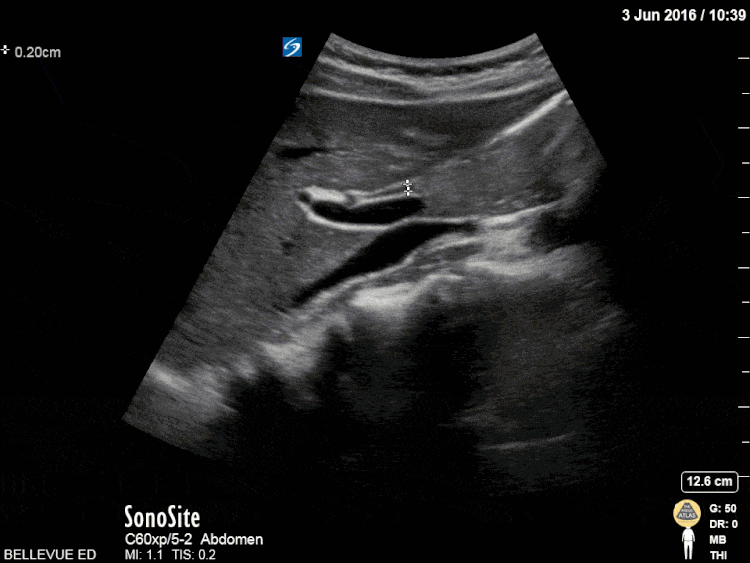
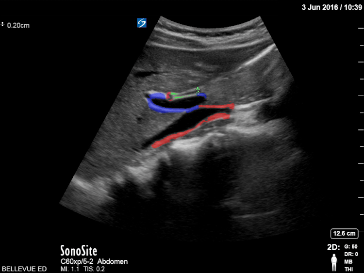
Name all the structures in the image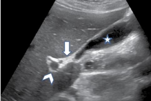
Star = gallbladder
Arrowhead = portal vein
Arrow = Main lobar fissure
100
Sonographic features of a gallstone
Hyperechoic focus
Posterior acoustic shadowing
Dependent area
100
Threshold at which the gallbladder wall is considered thickened.
3 mm (though I, Dr. G, personally don't pay attention until it's 4 mm)
100
Name the pathological finding in this image
Pericholecystic fluid
200
Name this finding
Edge artifact
200
A patient presents with upper abdominal pain. You place the transducer on the patient's abdomen to initiate a RUQ scan, but the patient cannot tolerate the pressure. What can you do to perform the study.
Perform the exam via the intercostal window (aka "X minus 7")
200
The accepted definition of a positive sonographic Murphy sign.
Maximal pain eleicited by pressing over the fundus of the gallbladder with an ultrasound transducer.
200
Name this normal variant.
phrygian cap

300
Name this sign and explain why it's helpful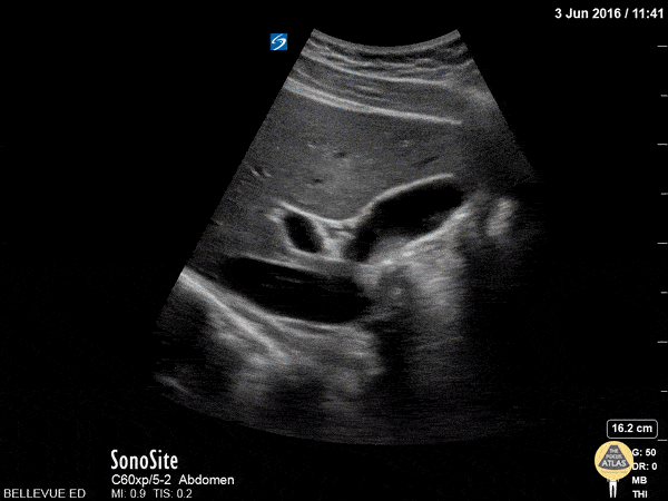
Exclamation point sign. Helps orient you to the location of the gallbladder and or portal triad.
300
Structures that form the portal triad
Hepatic artery
CBD
Portal Vein
300
The 5 sonographic findings consistent with acute cholecystitis
Gallbladder wall thickening
Pericholecystic fluid
Hydropic gallbladder
Sonographic Murphy
300
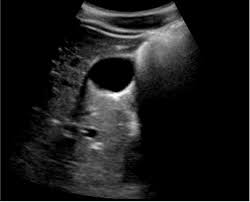
A patient presents with abdominal pain. You obtain the following POCUS image. What is the next best step in evaluation?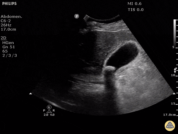
Turn the patient (e.g. left lateral decubitus) and re-scan to determine if it is an impacted gallstone.
400
What is the name of this finding?
Wall Echo Shadow (WES)
400
Structure that visually links the gallbladder to the portal triad
Main Lobar Fissure
400
A common bile duct that is this diameter (or greater) is considered dilated in the general population
4 millimeters (an additional millimeter is allowed for each additional decade of life after 40)
400
Name the pathological artifact and nonpathological artifact found in this image.

posterior acoustic shadowing
side lobe artifact
500
What is the name of this landmark sign?
Olive Sandwich
500
A limitation of the intercostal approach for evaluating the gallbladder
Cannot elicit sonographic murphy
500
A common bile duct that is this diameter (or greater) is considered dilated in postcholecystectomy patients
1.0 cm
500
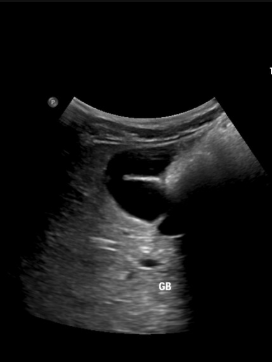
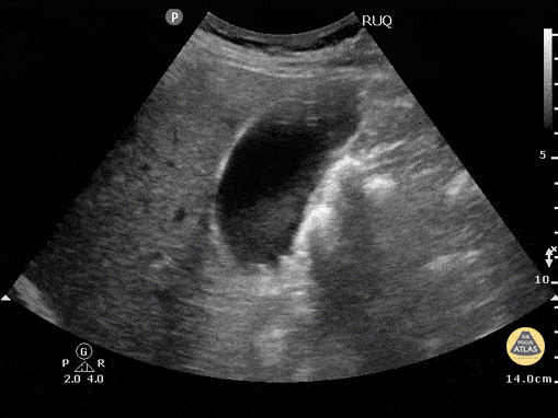
A 42 year old female with no known PMH presents with abdominal pain. The following image is obtained of her gallbladder. What is your differential diagnosis?
![]()
The patient has a (likely) thickened gallbladder wall with pericholecystic fluid.
Ddx thickened gallbladder wall includes:
Acute cholecystitis
Chronic cholecystitis
Malignancy
Postprandial state
Heart/Renal/Liver failure