Upper Extremity Blood Supply
Joints of the Shoulder Complex
Muscles of the Shoulder
Muscles of Shoulder cont'd
Muscles of shoulder cont'd
Muscles of Shoulder and Articulations
Arm/Forearm/
hand
hand
Forearm and Hand
Hand and Lower Extremity
Lower Extremity
Muscles of Lower Ext
Enter Category Name
Enter Category Name
100
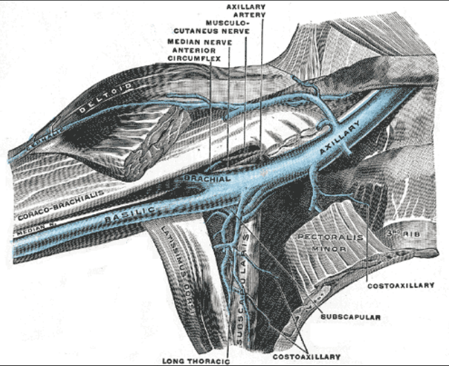
Continuous with the Subclavian Artery begins at the lateral border of the first rib and is continuous with the Brachial artery at the inferior border of the teres major?
What is Axillary Artery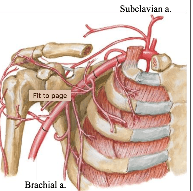
100
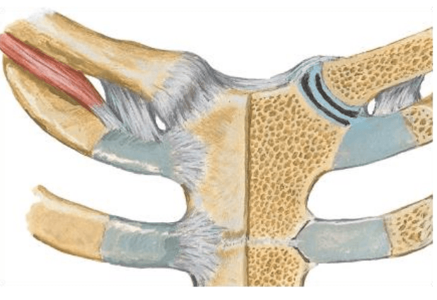
Sternal End of the Clavicle and Manubrium of the Sternum and type Saddle Synovial Joint.
What is the Sternoclavicular Joint
100
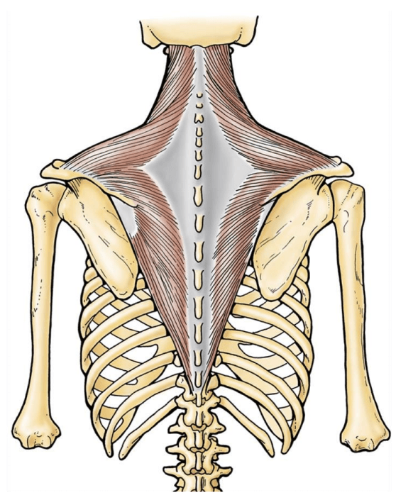
Attaches at the base of the skull, spinous process of C7-T12 and scapula spine, acromion, lateral 1/3 clavicle.
What is the Trapezius Muscle
100
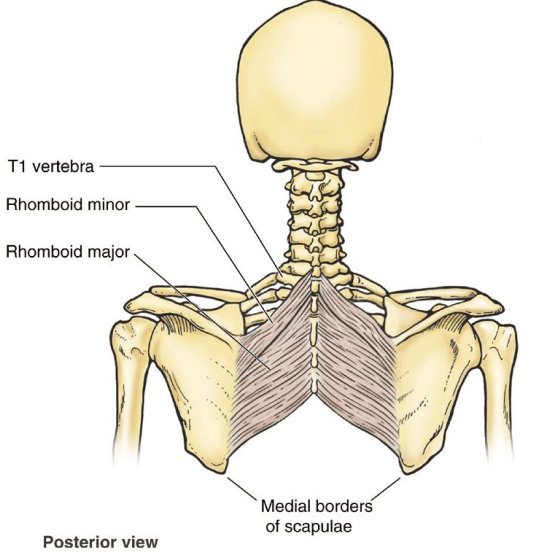
The action of the Rhomboid Major and Minor
What is aDduction/retraction and downwardly rotate the scapula
100
Action of the Serratus Anterior
What is: holds scapula against the thoracic wall, protracts, and upwardly rotates the scapula
100
The action of the Rotator Cuff Muscles
What is aBudction and rotation of the shoulder
100
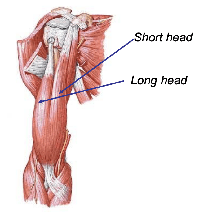
The short head attaches to the coracoid process of the scapula, the long head attaches to the supraglenoid tubercle and attaches distally to the radial tuberosity
What is the Biceps Brachii
100
Innervation of the Extensor Compartment (Wrist Extensors, extensor Digitorum, extensor Indicis proprius, Extensor Digiti Minimi)
What is the Radial Nerve
100
Innervation of the Hypothenar Group
What is the Ulnar Nerve
100
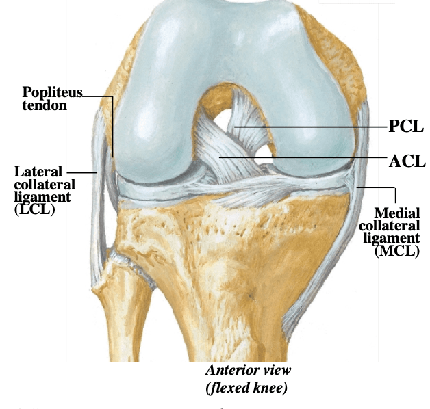
Attaches Distal femur to the fibula and other attaches Distal Femur to the tibia, respectively they are known as...
What is the LCL (lateral Collateral Ligaments) and MCL (Medial Collateral Ligaments)
100
The Vastus Medialis, Vastus Lateralis, Vastus Intermedius, and Rectus Femoris (2 movements) are responsible for this movement
What is knee extension
100
Name the labeled muscles 1-3
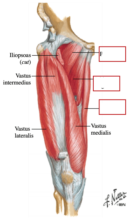
What is Pectineus, ADductor longus, Gracilis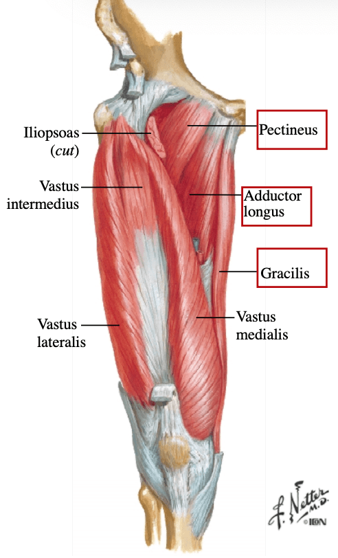
100
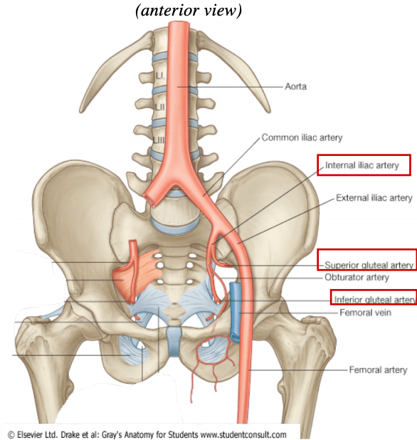
Gives rise to two branches that supply the gluteal region
What is Internal Iliac Artery
200
The Axillary Artery Comes from
What is the Aorta -->BT-->Subclavian artery -->Axillary Artery
200
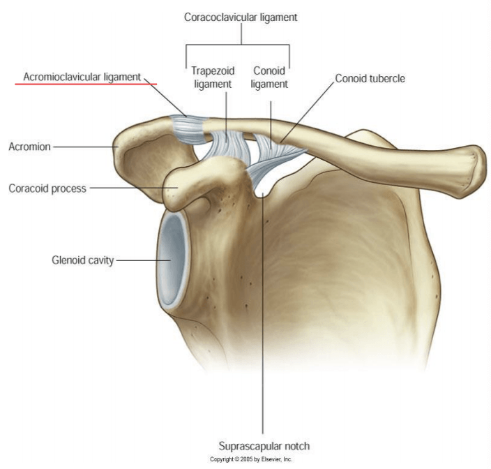
Joint at the Acromial End of the clavicle and acromion process of scapula, plane type synovial joint
What is the Acromioclavicular Joint
200
Innervation of the Rhomboid Minor and Major
What is the Dorsal Scapular Nerve
200
The action of the Latissimus Dorsi Muscle
What is shoulder extension, aDduction and internal rotation (IR)
200
Attaches to the coracoid process and the middle 1/3 of anterior humerus
What is the Coracobrachialis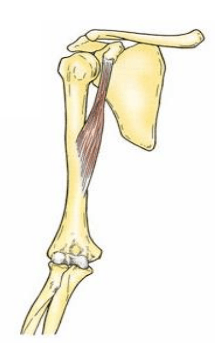
200
The innervation of the Deltoid Muscle
What is the Axillary nerve
200
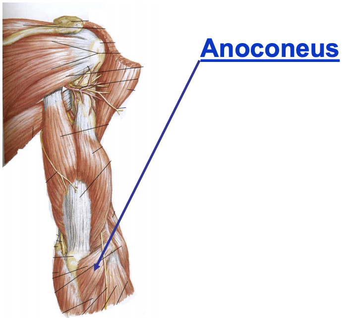
Attaches to the lateral epicondyle of the humerus and olecranon of the ulna
What is the Anoconeus Muscle
200
Innervation of the Intrinsic Thenar Group
What is the Median Nerve
200
Innervation of the Flexor Compartment (Flexor Digitorum Profundus, Flexor Digitorum Superficialis)
What is the Median and Ulnar Nerve
200
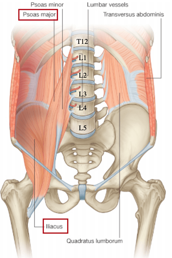
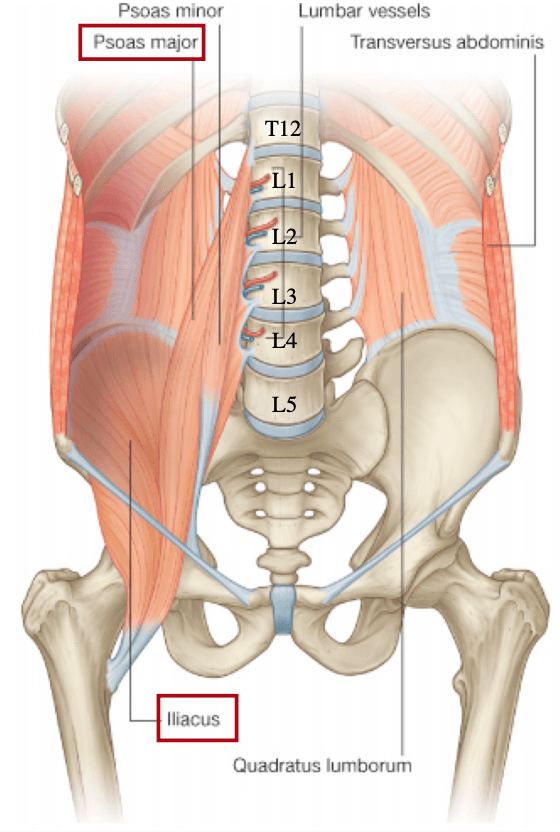
Innervation of the Psoas Major
What is the Lumbar Spinal Nerves
200
Attaches at the iliac fossa to lesser trochanter
What is the Iliacus
200
Attaches at the posterior ilium and greater trochanter (2 answers)
What is the Gluteus Medius and Gluteus Minimus
200
Innervation for the Gastrocnemius, Soleus, Plantaris, Flexor Hallucis, Tibialis Posterior, Flexor Digitorum Lonugs and Popliteus muscles
What is the Tibial Nerve
300
The major branch from the brachial artery (deep artery of the arm)
What is the The Brachial Profunda Artery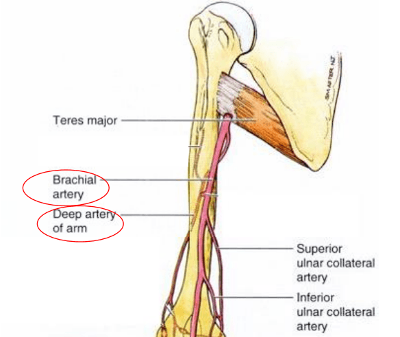
300
Type of movement provided by the Acromioclavicular joint
Elevation/Depression, Gliding between clavicle and scapula
300
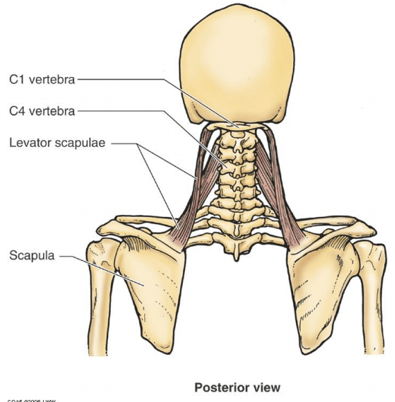
Attaches to the Transverse processes of C1-C4 and superior medial border of the scapula
What is the Levator Scapulae Muscle
300
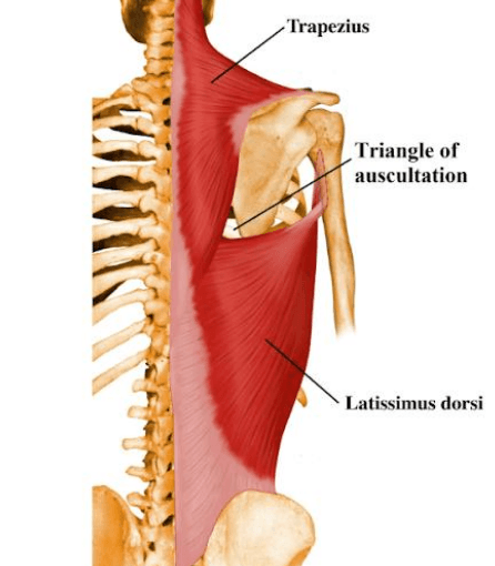
Boundaries for the site on the back where breath sounds may be easily heard through stethoscope
Latissimus dorsi, trapezius, medial border of scapula
300
Attaches the inferior, posterior surface of the scapula and intertubercular groove of humerus
What is the Teres Major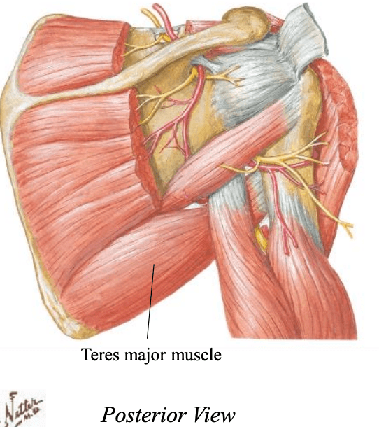
300
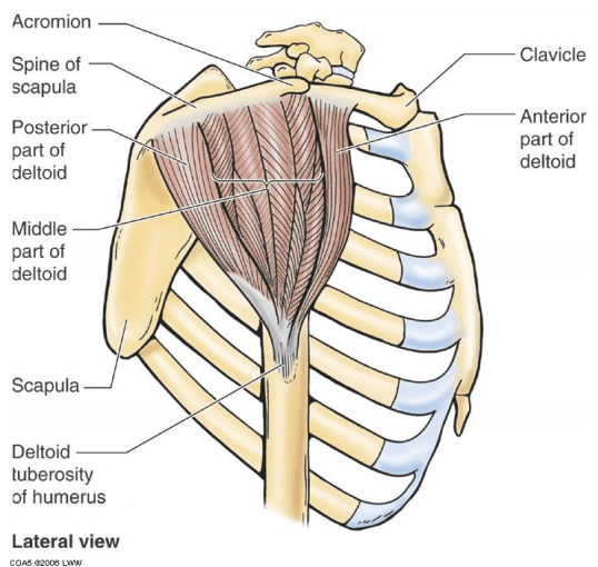
Attaches to the spine of scapula, acromion, lateral 1/3 clavicle, deltoid tuberosity of humerus
What is the Deltoid
300
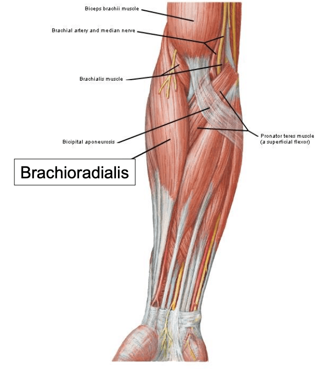
Attaches to the lateral, distal humerus
What is the Brachioradialis Muscle
300
The Joints responsible for Flexion and Extension of the digits 2-5
What is the MCP (Metacarpophalangeal), PIP (Proximal Interphalangeal) and DIP (Distal Interphalangeal) joints 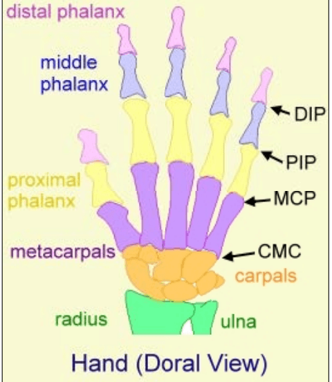
300
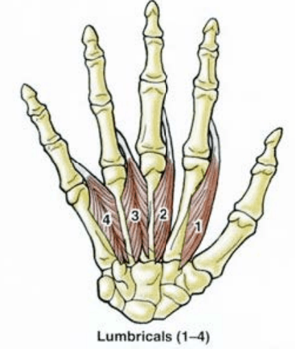
Attaches to the flexor tendons in palm and dorsum of proximal phalanges and cross on the anterior side of the MP joint
What is the Lumbricals
300
Articulation between the medial and lateral femoral condyles and medial and lateral tibial condyles
What is the Tibiofemoral joint
300
The Sciatic nerve gives rise to...
What is the tibial nerve and common fibular nerve
300
Attaches at the ASIS to the medial surface of the proximal tibia (pes anserinus) and is responsible for hip/knee flexion, aDduction, and external/lateral rotation
What is the Sartorius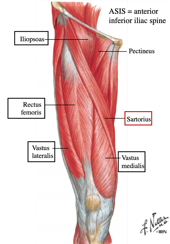
300
Innervation of the Lateral Compartment of leg: Fibularis (Peroneus Longus, Peroneus Brevi)
What is the Superficial Fibular (a branch from the common fibular nerve)
400
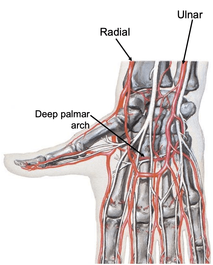
At the level of the Elbow, the brachial artery divides into these two blood supplies
What is the Radial Artery and Ulnar Artery 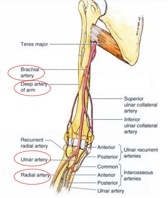
400
The movements of the Sternoclavicular Joint
What is Elevation/depression and protraction/retraction of the shoulder region
400
Nerve that innervates the Latissimus Dorsi Muscle
What is the Thoracodorsal Nerve
400
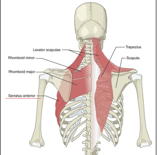
Attaches to the external surfaces of lateral parts of ribs 1-8, anterior surface of medial border of scapula
What is the Serratus Anterior
400
The innervation of the Coracobrachialis Muscle
What is the Musculocutaneous nerve
400
The action of the Anoconeus Muscle
What is extends the forearm
400
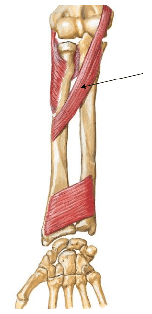
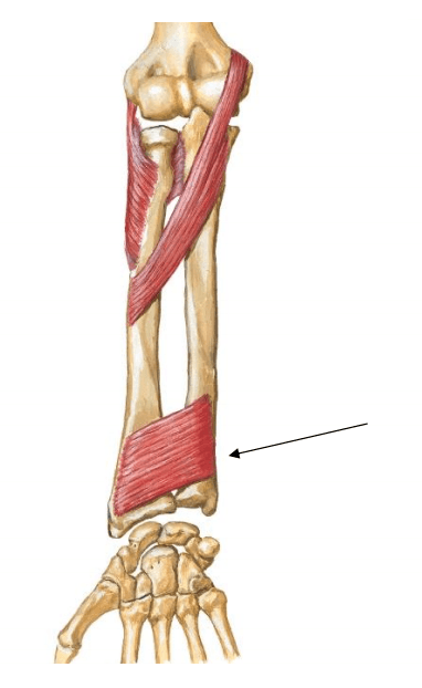
Attaches to the medial epicondyle, lateral surface of radius
What is the Pronator Teres
400
Attaches at the medial epicondyle to the middle phalanx of digits 2-5
What is the Flexor Digitorum Superficialis
400
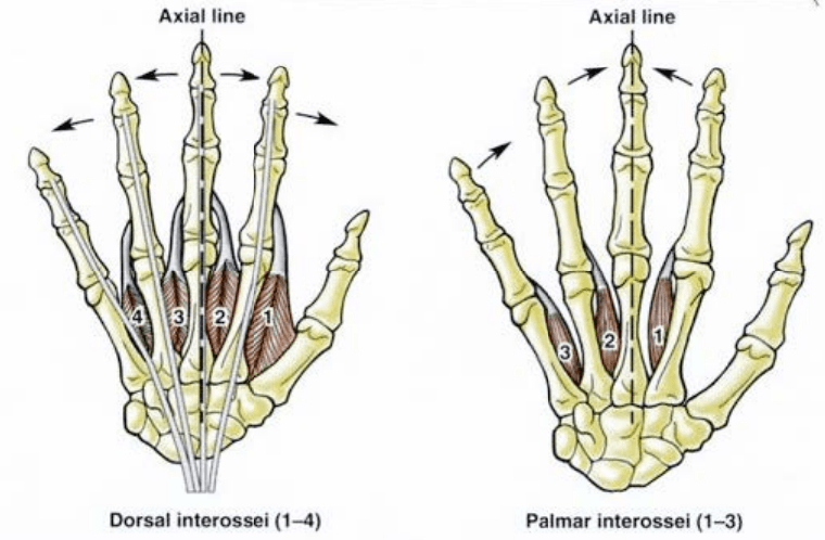
Attaches at the Metacarpals to adjacent proximal phalanges
What is the Dorsal and Palmar Interossei
400
Both attach from the femur to the tibia
What is the Anterior and Posterior Cruciate Ligament (ACL and PCL) 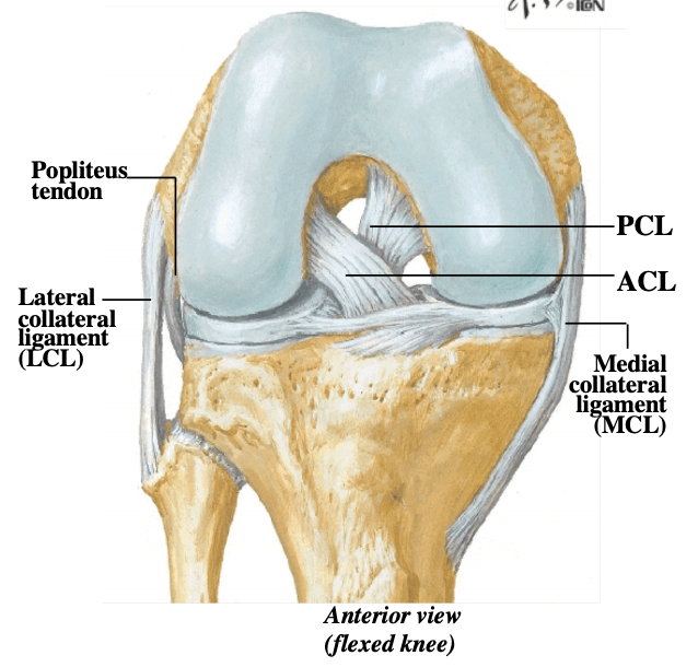
400
The Psoas Major, iliacus, Rectus Femoris (2 movements) and Sartorius (4 movements) are responsible for this movement
What is Hip Flexion
400
The innervation for the Gluteus Medius, Gluteus Minimus and Tensor Fascia latae
What is the Superior Gluteal Nerve
400
Innervation that provides all motor below the knee
What is the Tibial and common fibular nerves
500
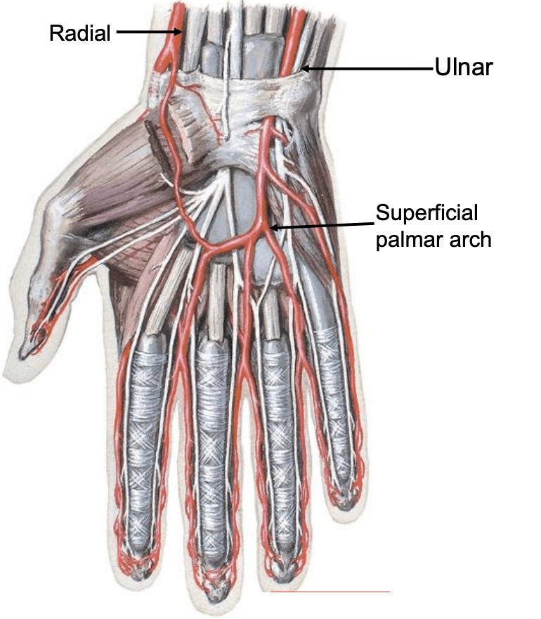
The Ulnar Artery Continues into the hand and is the main supply to the...
What is the Superficial Palmar Arch
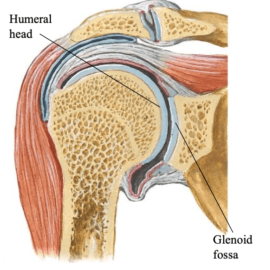
500
The Joint that articulates the humeral head and glenoid fossa, ball and socket type synovial joint
What is the Glenohumeral Joint
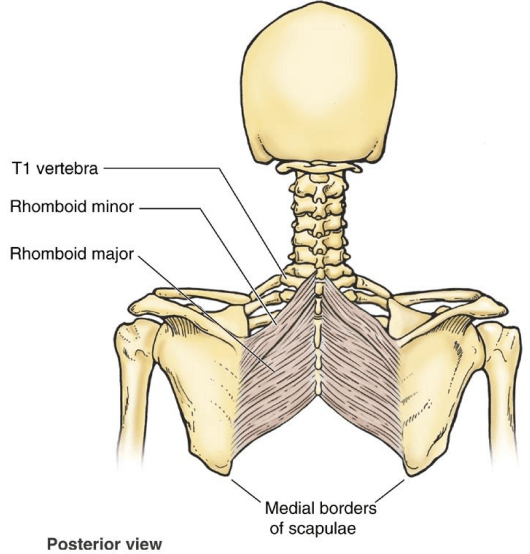
500
Attaches to the spinous processes T2-T5, the medial scapular border from the spine to the inferior angle
What is the Rhomboid Major
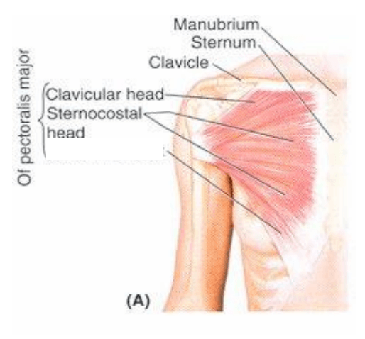
500
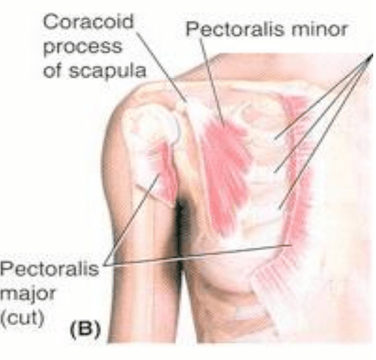
Attaches to the clavicle head: medial half of clavicle, sternocostal head: sternum, upper six 6 ribs both heads: intertubercular groove of humerus
What is the Pectoralis Major
500
The innervation of the Rotator Cuff muscles
What is varies
500
The innervation for the Biceps Brachii and Brachialis
What is the Musculocutaneous Nerve
500
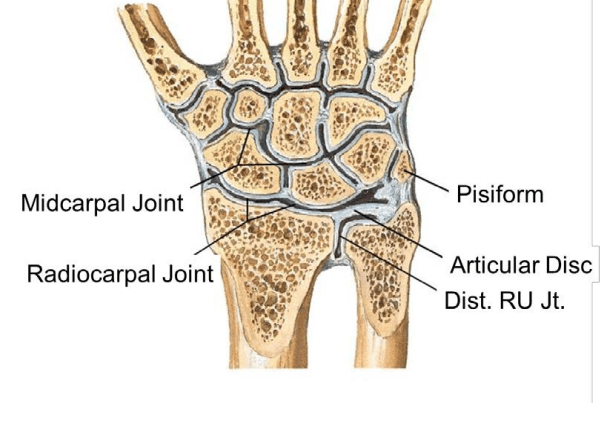
The condyloid type joint between the distal radius and proximal carpals
What is the Radiocarpal

500
Action of the Dorsal andn Palmar Interossei
What is ABduction (Dorsal) and ADduction (Palmar)
500
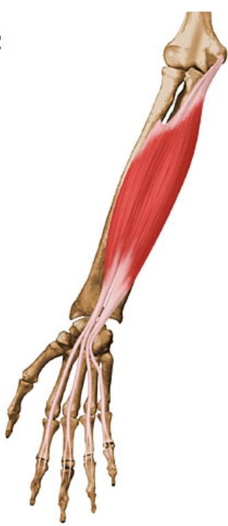
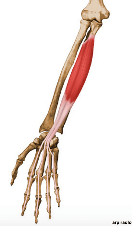
Name the muscle in the image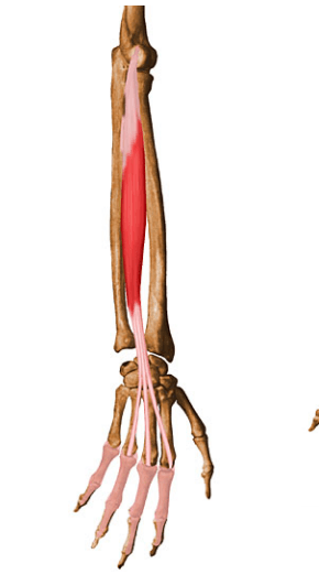
What is the Extensor Digitorum
500
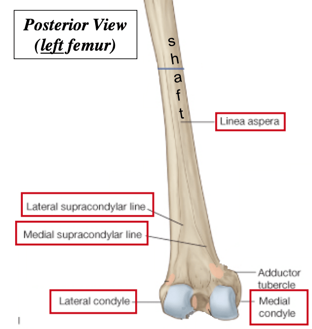
Label the distal Femur starting from 1-5
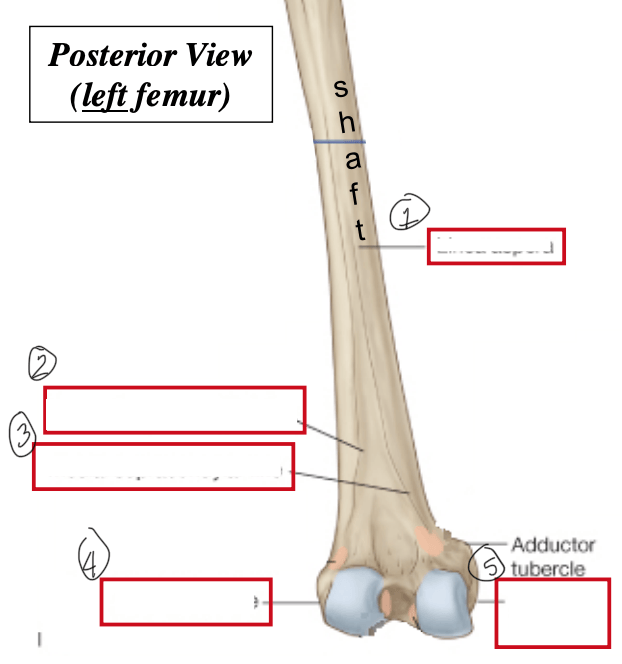
What is the Linea Aspera, Lateral Supracondylar Line, Medial Suprachondylar line, Lateral Condyle, and Medial Condyle
500
Innervation for the Gluteus Maximus
What is the Inferior Gluteal Nerve
500
The site of attachment for the Psoas Major
What is the lumbar vertebra to the lesser trochanter
500
Innervation of the Anterior Compartment of leg: Tibialis Anterior, Extensory Hallacucis Longus, Extensor Digitorum Longus
What is the Deep Fibular nerve (coming from the common fibular nerve)
600
The Radial Artery Continues into the hand and is the main supply to the...
What is the Deep Palmar Arch
600
Movement of the Glenohumeral Joint
What is Flexion/Extension, ABduction/ADduction, Internal and External Rotation
600
The action of the Trapezius muscle
What is (upper) elevates and upwardly rotates scapula, (middle) aDducts scapula, (lower) depresses and upwardly rotates scapula
600
The action of the Pectoralis Minor
What is Stabilizes scapula
600
The action of the Deltoid Muscle
What is flex shoulder, aBduct shoulder, extend shoulder
600
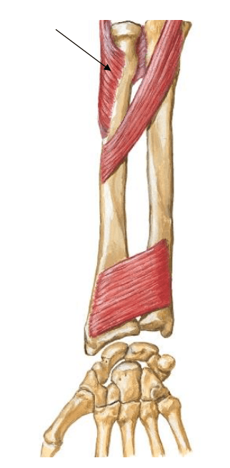
Attaches to the lateral epicondyle of humerus and proximal ulna
What is the Supinator Muscle
600
The plane synovial joint between the proximal and distal row of carpal bones
What is the midcarpal joint
600
Name the muscle in the image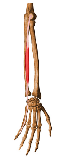
What is the Extensor Digiti Minimi
600
The Innervation for the Lumbricals
What is the Median and Ulnar Nerve
600
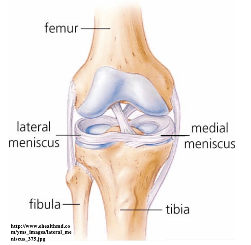
Crescent-shaped Fibrocartilagenous discs that increase joint congruency and stability
What is the Medial and Lateral Menisci
600
Innervation for the Semitendinosus, Semimembranous, and Biceps Femoris (long head)
What is the Tibial Nerve
600
The Gluteus Medius, Gluteus Minimus, Tensor Fascia Latae are responsible for which type of movement
What is Hip ABduction
600
Responsible for dorsiflexion of the foot
What is Tibialis Anterior (2), Extensor Hacucis Longus (2), Extensor Digitorum Longus (2)
700
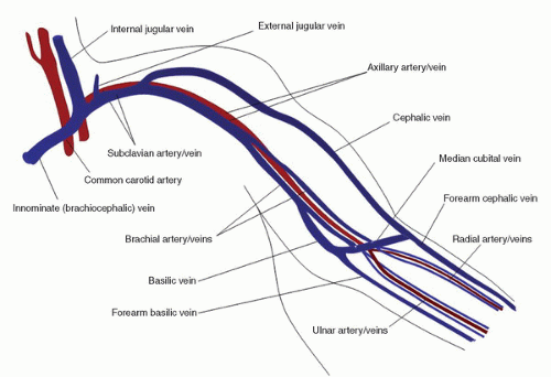
Drains the Superficial and Deep Venous Palmar Arches, is superficial to the metacarpus and prolonged proximally on the lateral side as Cephalic Vein and on the medial side as the Basilic vein.
What is the Dorsal Venous Network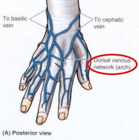
700
The Fibrocartilaginous ring attaches to the margin of the glenoid fossa, deepening the cavity and contributes to the stability of the GH (Glenohumeral Joint)
What is the Glenoid Labrum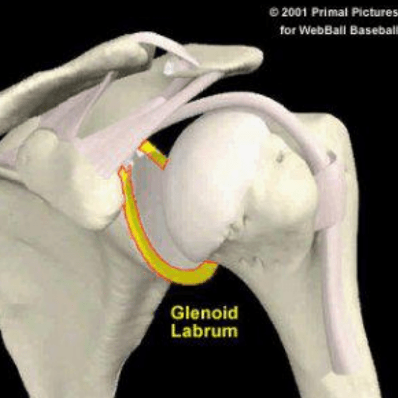
700
Nerve that innervates the Trapezius Muscle
What is the Cranial Nerve XI (Accessory Nerve)
700
The innervation for the Pectoralis Major
What is the Lateral and Medial Pectoral Nerves
700
The innervation of the Teres Major
What is the lower subscapular nerve
700
The action of the Biceps Brachii
What is elbow flexion, weak shoulder flexion
700
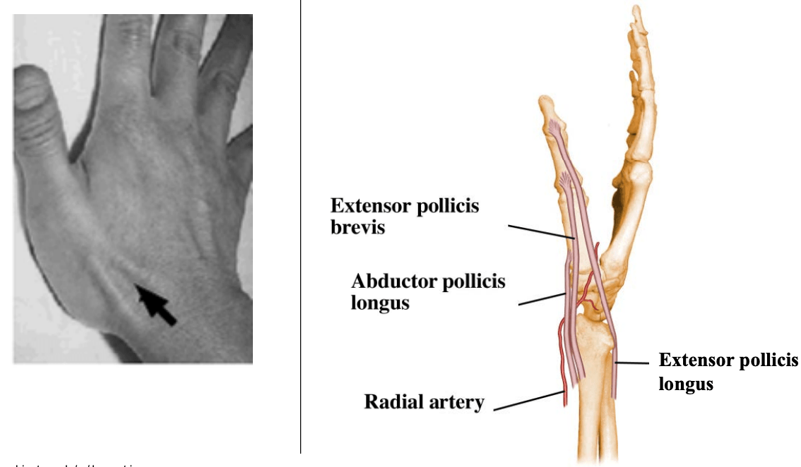
Structures included in the Anatomical Snuff Box
What is the Radial artery, Extensor Pollicis Brevis, ABductor Pollicis Longus and the Extensor Pollicis Longus
700
The action of the Lumbricals
What is Flex at MP and Extend at PIP/DIP
700
The joint responsible for ABduction and ADduction of the digits
What is the Metacarpophalangeal joints
700
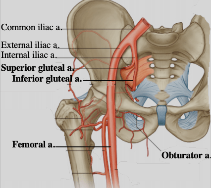
The Arterial Supply for the Lower Extremity
What is the External Iliac Artery (Femoral a), Internal Iliac Artery (Superior gluteal a., inferior gluteal a., obturator a,)
700
Label the image starting from 1-3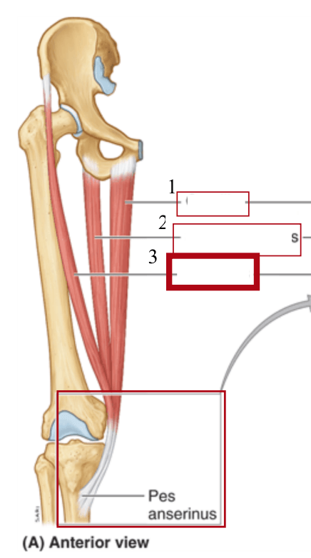
What is Gracilis, Semitendinosus, and Sartorius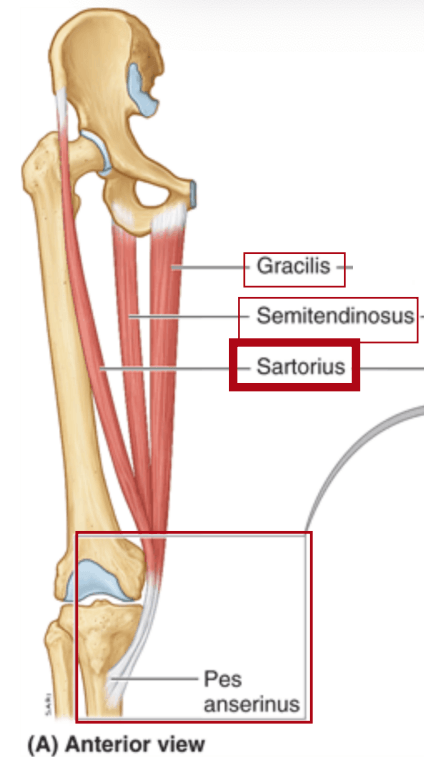
700
The action of the Gluteus Maximus
What is Hip extension and external/lateral rotation
700
Common action of the Soleus, Flexor Hallucis Longus (2), Tibalis Posterior (2), Flexor Digitorum Longus (2), Fibularis (Peroneus Longus and Peroneus Brevi) (2)
What is Ankle Plantarflexion
800
The communication between the basilic and the cephalic veins in the cubital fossa
What is the Median Cubital Vein

800
Attached to the glenoid rim and proximal humerus
What is Glenohumeral Ligaments
800
The Action of the Levator Scapulae Muscle
What is elevation and downwardly rotates scapula
800
Attaches to ribs 3-5, coracoid process of the scapula
What is the Pectoralis Minor
800
The action of the Coracobrachialis
What is Flex and aDduct shoulder
800
The innervation for the Triceps Brachii, Anoconeus, Supinator, and Brachioradialis
What is the Radial Nerve
800
The action of the Radioulnar Joint
What is supination/pronation 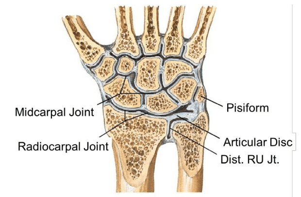
800
Attaches to the Proximal Ulna to the distal phalanx of digits 2-5
What is the Flexor Digitorum Profundus
800
Name the muscle in the picture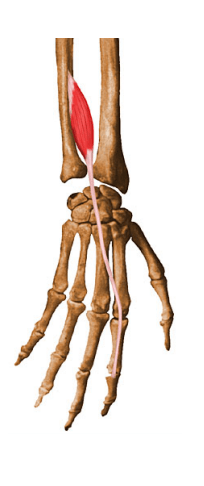
What is the Extensor Indicis Proprius
800
Innervation of the Anterior Compartment of the thigh: iliacus, Rectus Femoris, Vastus Medialis, Vastus Lateralis and Vastus Intermedius, Sartorius
What is the Femoral Nerve
800
Innervation of the Biceps Femoris short head
What is the Common Fibular Nerve
800
Innervation that passes through the gluteal region, but does not innervate any of the gluteal muscle
What is the Sciatic Nerve
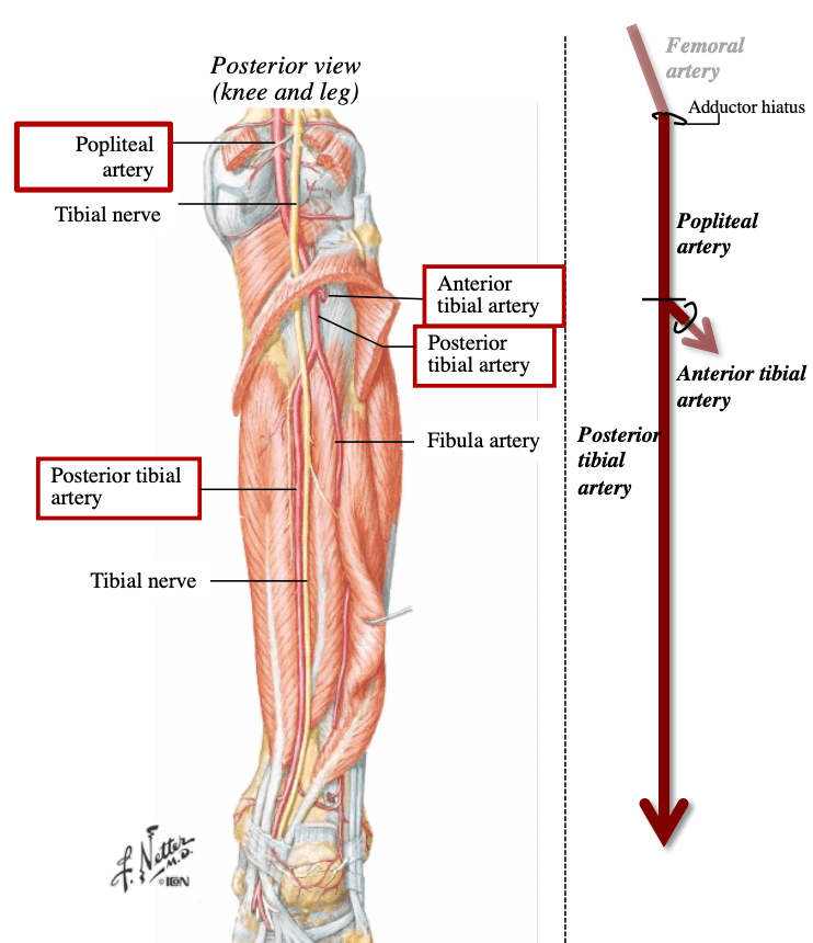
800
Change of name from the femoral artery after transversing the adductor hiatus
What is the Popliteal artery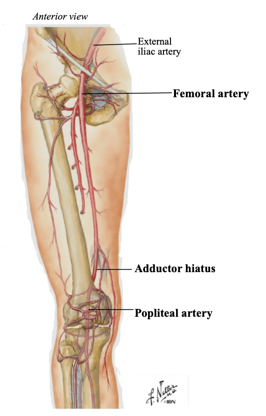
900
the Ultimate Drainage of the Basilic and Cephalic Veins
What is the Axillary Vein

900
A Physiological Joint in which movement occurs between musculoskeletal structures
What is the Scapulothoracic (ST) Joint
900
The innervation of the Pectoralis Minor
What is the Medial Pectoral Nerve
900
The action of the Pectoralis Major
What is aDducts and medially rotates humerus
900
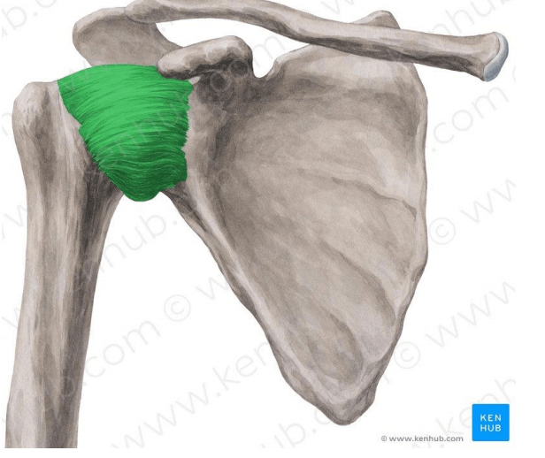
The muscles that are involved in the "Rotator Cuff" Muscles
What is supraspinatus, infraspinatus, teres minor, subscapularis muscle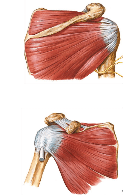
900
The action of the Brachialis
What is flexes forearm
900
Joints that are responsible for wrist flexion/extension and AB/AD
What is the Radiocarpal and Midcarpal joints
900
Innervation of the Dorsal and Palmer Interossei
What is the Ulnar Nerve
900
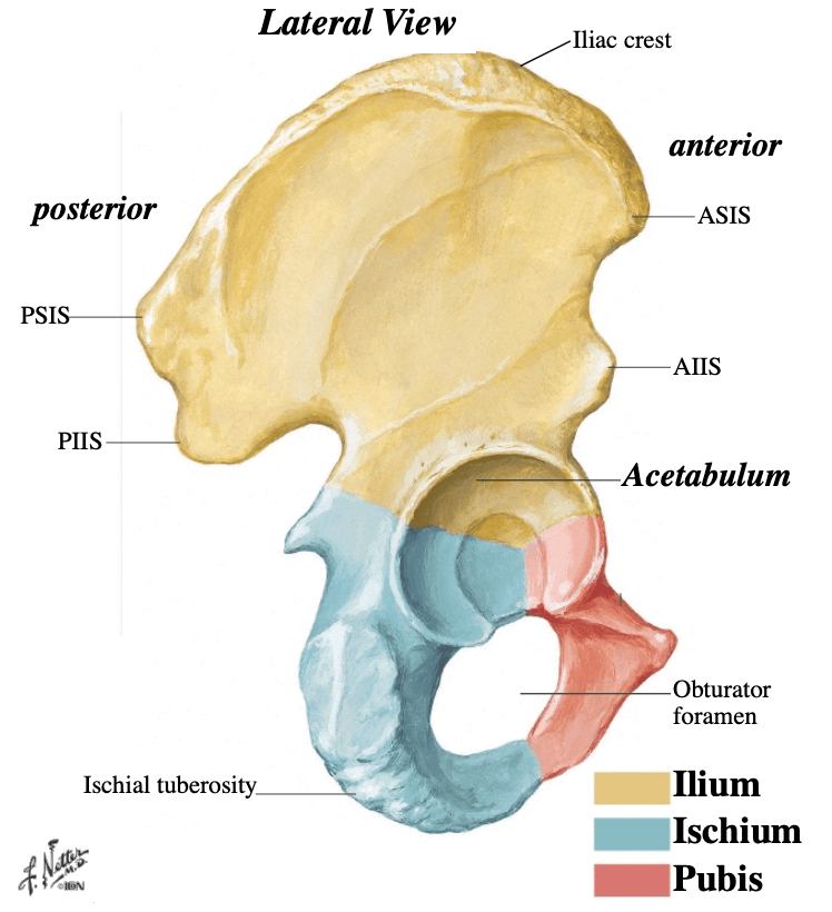
The 3 bones of the hip unite at this site (location of femoral head articulation)
What is the Acetabulum
900
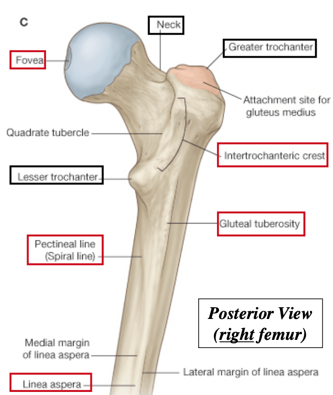
Label the Proximal and Body of the Femur from 1-5 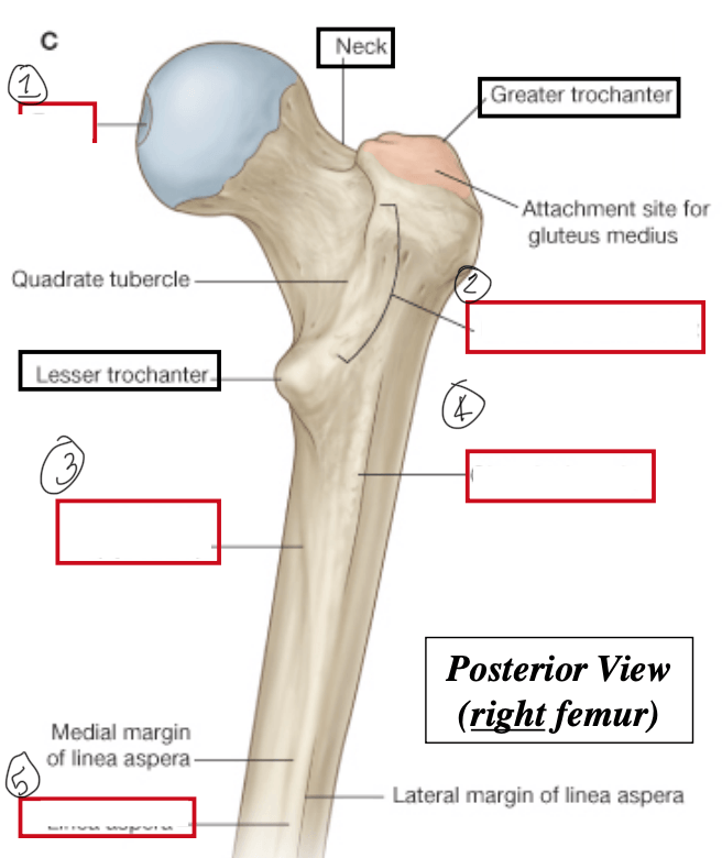
What is the Fovea of the head, Intertrochanteric Crest, Pectineal Line, Gluteal Tuberosity, and Linea Aspera
900
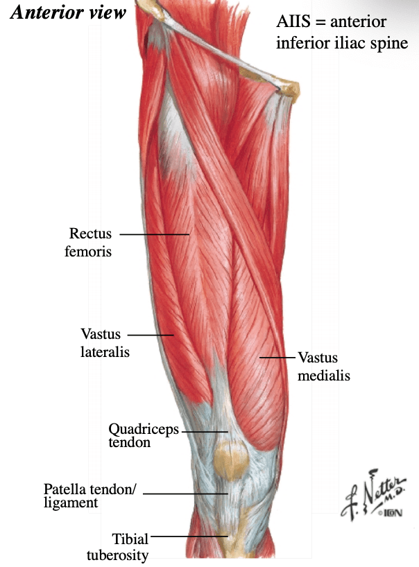
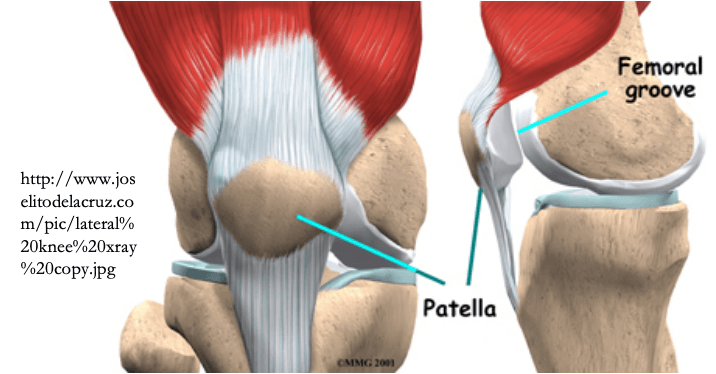
Label the image from 1-6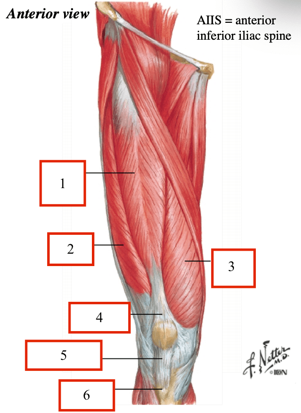
What is the Rectus Femoris, Vastus Lateralis, Vastus Medialis, Quadriceps Tendon, Patella Tendon/ligament, and Tibial Tuberosity
900
List the structures starting from 1-5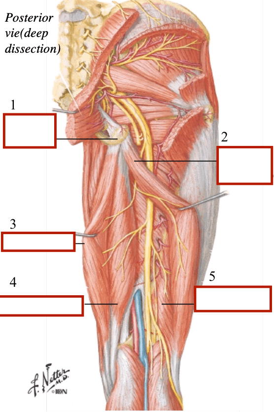
What is the Ischial Tuberosity, Biceps Femoris (long head), Semitendinosus, Semimembranosus and Biceps Femoris (short head)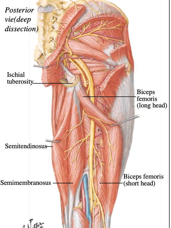
900
Starting from 1-4 name the structures of the image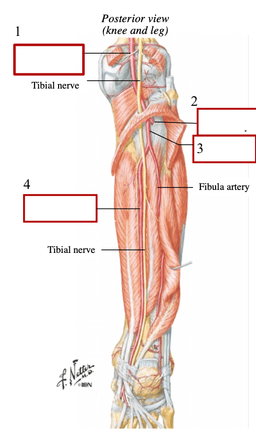
What is the Popliteal Artery, Anterior tibial artery, Posterior tibial artery, Posterior Tibial Artery
1000
Deep Vessels which course with the brachial artery
What is the Brachial Veins
1000
Movements of the Scapulothoracic Joint
What is Elevation/Depression, ADduction(retraction)/ABduction(protraction), Upward Rotation, Downward Rotation
1000
The innervation of the Levator Scapulae Muscle
What is the Dorsal Scapular Nerve
1000
Attaches to the spinous processes C7 and T1, medial scapular border at the scapular spine
What is the Rhomboid Minor
1000
Attaches to the scapular surface to greater and lesser tubercles of the humerus
What is the Rotator Cuff Muscles
1000
The action of the Triceps Brachii
Extends elbow long head-extends shoulder
1000
Attaches to the distal anterior ulna and distal anterior radius
What is the Pronator Quadratus
1000
Innervation for the Pronator Teres and Pronator Quadratus
What is the Median Nerve
1000
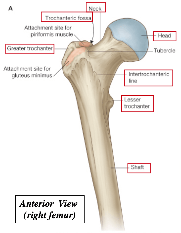
Label the Proximal Femur Starting from 1-5
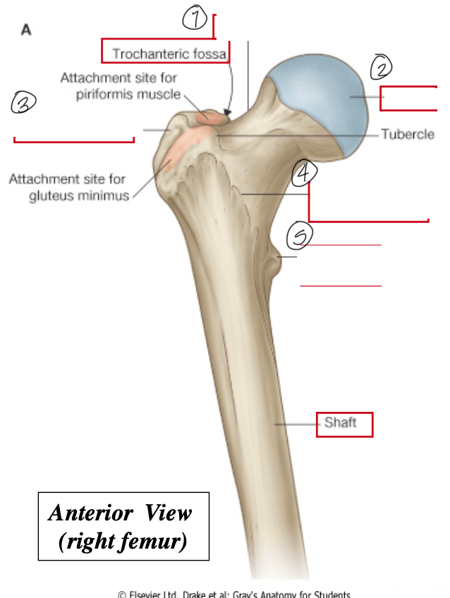
What is the Femoral Neck, Femoral Head, Greater Trochanter, Intertrochanteric Line, and Lesser Trochanter
1000
Innervation for the ADductor Longus, ADductor Brevis, and Gracilis
What is the Obturator Nerve
1000
The action of the Semimembranous, Semitendinosus, and Biceps Femoris (long head)
What are Hip Extension and knee flexion
1000
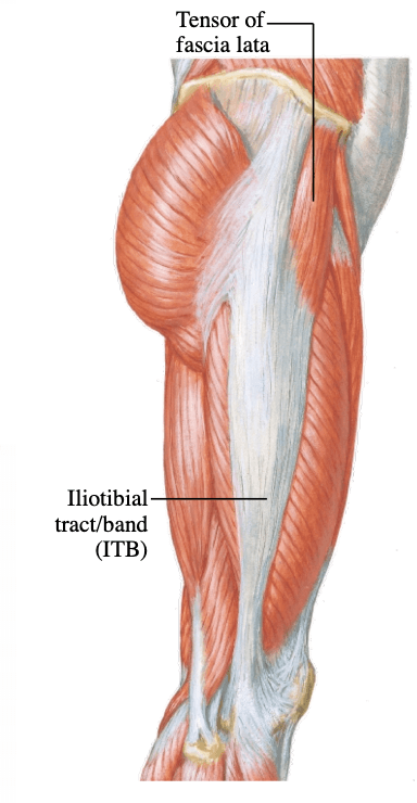
Attaches at the ASIS, anterior iliac crest to the ITB
What is the Tensor of Fascia lata
1000
From 1-4 name the muscles in the image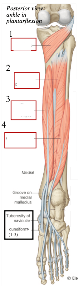
What is the Popliteus, Tibialis Posterior, Flexor Digitorum longus and Flexor Hallucis Longus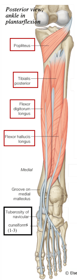
1100
Formed at the inferior border of the teres major muscle by the union of the brachial veins and the basilic vein
What is the Axillary Vein
1100
Each joint makes contributions in a specific and consistent pattern known as the...
What is the Scapulohumeral Rhythm
1100
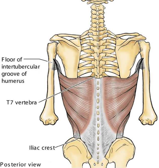
Attaches to the Spinous Process T7-T12 and intertubercular groove of the humerus
What is the Latissiums Dorsi Muscle
1100
The Innervation of the Serratus Anterior
What is the long thoracic nerve
1100
The action of the Teres Major
What is shoulder medial/internal rotation (IR)
1100
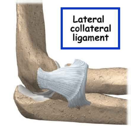
Protects against medial deviation of the forearm and is lateral epicondyle to the annular ligament of radius
What is the Lateral Collateral Ligament
1100
Attaches to the lateral epicondyle of the humerus and the base of metacarpals
What is the Extensor Compartment
1100
In order from left to right name the muscles in the image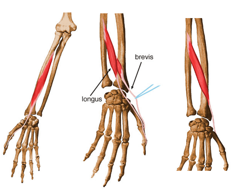
What is the Flexor Pollicis Longus, Extensor Pollicis Longus and Brevis, and Abductor Pollicis Longus
1100
Has a medial and lateral articular facet that articulates with the femoral condyles
What is the Patella
1100
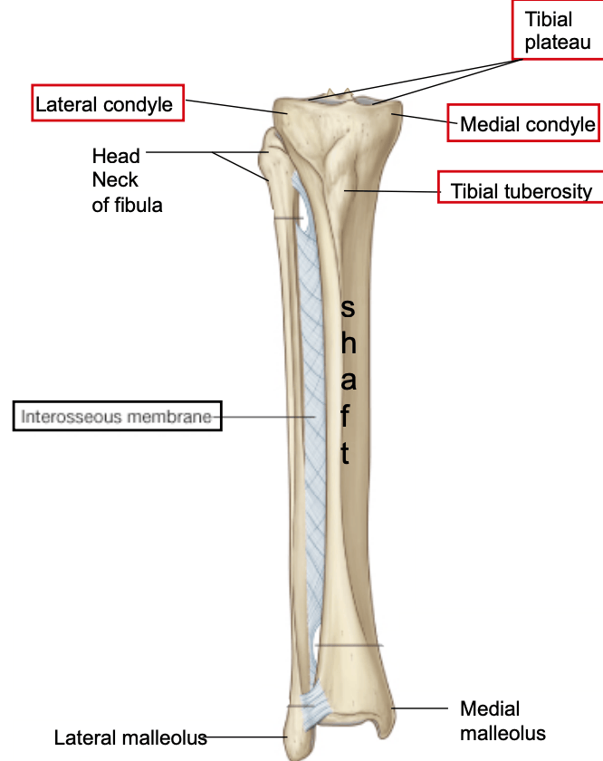
Name the parts of the Tibia from 1-5
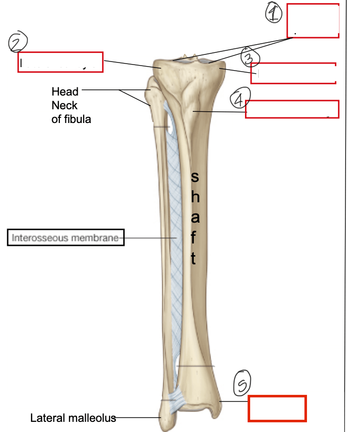
What is the Tibial Plateau, Later Condyles, Medial Condyles, Tibial Tuberosity, and the Medial Malleolus
1100
The action of the Pectineus, ADductor Longus, ADductor Brevis, Gracilis (2 movements), ADductor Magnus
What is Hip ADduction
1100
The following muscles from 1-7
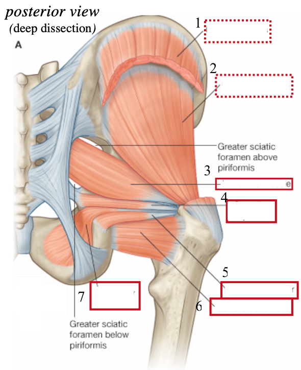
What is the Gluteus Media, Gluteus Minimus, Piriformis muscle, Gemellus Superior, Gemellus Inferior, Quadratus Femoris, Oturator Internus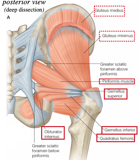
1100
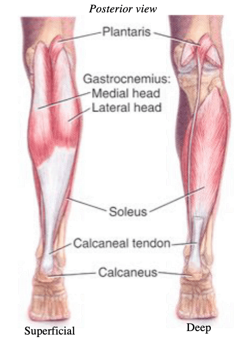
The following structures from 1-3 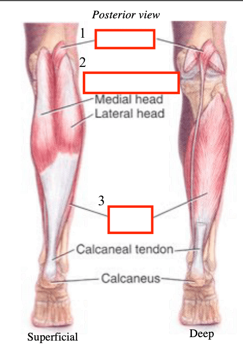
What is the Plantaris, Gastrocnemius and Soleus