Pathologies
Inner Ear Anatomy
Inner Ear Physiology
CANS/CNVIII Anatomy
CANS/CNVIII Physiology
100
Hi, I’m Tyler. I just went to a rock concert—it was wild! Afterward, my ears were ringing, and I had trouble hearing high pitches while working on my songs. Now, two weeks later, everything seems back to normal. What do you think that could have been?
Temporary Threshold Shift
100
What are the floor and ceiling of the Organ of Corti called?
Basilar and Reissner Membrane
100
The part of the cochlea that maintains ion imbalance within the system is called the _____?
Stria Vascularis
100
What is the root entry zone?
where auditory nerve fibers bifurcate as they enter the cochlear nucleus [between the anterior cochlear nucleus (ACN) posterior cochlear nucleus (DCN)]

100
The SOC neurons are extremely sensitive to the _____ of arrival and the ______ levels of the original auditory signals that have arrived at the two ears.
time (interaural time difference)
intensity (interaural intensity difference)
200
What causes hidden hearing loss/ what measure shows lasting damage?
Cochlear neuropathy or temporary threshold shift (TTS) causes perceptual hearing loss. High-SR (Spontaneous Firing Rates) fibers recover in 2–3 days, but low-SR fibers sustain lasting damage. Hearing thresholds may return to normal, but suprathreshold responses (amplitude of ABR waveforms) remain permanently reduced.
200
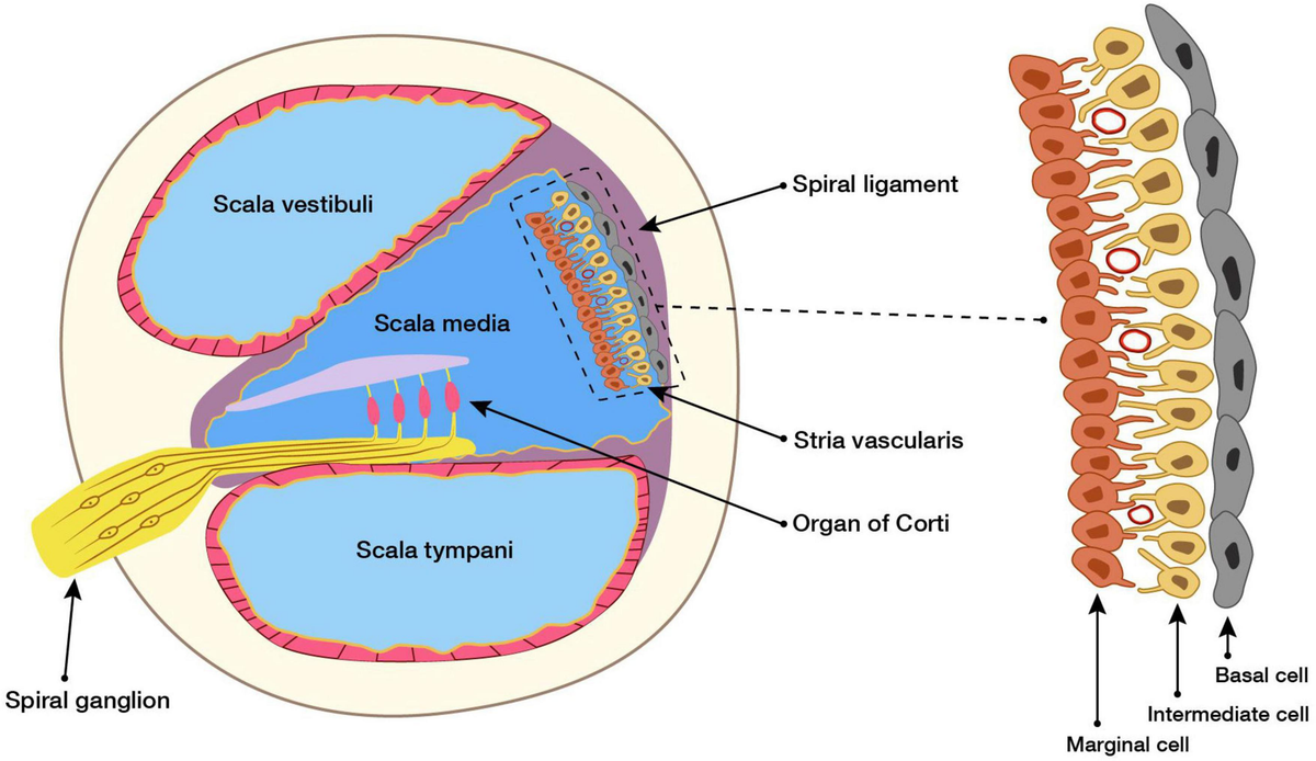
Name the starred structures from superior to inferior.

Scala Vestibuli
Scala Media/ Cochlear duct
Scale Tympani

200
These sensory structures in the vestibular system detect rotational head movements (dynamic equilibrium) and linear acceleration (static equilibrium).
Cristae ampularis within semicircular canals --> dynamic equilibrium (head rotations/rotational angular acceleration)

Maculae in the utricle and saccule --> static equilibrium/ linear acceleration (horizontal/vertical)

200
As fibers leave the internal auditory meatus, the auditory, facial, and vestibular nerves course
through this recess
Cerebellopontine angle (CPA)

200
What is the difference between Schwann cells and oligodendrocytes?
Both are supporting cells that coat axons with myelin; however, the Schwann cells are in the peripheral nervous system, whereas the oligodendrocytes are in the central nervous system.
300
Hello, we are a group of new mothers—Taylor, Olivia, Rebecca, Casey, and Hannah. Our babies have all been diagnosed with exogenous congenital hearing loss related to prenatal factors. What are the most likely causes of this?
TORCH:
Taylor - Toxoplasmosis
Olivia- Other
Rebecca- Rubella
Casey - CMV (cytomegalovirus)
Hannah - Herpes simplex virus
300
Ductus Reuniens of Hensen connects what two structures
saccule of the vestibule and cochlear duct (scala media)

300
Which cochlear structure contains the spiral ganglion neurons, and through which structure do auditory nerve fibers pass to reach them?
A. Rosenthal’s canal; habenula perforata
B. Habenula perforata; Rosenthal’s canal
C. Osseous spiral lamina; scala media
D. Spiral ligament; modiolus
A. Rosenthal’s canal; habenula perforata (perforations/holes in the ossesous spiral lamina)

300
What is the large white matter pathway that runs from the subcortical areas to the cortex and is considered the bottleneck of the auditory pathway?
internal capsule

300
_______ is the first synapse point within the auditory brainstem, where _______ (afferent vs. efferent) Type I _______ (axon/dendrite) terminals make contact.
cochlear nucleus
afferent (signals to the brain; see YMC"A")
axon (away from the cell)

400
Hello, I’m Oscar. My doctor said I have high-frequency hearing loss, possibly from the medications I’m taking. He mentioned a specific term and some drugs that could damage my ears, but I can’t recall the word...or the medications. Can you help me figure it out?
Ototoxicity:
1. Aminoglycosides (antibiotics)
2. Diuretics (waterpill)
3. Salicylates (Pain relievers/ antiinflammatories)
4. Cisplatin/Cisplatinum (Cancer Medication)
400
What are the green cells in the picture called and what is their function?

Deiters' cells
Rows of long supporting cells that extend from the basilar membrane and help form the reticular lamina via their phalangeal processes

400
Purpose of the stria vascularis
Vascularized epethilial structure that regulate the chemical composition of endolymph by pumping potassium ions into the scala media, creating endocochlear potential.
400
The medial fibers of the auditory nerve correspond to ____ frequencies, while the outer fibers correspond to _____
medial = low frequencies
outer = high frequencies
(Think of how the cochlea is organized from base to apex/ what type of frequencies people typically "lose" first)

400
When looking at the first three waves of an ABR, what structures generate these averaged synchronous neural responses?
Wave 1 & 2 --> auditory nerve
Wave 3 --> cochlear nucleus
500
Hello, my name is Marvin. I have fluctuating low-frequency hearing loss, tinnitus, vertigo/drop attacks, and aural fullness. My doctor found excess endolymph in my cochlea, which makes me feel unbalanced, and my Electrocochleography showed a summating potential (SP) greater than 50% of my action potential (AP). Dietary changes like reducing salt didn’t help, so I may need diuretics, surgery, or transtympanic injections. What pathology do you think I have?
Meniere’s Disease
500
Compare and Contrast the features and functions of the inner and outer hair cells.

500
Describe the process of a neuron firing (action potential).

1. Resting Potential – Neuron is at -70 mV, ready to fire.
2. Threshold – Stimulus reaches -55 mV, triggering action potential.
3. Peak – Inside becomes positive (~ +30 mV) as Na⁺ floods in.
4. Repolarization – K⁺ exits, restoring negativity.
5. Hyperpolarization – briefly more negative than at rest, entering a refractory period.
6. Reset – Ion balance restored; neuron ready to fire again.
500
Name all the stops on the Central Auditory Nervous System Pathway (afferent) from the cochlea to the final stop.
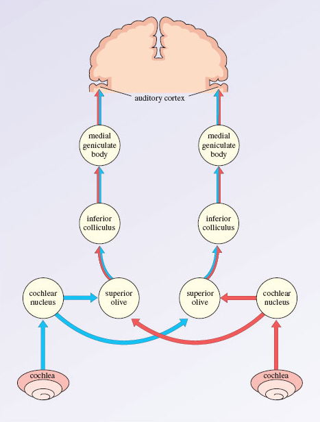
Cochlea
Auditory Nerve (Cranial Nerve VIII)
Cochlear Nucleus
Superior Olivary Complex (SOC)
Lateral Lemniscus
Inferior Colliculus
Medial Geniculate Body (MGB)
Primary Auditory Cortex (Heschl’s gyrus)
500
Describe the auditory reflex arc involved in protecting the cochlea from loud sounds (the entire loop).

Stimulus Detection: Loud sound detected by cochlear hair cells.
Afferent Pathway: Auditory nerve (CNVIII) transmits signal to the cochlear nucleus.
Central Processing: Signal relayed to the superior olivary complex.
Efferent Pathway: Facial nerve (CNVII) carries motor output.
Motor Response: Stapedius muscle contracts to dampen movement of the stapes and reduce sound transmission.