HemeOnc
Lung & Heart 1
Lung & Heart 2
Lung & Heart 3
Heart Rhythms
100
A 6-year-old boy is brought to the office due to pallor and decreased energy. For the past 2 days, the patient has been less active, uninterested in playing, and sleeping throughout the day. On review of systems, the parents note that he developed a fever, emesis, and diarrhea 10 days ago, but his symptoms resolved without intervention after a few days. Temperature is 37.1 C (98.8 F), blood pressure is 120/70 mm Hg, and pulse is 145/min. Physical examination shows a tired-appearing boy with pale lips. Rest of exam is normal. Laboratory results are as follows:
Complete blood count: Hemoglobin 8.4 g/dL, Platelets 30,000/mm3, Leukocytes, 15,200/mm3
Serum chemistry: Blood urea nitrogen 32 mg/dL, Creatinine 2.4 mg/dL
What does he have and what is the mechanism of injury?
HUS via Renal vascular occlusion.
He shows the classic triad of microangiopathic hemolytic anemia (Hb 8.4), thrombocytopenia (30,000), and acute kidney injury (Cr 2.4, BUN 32). It came about 10 days after a self-limited gastroenteritis, which fits the typical lag of about 7 days between diarrhea and HUS.
100
A 26-year-old white woman is brought to the ER with complaints of a 2-day history of dyspnea, productive cough, and high-grade fever with chills. She has been feeling extremely fatigued for the past few days. She has a 10 pack-year hx of smoking and IVDU. Vitals are as follows:
HR 110/min, Temp 101 degrees F, BP 110/70 mmHg, and RR 26/min. On exam, the JVP is raised and mild hepatomegaly is present. Cardiovascular exam reveals a pansystolic murmur at the lower left sternal border.
What is the next best step in diagnosis, and what is the suspected condition? How would you treat it?
Next best step: draw 3 sets of blood cultures from separate venipunctures before giving antibiotics, then get a transthoracic echocardiogram (TTE). Start empiric IV antibiotics as soon as the cultures are drawn.
S. aureus IE.
Empiric abx include Vanc (MRSA), Cefepime/CTX (G- organisms), and PsA coverage via Cefepime or Zosyn (if injection of non-sterile water) for 4-6 wks. Definitive therapy once susceptibilities return include Nafcillin (MSSA), Vanc (MRSA).
100
A 65-year-old white man has a 10-year history of angina which has, up until now, been treated effectively with nitroglycerin. Lately, he has been noticing that he is having chest pain more frequently. He no longer is able to complete his morning walk without the development of pain, whereas he previously had been able to complete the walk without a problem. He also notices that light gardening is starting to produce chest pain. He feels that the chest pain is more severe and lasts longer. As a consequence of these changes, he has to take increasing amounts of nitroglycerin to control the pain. What does he have, and what is the next step in evaluation?
Unstable Angina, evaluate with EKG and troponin (to distinguish from NSTEMI).
100
A 57-year-old Caucasian man presents with worsening shortness of breath. While obtaining his history, you uncover that he has noted increasing shortness of breath with minor exertional activity and a persistent but non-productive cough. The patient admits to being a former smoker with a 34 pack-year history, admitting to cessation at age 50. He denies any known caustic occupational exposures and states he worked in an office his whole life. He admits to an uncle having some kind of breathing issues, although he is unsure of a definite diagnosis. Patient denies weight loss, fever, or significant recent illness. Physical examination is pertinent for significant clubbing of the fingers, fine inspiratory crackles. What is the diagnosis and treatment(s)?
Idiopathic pulmonary fibrosis. Why? No infx sx, no environmental exposure.
Treatment centers on antifibrotic therapy (nintedanib or pirfenidone), oxygen, pulmonary rehabilitation, evaluation for pulmonary hypertension, and early lung transplant referral.
100
35 year old man who had presented with a six month history of chest pain and lightheadedness on exertion. What is the dx?
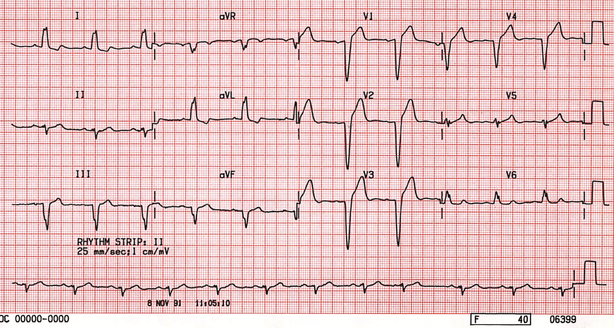
LBBB.
Why? In LBBB, conduction delay means that impulses travel first via the right bundle branch to the RV, and then to the LV via the septum... Septal activation is thus reversed eliminating lateral Q waves... The overall depolarisation vector from the right to left ventricle produces tall R waves in lateral leads (I, V5-6) and deep S waves in the right precordial leads (V1-3). The delay between activation of the RV and LV produces the characteristic “M-shaped” R wave seen in lateral leads... PLUS, Delayed overall conduction time to the LV extends the QRS duration to ≥ 120 ms
200
A 4-year-old boy is brought to the office for evaluation of a rash. His mother states that "pinpoint" bruises have appeared all over his body during the past 24 hours. The patient has had no bleeding or recent trauma. Three weeks ago, he had an upper respiratory tract infection that resolved uneventfully. The patient takes no medications, he has no allergies, and his immunizations are up to date. There is no family history of bleeding or clotting disorders. Vital signs are normal for age. Physical examination shows a cooperative, well-appearing child with scattered petechiae on the trunk and extremities. Oral mucosa appears normal. There is no lymphadenopathy or splenomegaly. Peripheral smear shows few platelets; the platelets are of normal size and morphology. Laboratory results are as follows:
Hemoglobin 13.5 g/dL, Platelets 40,000/mm3, Leukocytes, 7,000/mm3
What is the diagnosis and treatment?
ITP. Tx: observation with treatment, because he only has minor bleeding. If he develops significant bleeding, can try a short course of steroids or IVIG. Most children remit on their own within 12 months.
200
A 58-year-old woman with a past medical history of hypertension, hyperlipidemia, breast cancer, hip fractures, and coronary artery disease is being evaluated for acute-onset severe left-sided pleuritic chest pain over the course of the last 2 hours. The pain is associated with feelings of anxiety, hemoptysis, shortness of breath, and nausea. She "feels warm" but denies chills, palpitations, wheezing, edema, vomiting, abdominal pain, abnormal bowel habits, or dietary intolerances. She admits to a 30 pack-year smoking history but denies drug or alcohol use. Upon physical exam, she is found to be febrile, hypotensive, tachycardic, tachypneic, diaphoretic, and in acute painful distress. There are perioral cyanosis and a pleural friction rub to the left lung fields; the remainder of the exam is normal. What is the suspected diagnosis, and what is the treatment?
In a patient with likely pulmonary embolism, immediate anticoagulation via UFH prior to diagnostic testing. Immediate tx with UFH; evaluate for TPA.
TPA: within 4.5hr, no prior hemorrhagic stroke, or ischemic stroke within 6mo, CNS neoplasm.
Modified Wells criteria for pretest probability
of pulmonary embolism
+3 points
- Clinical signs of DVT
- Alternate diagnosis less likely than PE
+1.5 points
- Previous PE or DVT
- Heart rate >100/min
- Recent surgery or immobilization
+1 point
- Hemoptysis
- Cancer
Total score
- ≤4 = PE unlikely
- >4 = PE likely
200
A 68-year-old woman presents with shortness of breath, fatigue, dry cough, and ankle swelling. Symptoms started 6 weeks ago; she dismissed them as stress-related, but they have worsened in intensity and frequency. She becomes short of breath with any exertion, such as climbing stairs. She feels like she may pass out and has to sit when she gets lightheaded. Her breathing is worse when lying down. She denies productive cough, fever, or chest pain. She notes she has had no medical care in the past few years and is post-menopausal. She denies any other past medical or surgical history. Social history is unremarkable. Vitals are notable for pulse 101, BP 158/98, and BMI 28.5. Exam reveals bibasilar crackles and 2+ pitting edema of the lower extremities. What does she likely have and what are treatment options?
CHF; ACE/ARB, b-blocker, spironolactone. ARNi, SGLT2.
200
A 56-year-old woman presents with fatigue, cough, and breathlessness on exertion for the last 2 months that have progressively increased. She gets short of breath after walking 2 blocks or climbing several flights of stairs. In addition, she gives a history of hemoptysis on 2 recent occasions and has noticed some recent ankle swelling. Her past medical history is significant for rheumatic fever 30 years ago. On examination, her blood pressure is 128/80 mmHg, RR 22/min, pulse 70 bpm. Cardiac auscultation reveals a very loud S1, presence of an opening snap, mid-diastolic murmur heard at the apical region, and a loud P2. What does she have, and what does the pressure-volume loop look like?
Mitral stenosis

200
64yoF presents with 2hr of epigastric pressure, n/v, and fatigue. What is the dx? And what is the next best step(s) in management?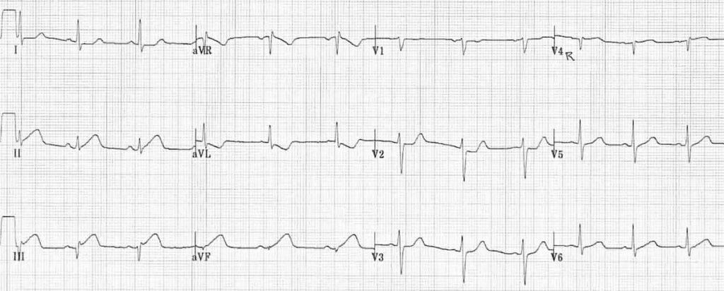
Inferior STEMI. Aspirin load, P2Y12inh, UFH, HOLD NITRATES d/t Right side infarct, Cath lab activation.
What artery is affected?
300
A 5-year-old girl is brought to the office by her mother and the mother's boyfriend due to leg pain. One week ago, the boyfriend saw the patient fall onto the living room floor after climbing on a couch. Since the incident, she has had persistent leg pain that appears to be worse at night, and for the last 2 days she has been refusing to walk, asking her mother to carry her.
Temperature is 38.3 C (100.9 F). All other vital signs are normal. Examination shows a pale child. The abdomen is nontender and the liver is palpated 3 cm below the costal margin. Palpation of the bilateral anterior proximal tibias elicits pain. There are a few scattered bruises across the chest and back.
Complete blood count results are as follows: Hemoglobin 8 g/dL, Platelets 30,000/mm3, Leukocytes 3,000/mm3.
What is the diagnosis?
ALL
Classic features:
Pancytopenia (Hb 8, platelets 30,000, WBC 3,000)
Bone pain worse at night, with tibial tenderness and refusal to walk
Fever, pallor, bruising
Hepatomegaly
300
An 18-year-old girl comes to the office with 3 days of fever, nonproductive cough, dyspnea, and fatigue. The symptoms started after she returned home from a summer school trip to Europe with friends. Her roommate had similar symptoms. The patient has no prior medical issues, and her vaccinations, including COVID-19 vaccination, are up to date. She is not sexually active. She does not use tobacco, alcohol, or recreational drugs. Temperature is 38.3 C (101 F), blood pressure is 120/80 mm Hg, pulse is 88/min, and respirations are 16/min. The patient does not appear ill. Faint bilateral crackles are present. SARS-CoV-2 testing is negative. Chest x-ray reveals bilateral diffuse infiltrates. What is the diagnosis and treatment?
Mycoplasma pneumonia, macrolides (e.g., azithromycin).
300
A 45-year-old man presents with a 6-hour history of retrosternal chest pain. He describes it as sharp and episodic. It is relieved by sitting upright and worsened by lying down. He denies any trauma to the chest. ECG reveals diffuse ST elevations in inferior and lateral leads with depression of the PR segments. What does he have? How do you treat it?
Pericarditis, treated with NSAIDs/aspirin and Colchicine as an adjunct.
300
A 2-year-old boy is brought to the emergency department due to respiratory distress. For the past 4 days, the boy has had rhinorrhea, nasal congestion, and low-grade fever. Today he appears increasingly tired, has decreased urine output, and his breathing is labored. The patient has no chronic medical conditions and is up to date with recommended vaccinations. Temperature is 38.2 C (100.7 F), pulse is 155/min, respirations are 48/min, and blood pressure is 60/40 mm Hg. Pulse oximetry on room air shows an oxygen saturation of 91%. Scattered, mobile, subcentimeter lymph nodes are palpable in the anterior cervical chain bilaterally. Cardiac examination shows tachycardia, gallop rhythm, and a grade 3/6 holosystolic murmur loudest at the cardiac apex. Brachial and femoral pulses are 1+. Pulmonary examination reveals tachypnea, nasal flaring, and subcostal retractions. Scattered rales are present at the lung bases. What is the most likely diagnosis in this patient?
Viral Myocarditis with evidence of cardiogenic shock (hypotension (60/40), 1+ brachial and femoral pulses, lethargy, and oliguria).
300
Describe the changes over time of a STEMI.
Seconds: hyperacute T waves, +/- ST changes
Minutes: ST elevation, J pt elevation
Hours: Q wave begins, STE remain, decreased R wave
Day 1-2: T wave inversion, deeper Q wave
Days-Weeks: ST normalizes, T wave changes remain
Chronic: Q wave persists
400
A 15-month-old boy is brought to the emergency department for evaluation of ankle swelling. The symptoms started yesterday evening after he played at a park, and today he has refused to walk. Since he began walking at age 12 months, he has developed bruises following minor trauma. The patient has had no fever or recent illness. Vital signs are normal. Examination shows an edematous and tender left ankle with decreased range of motion. Skin examination reveals large ecchymoses on both thighs from recent vaccinations. The nares and oropharyn are clear. The abdomen is soft with no organomegaly. Plain radiographs reveal no acute fracture or dislocation at the left ankle.
What does he have and Laboratory testing in this patient is most likely to reveal which of the following in terms of Platelet count, PT, PTT, Platelet aggregation?
Hemophilia (A is much more common than B), with characteristic symptoms of bruising beginning with crawling/walking, hemarthrosis afeter minor trauma, and large ecchymoses at vaccination sites.
Platelet count: normal
PT: normal
PTT: prolonged
Platelet aggregation: normal
400
A 54-year-old man comes to the office due to shortness of breath at night. He has also felt weak but has had no dyspnea on exertion or leg swelling. His wife adds that the patient has had occasional cough while eating and that his speech is slightly slurred. He has no chronic medical conditions and is a lifelong nonsmoker. Vital signs are within normal limits. BMI is 25 kg/m?. On physical examination, the soft palate and uvula are completely visualized. The tongue is mildly atrophic with visible fasciculations. The lungs are clear on auscultation and heart sounds are normal. The abdomen is flat and moves outward during expiration. There is no extremity edema. Chest x-ray shows elevated hemidiaphragms but no parenchymal opacities. Compared to a healthy individual's tests, this patient's pulmonary function testing in terms of FVC, Maximal inspiratory pressure, DLCO? What is his diagnosis?
ALS.
FVC -, MIP -, DLCO normal.
400
A 65-year-old woman with a 40 pack-year smoking history (still smoking) presents with 3 months of worsening cough, dyspnea on exertion, and blood-streaked sputum. Over the past 2 weeks her voice has become hoarse. She has lost 25 lb unintentionally and reports fatigue, trouble climbing stairs, and new ankle swelling. Her family has noticed she is more irritable and emotional. What is the diagnosis, treatment, and prognosis?
Small cell lung cancer with mediastinal spread (hoarseness - L vocal cord paralysis) and ectopic ACTH (Cushing) syndrome.
Other considerations: squamous cell carcinoma (d/t smoking hx), although its paraneoplastic syndrome is traditionally hypercalcemia of malignancy (secretion of PTHrP). In that case, her sx may include polyuria, polydipsia, n/v, mental status changes, AKI from dehydration.
400
A 55-year-old man, with a 30-pack-year history of cigarette smoking, presents with a 3-nonth history of cough productive of blood and sputum. The patient admits a weight loss of 25 pounds over the past year. The patient also notes diffuse 'bone pain', abdominal pain, polydipsia, polyuria, and anxiety over the past month; all are unusual symptoms for him. The patient denies recent travel. The patient was treated in the emergency room twice over the past 2 months for nephrolithiasis. On chest radiography, a 3 cm cavitary lesion with an air-luid level is noted centrally in the left upper lung field; it is considered suspicious for a sulmonary abscess. The patient also exhibits clubbing on physical examination. The patient's PD is negative, and there is no history of anti-tuberculosis medication use in this patient's mistory. The patient's laboratory values are as follows:
Calcium: 13.2 (normal: 8.5 - 10.8 mg/dL), Phosphorus: 1.8 (normal: 2.5 - 4.5 mg/dL), Urine cyclic adenosine monophosphate (CAMP): 9.2 nmol/mL (normal: 1.6 - 6.2 nmol/mL)
Squamous cell carcinoma.
Its paraneoplastic syndrome is traditionally hypercalcemia of malignancy (secretion of PTHrP). This aligns with his sx of include polyuria, polydipsia, n/v, mental status changes, AKI from dehydration.
400
A 74-year-old woman with hypertension and paroxysmal atrial fibrillation comes to the ED after two episodes of near-syncope and one witnessed syncope while gardening. She has had 6 months of fatigue, reduced exercise tolerance, and intermittent palpitations, and episodes of lightheadedness that come right after a burst of palpitations. Her AF was diagnosed 1 year ago. She takes metoprolol succinate 100 mg daily, apixaban, and amlodipine. She denies chest pain, seizure activity, and incontinence. Vitals: BP 108/64, HR 42 and irregular, RR 16, SpO₂ 97% RA. What is your leading diagnosis, and what might you expect on ECG?
Leading diagnosis: sick sinus syndrome (sinus node dysfunction). FYI - probable subtype of tachy-brady.
Three features point there:
1) Lightheadedness that comes right after a burst of palpitations. This is a classic conversion (post-tachycardia) pause. 2) Known paroxysmal AF in an older patient. Tachy-brady physiology is present in about 50% of patients with SND and raises the risk of stroke and death. 3) Syncope, which is reported in about 50% of patients who get a pacemaker for SND.
Expected ECG: Sinus bradycardia below 50 bpm, with an identical P wave before each QRS. Potentially paroxysmal AF or flutter alternating with bradycardia.
Also consider dx like medication-induced bradycardia, intermittent AV block, orthostatic hypotension.