Immunology terms
Staining Methods
Name that Fixative
Troubleshooting IHC
Name that tissue
100
This is a host protein produced in response to the presence of foreign molecules, organisms, or other agents in the body
Antibody
100
In this method, the primary antibody is followed by a biotinylated secondary antibody
Avidin-biotin methods
100
Helly Solution
Mercuric chloride, Potassium dichromate, sodium sulfate, formaldehyde, distilled water
100
If the specimen and control are unstained what might not have been applied
The primary antibody
100
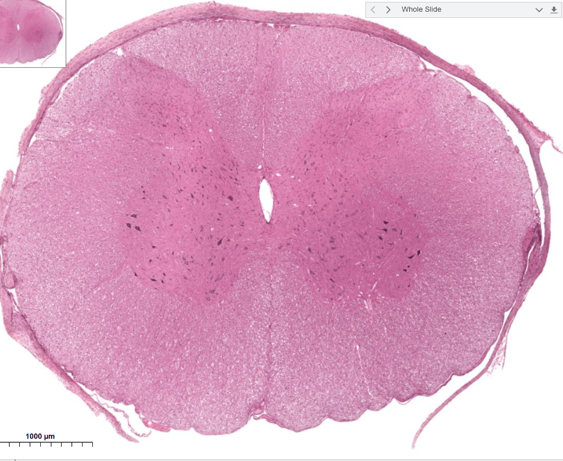
Spinal cord
200
An immunoglobulin is a Y shaped protein molecule that is composed of this
Heavy and light chains
200
This is a 3-step method using primary antibody, secondary antibody and soluble enzyme-antienzyme complex
Unlabeled or soluble enzyme immune complex method
200
Picric acid, formaldehyde, acetic acid
Bouins
200
If the specimen and control have excessive background staining and the slides are not washed well, this is how you fix this
Increase the buffer washes
200
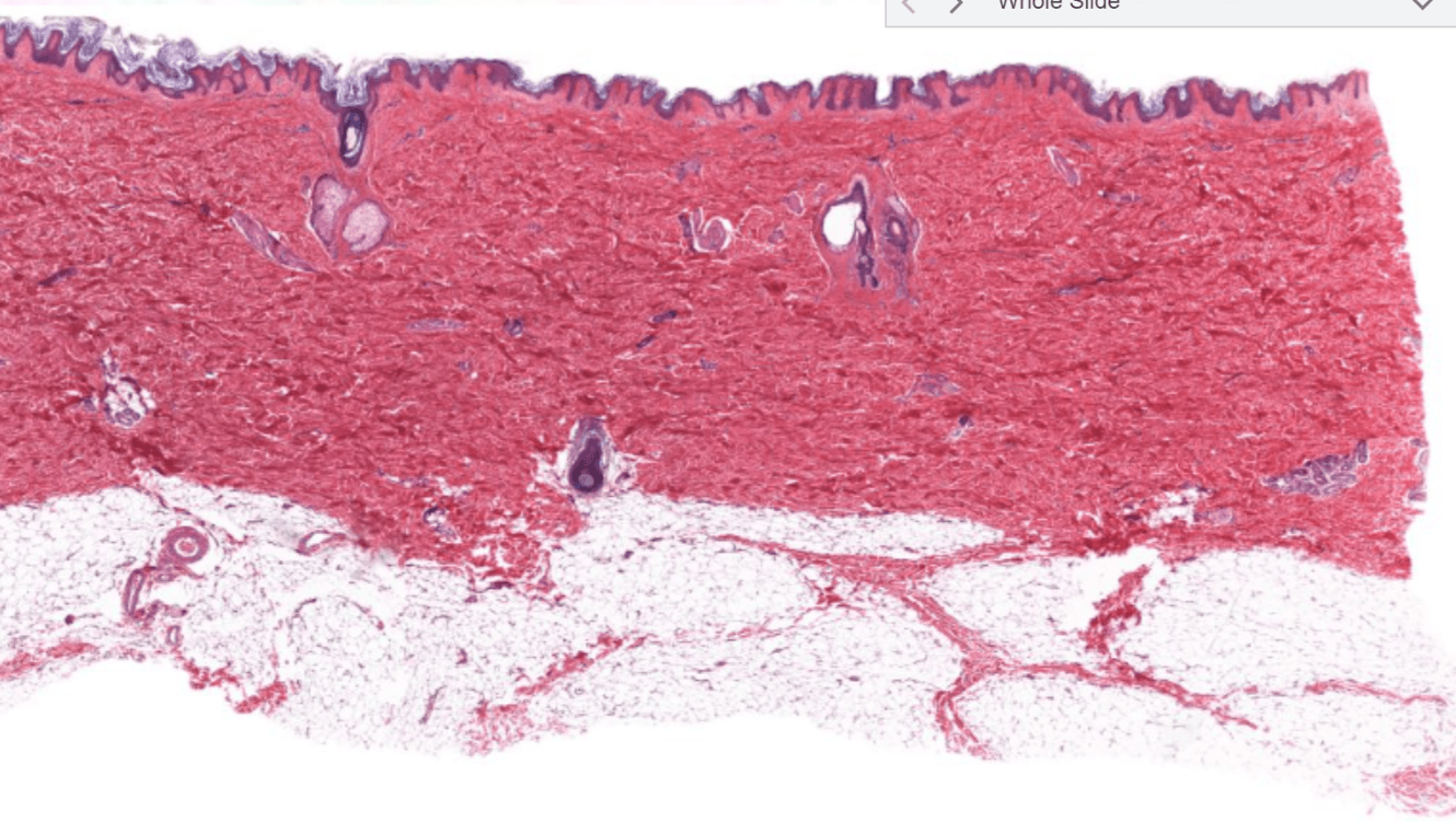
Skin
300
Monoclonal antibodies are prepared by injecting this animal with an antigen
Mice
300
The staining steps for this method are antibody, polymer and chromogen. TAT have improved with the serum and avidin-biotin blocking steps eliminated
Polymeric detection methods
300
B-5 solution
Mercuric chloride, sodium acetate, formaldehyde, distilled water
300
If there is excessive background staining in tissue, the concentration could be too high for this
Primary antibody, link reagent or label reagent
300
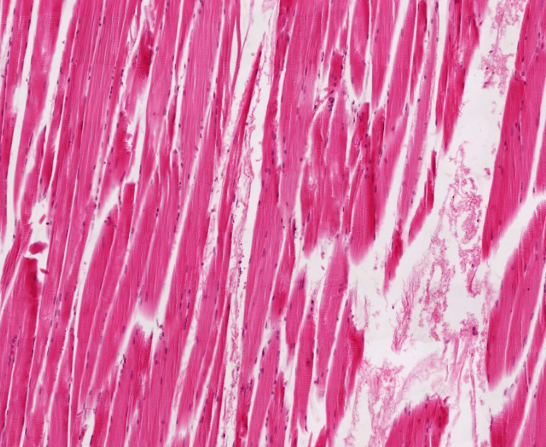
Skeletal muscle
400
This is the simplest form of antigenic determinant present on a complete antigenic molecule; the site at which the antibody attaches to the tissueE
Epitope
400
In this method a labeled antibody of known specificity is used to identify antigens in the patient's tissue
Direct method
400
Absolute alcohol, glascial acetic acid
Clarke fluid
400
If the specimen has weak staining but the control is stained this could be the cause
The antigen present is in low concentration or masked during fixation
400
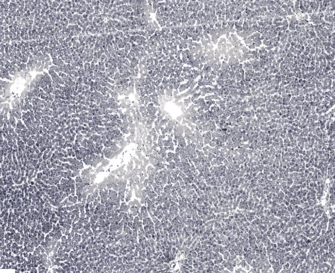
Liver stained with iron hematoxlyin
500
Polyclonal antiserum is highly this because it binds to multiple epitopes, but not as as this, resulting in non-specific staining
Polyclonal antiserum is highly sensitive, but not as selective
500
In this method, patient serum is added to tissue sections containing known antigens to test the patient for the presence of antinuclear antibodies
Indirect method
500
Copper acetate, picric acid, formaldehyde, acetic acid, distilled water
Hollande solution
500
If the specimen has excessive background staining and the control has no background, this could be the cause
Necrosis, autolysis or degeneration of tissue
500
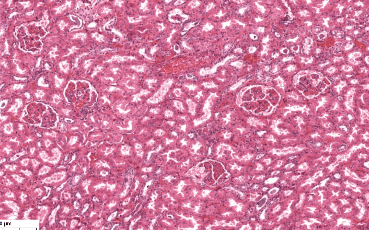
Kidney