Dendritic Cells. They aren't as scary as they look!
Bad R..ec..ep..tio..n
Drake vs. Pusha T (cell)
Is this Affecting You? I mean Effecting!
Let me Express My(cell)-f!!
100
Which vessel do dendritic cells use to enter the lymph node?
Dendritic cells enter the lymph node via the afferent lymphatic vessel.
100
True or false:
B7 is not expressed all the time.
TRUE!
B7 is NOT expressed constitutively (constantly).
B7 is only expressed as a consequence of infection. When a person has an infection, B7 is expressed because it is signaled by molecules such as TLRs (Toll-like receptors).
100
What does CD stand for?
Cluster of Differentiation.
100
This T-cell functions to activate the cellular and antibody response(s) to parasites
What is TH2 cell?
100
True or false:
T-cells can work together with B-cells ONLY if they have the SAME epitope and the SAME antigen.
FALSE!
T and B cells can work together even if they have different epitopes of the same antigen.
Epitope = part of an antigen that binds to an antibody
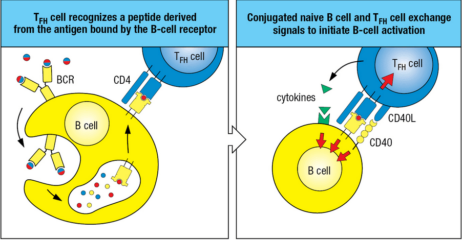
LINKED RECOGNITION
200
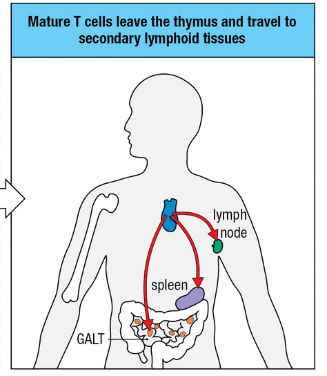
Dendritic cells carry antigens from sites of infection to secondary lymphoid tissues. Please list the secondary lymphoid tissues.
What are lymph nodes, MALT (mucosal-associated lymphoid tissue, spleen
200
This interferon prevents viral replication of infected cells, increases the processing of viral antigens, and increases the presentation of viral antigens by MHC Class 1.
What is IFN-γ?
200
True or false: Dendritic cells are the only cells that can activate both CD4 and CD8 T-cells? Why?
FALSE!
While it is true that dendritic cells are the only cells that can activate CD8 T-cells, CD4 T-cells can also be activated by macrophages.
200
TGF-β (transforming growth factor beta) s a cytokine that aids in proliferation and differentiation, among other processes.
TGF-β induces differentiation in which T-cell(s)?
What are TH17 and Treg cells?
200
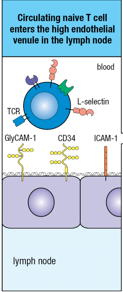
This cell surface protein on T-cells allows it to bind to corresponding molecules on other endothelial cells.
What is L-selectin?
Specifically, l-selectin binds to CD34 and GlyCAM-1 on the surface of the HEVs of the lymph nodes.
This binding causes the T-cell to slow down its motion and bind to the high endothelial venule.
After the T-cell has been attached to the HEV, LFA-1 on the T-cell interacts with ICAM-1 to strengthen the interaction between the T-cell and the HEV.
300
True or false:
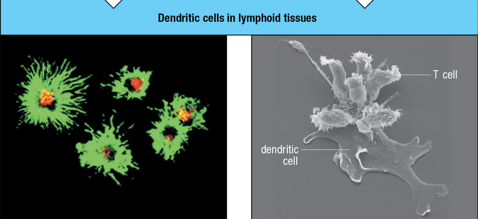
Immature dendritic cells have highly-elaborated fingerlike processes.FALSE:
On maturation, the dendrites become highly elaborated.
immature dendritic cell:
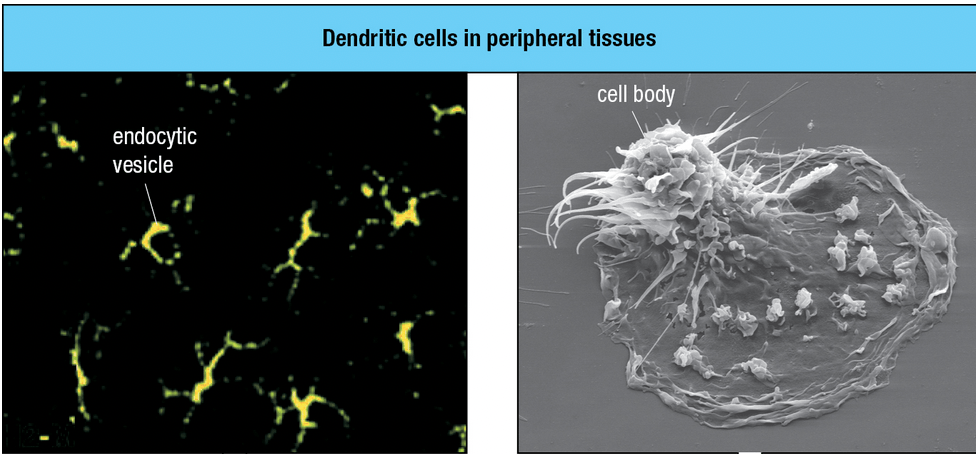
completely mature dendritic cell:
Dendritic cell life cycle and development:
1. dendritic cells arise from hematopoietic stem cells from the bone marrow.
2. immature dendritic cells use the bloodstream to migrate to peripheral tissues and lie waiting for pathogens
3. The PRRs (pattern recognition receptors) on dendritic cells allow them to recognize many pathogens
4. The PRR binding to the pathogen causes the ACTIVATION of the dendritic cell.
5. The dendritic cell LEAVES the peripheral tissue and begins expressing immune stimulatory molecules. The activated dendritic cell migrates to lymphatic tissue
Video: https://youtu.be/JKqFOwudwEI
300
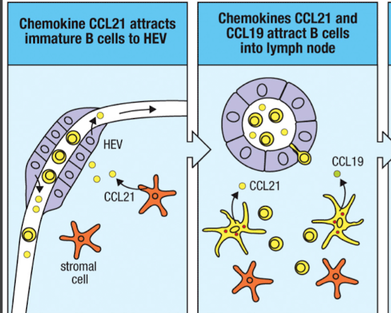
These 2 chemokines are secreted by stromal cells and dendritic cells of the lymph node.
They are also bound to the surface of HEVs (high endothelial venules) and aid in the homing process.What are CCL21 and CCL19?
By the way, CCL = C-C Ligand, belonging to the CC chemokine family (in case you were curious what CCL stood for)
300
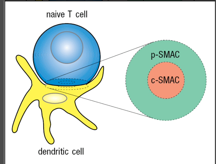
Please describe the immunological synapse.
The immunological synapse is the location of contact between two cell membranes.
Ex: A T-cell is in contact with a dendritic cell.
The MHC complexes on the dendritic cell are binding to the T-cell receptor on the T-cell.
B7 from the dendritic cell binds to CD28 on the T-cell.
Multiple adhesion molecules are also involved. (Different question)
300
Please describe the difference between apoptosis and necrosis.
Apoptosis is a programmed cell death that prevents pathogen replication and prevents the release of infectious bacteria or virus particles outside the infected cell. The apoptosed cell is then eaten by a 'scavenger' macrophage. Apoptosis can be induced within 5 minutes of contact with an effector CD8 T cell.
Necrosing cells are not neat and tidy- they undergo lysis and disintegration.
300
Fill in the blank:
Receptor mediated endocytosis increases efficiency of antigen presentation by more than ______x
10,000x!
400
Please list the contents of c-SMAC (central- Supramolecular Activation Complex)
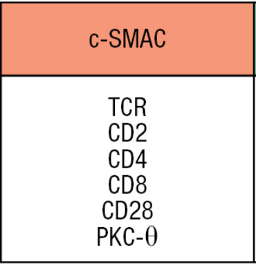
What is c-SMAC?
400
Lck is an example of a tyrosine Kinase. Lck is found next to the cytoplasmic tals of T-cell receptor complex components.
Since Lck is a tyrosine kinase, it will phosphorylate (add phosphate group(s) to molecules).
There are specific areas in the T-cell receptor complex to be phosphorylated. Please detail those areas.
Hint: Remember phosphorylation cascade
After the synapse forms (point of contact between T-cell and antigen-presenting cell), Lck phosphorylates ZAP70, another kinase. ZAP70 is now activated, and it binds to the phosphorylated tyrosines on the zeta chain. ZAP70 transmits the activation signal along the T-cell signaling pathway.
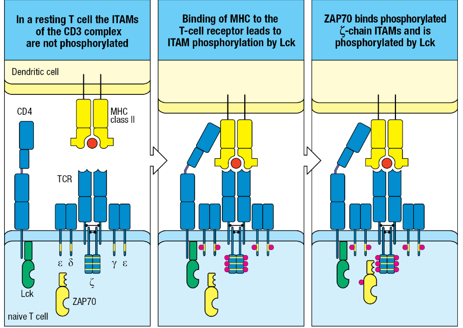
The yellow areas are sequences in the cytoplasmic domain (the tail part sticking into the cytoplasm) is a site to be phosphorylated.
ITAM= Immunoreceptor Tyrosine-based Activation Motif
400
These cells are characterized by:
high surface levels of CD25
make IL-4, IL,10, and TGF-B (all immunosuppressive and anti-inflammatory)
These cells can interact with dendritic cells and prevent them from activating naive T cells.
These cells also function by directly interacting with other effector T cells and preventing them from responding to the antigen.
What is Treg CD4?
400
This is the 'master regulator that commits naive T cells to becoming TH1 cells.
What is T-bet?
T-bet is a transcription factor that commits naive T cells into becoming TH1 cells. T-bet also turns on the T-cell's interferon-gamma (IFN-γ) to turn on, which increases the concentration of IFN-γ and causes more cells to become TH1 CD4 cells.
400
This cell-surface protein is not expressed in naive T-cells, but is expressed in effector (activated) T-cells.
This cell-surface protein allows effector T-cells to enter infected tissue. IT binds to VCAM-1 (Vascular Cell Adhesion Molecule 1)What is VLA-4?
(Very Late Antigen-4)
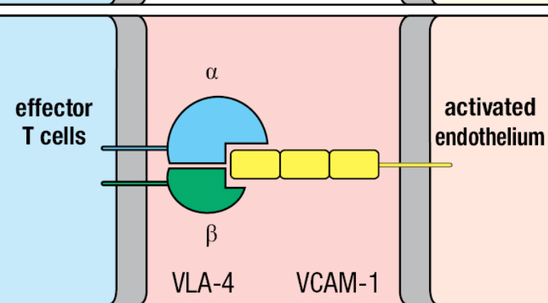 ('activated' endothelium= infected tissue)
500
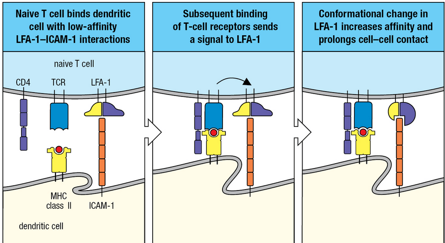
Please list and describe interactions taking place between naive T-cells and dendritic cells.
On the naive T-cell: LFA-1, TCR, CD4
On the dendritic cell: MHC Class 2 and ICAM-1
Binding of the T-cell receptor to MHC Class 2 sends a signal to LFA-1, and LFA-1 binds with ICAM-1. Conformational change occurs in LFA-1, affinity is increased between the two cells and cell contact is increased
500
Please list all the surface molecules on a T-cell.
T-cell receptor
T-cell Co-receptor ( CD4 or CD8)
CD28
CD3 complex
Signaling components (such as IL-2)

500
This condition is characterized by hypergammaglobulinemia, high infectivity, increased reddish macrophage growth, bone, cartilage, and nerve damage, as well as low or no T-cell responsiveness.
Please name and descrbe this condition.
What is Lepromatous leprosy?
Polarized towards TH2 cells. (TH2 cells = activate cellular + antibody response to parasites, secrete IL-4 + IL-5)
Their macrophages become overwhelmed by bacteria, in contrast to tuberculoid leprosy, (polarized towards TH1 cells) wherein the macrophages suppress bacterial growth and the consequence is chronically activated macrophages.
500
How does CD8 kill cells?
CD8 synthesizes lytic granules (vesicles full of cytotoxins). First, the CD8 binds to the target cell in a non-specific way using cell adhesion molecules (CAMs). Next, the antigen receptor causes the CD8 T cell to become polarized and the cytoskeleton at the contact site rearranges. The CD8 T cell aims its Golgi, lytic granules, and MTOC (MicroTubule Organizing Center) towards the target cell. The vesicle containing lytic granules fuses with the CD8 membrane and they are channeled towards a specific spot on the target cell membrane. The target cell then quickly deteriorates.
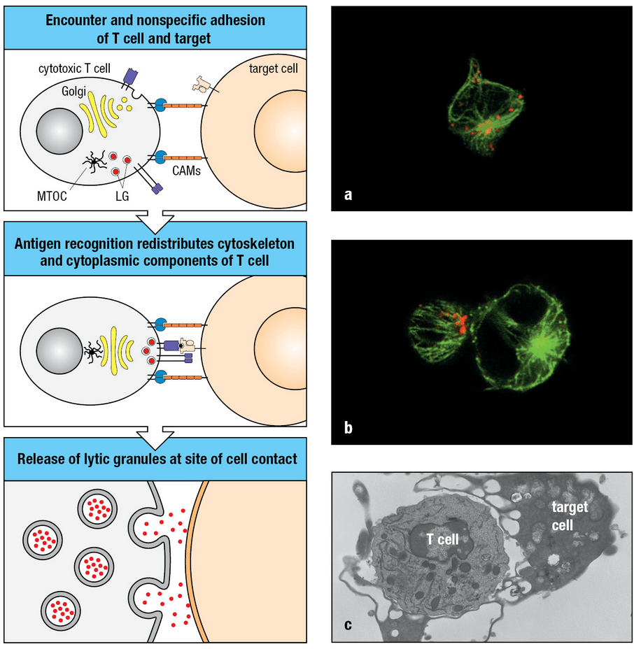
IN SUCCESSION: CD8 T-cell recognizes a peptide on the MHC Class 1 molecule of an infected cell and programs the cell to die by apoptosis. Next, the CD8 T-cell synthesizes more lytic granules and continues attacking.
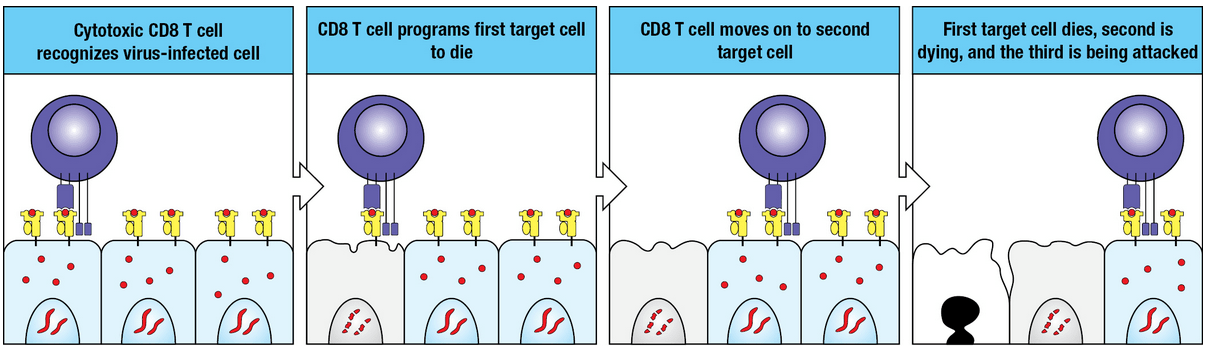
CD8 IS A SERIAL KILLER
500
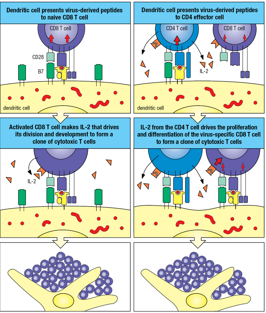
Please describe the 2 ways to activate a CD8 T-cell
1. A dendritic cell presents the peptide fragments from a virus to the naive CD8 T-cell. The (now activated) CD8 T-cell makes IL-2 and IL-2 drives the cell's development and division to form clones of cytotoxic T cells.
2. Dendritic cells present peptide fragments from a virus to CD4 effector cell. IL-2 is made on the CD-4 CELL and drives the proliferation and differentiation of the CD8 cells.