Osteology
Musculature
Vasculature
Nerves
100
These two bones make up the zygomatic arch and form the lateral border of the infratemporal fossa.
The zygomatic process of the temporal bone, and the temporal process of the zygomatic bone.
100
These muscles assist in lateral deviation of the mandible.
All four muscles of mastication.
100
The second portion of the maxillary artery may travel on either side of this muscle before passing between its two heads as it approaches the pterygomaxillary fissure.
The maxillary artery may pass either superficial or deep to the lateral pterygoid. As it approaches the pterygomaxillary fissure, it passes between the muscle’s superior and inferior heads.
100
This sensory branch of the primarily motor anterior division of V3 passes between the two heads of the lateral pterygoid and supplies the cheek mucosa, retromolar pad, and buccal gingiva of the mandibular molars.
The long buccal nerve is the sensory exception within the anterior division of V3. Do not confuse it with the buccal branch of CN VII, which is motor.
200
These are the five different openings found in the infratemporal fossa.
Bonus: What passes through each of these openings.
1. Foramen Spinosum
2. Foramen Ovale: V3, Accessory Meningeal, Lesser Petrosal Nerve
3. Pterygomaxillary Fissure: Maxillary artery, posterior superior alveolar artery
4. Posterior Superior Alveolar Foramina
5. Petrotympanic Fissure: Chorda Tympani
200
This muscle splits the heads of the Medial Pterygoid.
The lateral pterygoid inferior head.
200
This artery frequently passes between the two roots of the auriculotemporal nerve before entering the cranial cavity through the foramen spinosum.
The middle meningeal artery.
In the first-portion branch of the maxillary artery. Do not confuse it with the accessory meningeal artery, which enters through the foramen ovale.
200
This branch of V3 descends lateral and parallel to the lingual nerve, gives off a motor branch, and then enters the mandibular foramen deep to the sphenomandibular ligament.
Before entering the mandibular foramen, the inferior alveolar nerve gives off the mylohyoid nerve, which supplies the mylohyoid and anterior belly of the digastric. The inferior alveolar nerve then supplies the mandibular teeth and continues as the mental nerve.
300
Attaches to the posterior end of the mylohyoid line and connects two muscles. Represents a landmark in anesthetizing.
The pterygomandibular raphe. The superior constrictor and the buccinator attach here. This feature is an important landmark for an inferior alveolar nerve block.
300
These muscles act to protrude the mandible. A ligament nearby limits this action.
Bonus: this ligament also represents a boundary between two structures outside of the infratemporal fossa.
The Lateral Pterygoid, Medial Pterygoid, and Masseter all assist in protrusion. The stylomandibular ligament prevents excessive protrusion.
Bonus: the parotid and submandibular gland.
300
This third-portion branch of the maxillary artery travels backward through the pterygomaxillary fissure, crosses the infratemporal surface of the maxilla, and enters several small canals to supply the maxillary teeth, gingiva, and sinus.
The posterior superior alveolar artery.
Arises in the pterygopalatine fossa but returns to the infratemporal fossa. The important trick is that it is a third-portion branch even though it reenters the infratemporal fossa.
300
A nerve passes through the middle ear, exits through the petrotympanic fissure, and joins the lingual nerve. Damage to the lingual nerve after this union would eliminate which three functions?
General sensation and taste from the anterior two-thirds of the tongue, plus parasympathetic supply to the submandibular and sublingual glands.
The lingual nerve provides general sensation through V3. Chorda tympani contributes taste and preganglionic parasympathetic fibers from CN VII after joining the lingual nerve.
400
This bony structure prevents superior access from the infratemporal fossa into the middle cranial fossa. Two major foramina/canals in this specific bony landmark connect these two fossae.
The infratemporal surface of the greater wing of the sphenoid bone forms the roof of the infratemporal fossa, separating it from the middle cranial fossa above. Foramen ovale and foramen spinosum.
400
What muscle(s) originates at these two points (the red and blue dots) and what landmarks are they?
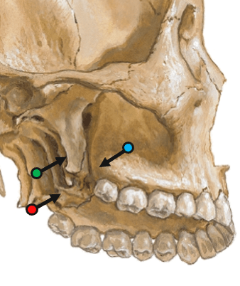
The medial pterygoid heads share an origin at the pyramidal process of the palatine bone (red point). The superficial head also originates at the maxillary tuberosity (blue point).
400
The pterygoid venous plexus communicates with these three structures, creating potential routes for the spread of infection from the infratemporal region.
The pterygoid plexus occupies much of the infratemporal fossa and communicates with the facial vein, ophthalmic veins, and cavernous sinus.
The cavernous sinus is technically a dural venous sinus, but it is one of the three venous connections emphasized in the slides.
400
Preganglionic parasympathetic fibers pass through the tympanic plexus, form a named nerve, and exit through a small opening between the foramina ovale and spinosum. Name the nerve, opening, ganglion, and V3 branch used to reach the parotid gland.
The Lesser petrosal nerve enters the infratemporal fossa through the canaliculus innominatus, synapses in the otic ganglion, postganglionic fibers then hitchhike along the auriculotemporal nerve to reach the parotid gland.
500
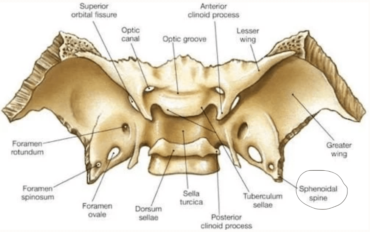
A bony projection represents the posterior superior boundary of the infratemporal fossa. This bony feature on the sphenoid bone is the origin site for a ligament we have discussed.
Spine of the Sphenoid. The sphenomandibular ligament originates here and inserts on the lingula of the mandible.
500
Between the superior border of the lateral pterygoid muscle and the infratemporal surface of the greater wing of the sphenoid exists a narrow horizontal gap. Two specific nerve branches exit this gap by turning sharply upward and outward to enter another fossa/region.
Deep temporal nerves (anterior and posterior) and the masseteric nerve.
They pass superior to the lateral pterygoid muscle, before turning laterally over the mandibular notch (masseteric) or passing up into the temporal fossa (deep temporals).
500
During dissection of the third portion of the maxillary artery, one branch reverses direction and passes back through the pterygomaxillary fissure, while another is the terminal branch that enters the nasal cavity. Name both arteries and the opening used by each.
The posterior superior alveolar artery passes backward through the pterygomaxillary fissure into the infratemporal fossa and then enters the posterior superior alveolar canals. The sphenopalatine artery passes through the sphenopalatine foramen into the nasal cavity.
500
Preganglionic parasympathetic fibers originating in the midbrain travel with a cranial nerve and synapse in an orbital ganglion. The postganglionic fibers then reach the sphincter pupillae through branches of V1. Identify the ganglion.
Ciliary ganglion.
Edinger–Westphal nucleus → CN III → ciliary ganglion → short ciliary nerves of V1 → sphincter pupillae