EnLYTEn us
Name that Disease
Next best Step
AKI or Not
Your kidney on
DRUGS
DRUGS
100
AKI involving the late distal nephron and collecting duct can cause what dangerous electrolyte abnormality?
Hyperkalemia
(Direct injury of cells responsible for K+ secretion)
100
The 2 most common pathophysiologic classifications of AKI include?
pre-renal or post-renal
100
A 62 y.o man is hospitalized for acute coronary syndrome. He undergoes coronary angiography which reveals stenosis of the mid to distal RCA. Drug eluting stent is placed with good post procedure flow. Two days later, he develops elevated sCr 1.6 (baseline 1) and Ua reveals muddy brown casts. How could his AKI have been prevented?
IVF prior to angiography
Contrast induced- AKI from coronary angiography.
~ Intraarterial contrast can induce CA-AKI, usually within 24-48 hrs. There are 2 main mechanisms. Direct effects causing prerenal vasoconstriction (FeNa <1), leading to hypoxia and cell injury. Direct cytotoxicity causing mild ATN (muddy brown casts).
100
What change in urine output would constitute AKI
Urine volume <0.5 mL/kg/hour over six hours
100
What Antibiotic(s) is/are most likely to cause AKI?
Aminoglycosides or Amphotericin
Kidney toxicity caused by vancomycin alone is rare. There are a handful of case reports describing acute interstitial nephritis and even fewer describing ATN due to vancomycin.
200
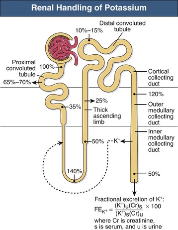
A 38-year-old Hispanic woman with a past medical history of diabetes and hypertension is admitted to the ICU with sepsis due to perforated sigmoid diverticulitis requiring an urgent sigmoid colectomy and colostomy. Postoperatively, she develops oliguria. Her laboratory evaluation includes a basic metabolic panel and the urine electrolytes listed below:
Serum values: sodium 145 mmol/L, potassium 4.2 mmol/L, chloride 100 mmol/L, creatinine 2.0 mg/dL
Urine values: sodium 120 mmol/L, potassium 25 mmol/L, chloride 100 mmol/L, creatinine 60 mg/dL
What is her fractional excretion of sodium (FENa)?
2.76%
The FENa is another important tool in identifying the underlying etiology of AKI. Urinary and serum sodium and urinary and serum creatinine are used in this calculation. FENa values less than 1% indicate prerenal causes for AKI, whereas values greater than 2% denote intrinsic renal pathology:
FENa = 100 × [(urinary sodium × serum creatinine)/(serum sodium × urinary creatinine)]
200
Patients with CHF will likely develop which type of AKI
Pre-renal
~Inadequate renal perfusion due to volume deficiency.
~Renal venous congestion. Increased central venous pressure (CVP) is transmitted into the efferent arteriole, reducing the pressure gradient and subsequently the GFR
200
A 19 y.o Football player presents to his PCP's office for generalized fatigue and muscle pain that does not improve with pain killers. He recently returned to the area 2 weeks ago for the pre-season football program. He states that he is enjoying getting back into shape and has been extremely sore. Labs ordered at this visit show a serum Cr 1.7 (baseline 1). What test might you order next to characterize his AKI
CPK
Rhabdomyolysis in setting of intensive exercise. This is a clinical syndrome caused by damage to skeletal muscle and release of its breakdown products into the circulation, (AKI) is a severe complication.
200
Most common cause of AKI in hospitalized patients and characterized by UA showing muddy-brown/ granular casts, epithelial cells, and epithelial casts on microscopy
ATN
Most common cause of intrinsic AKI. Results necrotic proximal tubule cells falling into tubular lumen, --> obstruction-->decreased GFR
200
This antibiotic class works by inhibiting polypeptide synthesis and can causes proximal tubular cell necrosis or ATN as they are reabsorbed into the proximal tubule cells, where they have a prolonged tissue half-life.
Aminoglycoside
300
A 42-year-old man with a history of alcohol abuse and pancreatitis presents to the emergency department with epigastric abdominal pain. The pain started about 6 days prior to presentation. He also describes nausea and poor oral intake. He is currently drinking 12 beers/day. He was treated for acute pancreatitis 3 months prior to this presentation and has a history of walled-off pancreatic necrosis that required surgical débridement. His vital signs at the time of presentation include a temperature of 37.2°C (99°F), blood pressure of 92/47 mm Hg, pulse of 135 beats/min, respirations of 22 breaths/min, and oxygen saturation of 97% on room air. His examination is notable for severe epigastric tenderness on palpation. Laboratory tests reveal a serum creatinine to be markedly elevated at 4.5 mg/dL.
Which of the following laboratory findings favors the diagnosis of acute tubular necrosis (ATN) over prerenal azotemia?
A. BUN:Cr 24
B. Urine osmolarity of 900 mOsm/kg
C. Urine sodium 14mEq/L
D. FeNa 0.3%
E. FeNa 2.6%
Correct E
It is important to distinguish prerenal from intrinsic renal failure. ATN is caused by a sustained decline in the glomerular filtration rate (GFR) that is triggered by an acute ischemic or nephrotoxic event. Prerenal azotemia can develop into ATN if the decline in the GFR is not reversed by volume expansion. It is helpful to evaluate urine volume, urine sediment, and other urinary and serum indices to help distinguish these two syndromes. Generally, a BUN-to-creatinine ratio greater than 20:1 favors a diagnosis of prerenal azotemia. In prerenal failure, hyaline and granular casts may be seen, whereas in ATN, urine sediment classically demonstrates muddy-brown casts. With tubular dysfunction seen in ATN, the ability to concentrate urine is lost and urine osmolarity is usually less than 450, as opposed to concentrated urine greater than 500 mOsm/kg seen in prerenal azotemia. Furthermore, urine sodium is low in renal azotemia (< 20 mEq/L) and is high in ATN (> 40 mEq/L). In ATN, the nephrons excrete a large fraction of their filtered sodium and water, resulting in higher fractional excretion of sodium, greater than 1% compared with less than 1% in prerenal azotemia.
300
A 72-year-old man is admitted to the hospital with fevers, weakness, and a productive cough. He was recently treated with a course of oseltamivir for influenza and was recovering until 1 day prior to presentation when he developed a fever at home to 38.9°C (102°F), along with a worsening cough and weakness. On admission, a chest x-ray reveals a right lower lobe consolidation, and he is started on ceftriaxone and azithromycin. On hospital day 2, blood cultures collected at the time of admission return positive for methicillin-sensitive Staphylococcus aureus. He is switched to a continuous nafcillin infusion. After recovering from a mild acute kidney injury from volume depletion on admission, on the fourth day of nafcillin treatment, his serum creatinine increased from a baseline of 1.2 to 1.8 mg/dL. The next day, his serum creatinine increases to 2.5 mg/dL. A urinalysis is positive for red blood cell and white blood cell casts as well as urine eosinophils.
Which of the following is the most likely cause of this acute renal failure?
A. prerenal azotemia
B. ATN
C. contrast-induced nephropathy
D. AIN
E. Acute urate nephropathy
Correct D
This patient initially presented with an acute kidney injury (AKI) associated with volume depletion in the setting of sepsis. This improved, but his kidney function acutely worsened 4 days into treatment with nafcillin. AIN is important to consider when a patient who is recovering from sepsis-induced AKI develops AKI while receiving antibiotics. Antibiotics are a major cause of AIN, especially with β-lactams, as seen in this patient receiving a continuous nafcillin infusion. AIN is suggested by a urinalysis with urine sediment findings of blood, protein, and white blood cells in clumps and casts. The classic clinical presentation includes a patient with a rash, joint pains, and a fever, although it is uncommon to have all three signs. Treatment is focused on removing the offending agent and is otherwise supportive. Prerenal azotemia is less likely at this point in the patient’s clinical course given that his initial kidney injury apparently responded to treatment. Acute tubular necrosis is a possibility but also less likely given his initial improvement and would be more likely in the setting of shock or hypotension from sepsis. There is no mention of crystals on the urinalysis to suggest urate nephropathy, and the patient did not receive intravenous contrast to suggest CIN.
300
A 35-year-old male presents with a 2-week history of lower extremity edema. He is in good health and does not take any medications. You note weight gain, and mild dyspnea with exertion. An examination is unremarkable except for 2+ to 3+ pitting edema of the lower extremities to his knees bilaterally. A CBC and metabolic panel are unremarkable except for a low albumin level. A urinalysis reveals 3+ protein on the dipstick with no microscopic findings. Which one of the following would be the most appropriate next step?
A) Urine microscopy to check for eosinophils
B) A spot urine protein/creatinine ratio
C) Renal ultrasonography
D) Echocardiography
E) Referral for a renal biopsy
Correct B
Individuals with nephrotic syndrome often present with edema and fatigue with no evidence of severe liver disease or heart failure. Hallmarks of this problem include heavy proteinuria, hypoalbuminemia, and peripheral edema, often with hyperlipidemia as well. While most of these cases are idiopathic, secondary causes such as diabetes mellitus, systemic lupus erythematosus, and medication reactions should be considered. To confirm proteinuria in the nephrotic range a spot urine protein/creatinine ratio is now suggested instead of a 24-hour collection of urine. Checking urine for eosinophils has been recommended in the past for evaluation for acute interstitial nephritis but subsequent studies have shown a lack of specificity and sensitivity. Renal ultrasonography would be indicated if the glomerular filtration rate were reduced. Echocardiography would be appropriate if heart failure were suspected. While a renal biopsy is often recommended, it is most useful in patients with suspected underlying systemic lupus erythematosus or similar disorders when a biopsy can guide management decisions and prognosis.
300
Urine output <100mL/day
Anuria
300
Also known as “Spice” or “K2,” this type of recreational drug has been associated with AKI due to ATN or AIN.
Synthetic THC
400
A 77-year-old woman is admitted to the hospital after being found down in her assisted living facility. She has a previous history of hypertension, hyperlipidemia, chronic kidney disease, coronary artery disease, and mild cognitive impairment. She was found down in her bathroom, and the duration between her fall and when she was found is unclear. She is alert but confused. Her initial vital signs are as follows: temperature 34.9°C (94.8°F), blood pressure 104/64 mm Hg, pulse 122 beats/min, respiratory rate 22 breaths/min, and oxygen saturation of 91% on room air by pulse oximetry. A chemistry panel reveals a sodium of 128 mmol/L, potassium of 7.4 mmol/L, blood urea nitrogen of 74 mg/dL, and creatinine of 4.1 mg/dL. A serum creatine kinase is elevated at 11,200 U/L.
Which of the following describes the typical order of electrocardiographic (ECG) changes associated with hyperkalemia, from earliest to latest?
A. prolonged PR, peaked T waves, widened QRS, sine wave pattern
B. Prolonged PR, peaked T waves, sine wave pattern, widened QRS
C. Peaked T waves, prolonged PR interval, widened QRS, sine wave pattern
D. Peaked T waves, widened QRS, prolonged PR interval, sine wave pattern
E. Sine wave pattern, peaked T waves, prolonged PR, widened QRS
Correct C
Like many electrolyte disturbances, hyperkalemia can causes several ECG changes and arrhythmias. Hyperkalemia is common with acute kidney injury, especially in rhabdomyolysis, as in this patient. The sequential ECG changes observed in hyperkalemia are peaked T waves, prolongation of the PR interval, widening of the QRS complex, and a sine wave pattern. ECG changes associated with hyperkalemia are life threatening if not emergently treated. The first step in treatment is the administration of calcium gluconate, which immediately antagonizes the effect of potassium on cardiac conduction. Other treatment options include insulin and glucose, inhaled beta agonists, intravenous bicarbonate, sodium polystyrene sulfonate, and dialysis.
400
A 26-year-old male patient is being evaluated in the hospital for progressively worsening kidney function. He was admitted five days ago and was diagnosed with methicillin-resistant staphylococcus aureus infection. Intravenous vancomycin was immediately started and has been adjusted as needed to maintain a level of 15 to 20 mcg/ml. His fever has resolved, but his serum creatinine has gradually increased. His past medical history includes two prior episodes of staphylococcal endocarditis treated with antibiotics. He has a history of occasionally injecting heroin for the past four years. His only medication currently is vancomycin. On physical examination, the temperature is 37.3 C (99.1 F), blood pressure is 110/70 mmHg, heart rate is 92/min, and respiratory rate is 18 breaths/min. His BMI is 22 kg/m2. Cardiac examination reveals a soft diastolic murmur along the left sternal border and trace lower extremity edema bilaterally. No skin rash or arthritis is seen. Labs show: C3 low, C4 normal, creatinine 2.8 mg/dL from 1.5 mg/dL on admission, cryoglobulins negative, urinalysis- 3+ blood; 2+ protein; 30-40 erythrocytes per high power field (HPF); 10-15 leukocytes/HPF; and erythrocyte casts. A transthoracic echocardiogram shows moderate aortic regurgitation without vegetations. Renal ultrasound shows normal-sized, mildly echogenic kidneys. Doppler studies of the renal arteries and veins were normal. What is the most appropriate management?
A. Initiate glucocorticoids
B. Schedule a kidney biopsy
C. Switch vancomycin to daptomycin
D. Continue current therapy
C.
Vancomycin-related tubular toxicity typically occurs after 7 to 10 days and usually has bland urine sediment. Antibiotic-associated AIN is typically seen with beta-lactams and can lead to leukocyte casts but not erythrocyte casts.
400
A 21-year-old white man sustains a crush injury to both lower extremities following a building collapse. On arrival, he has tea-colored urine and a serum creatine kinase (CK) level of 10,300 U/L.
Which of the following is the best treatment to prevent AKI?
A. Sodium bicarb infusion
B. Mannitol infusion
C. Vigorous hydration
D. Continuous Renal Replacement therapy
E. Intermittent HD
Correct C
The diagnosis of rhabdomyolysis begins with a high index of suspicion based on the patient’s clinical history. In the surgical patient, a history of trauma is the most common cause, followed by a history of recent orthopedic/vascular procedures with prolonged ischemia time or operative time. Clinical findings of rhabdomyolysis can include myalgias, muscle swelling, muscle weakness, and dark or tea-colored urine (due to myoglobinuria). The presence of all of these clinical findings is not required to make the diagnosis. A classic laboratory finding in a patient with rhabdomyolysis is a positive urine dipstick for blood but few red blood cells on a microscopic examination. This is due to the presence of the heme pigment in both myoglobin and hemoglobin. An elevated CK is the hallmark laboratory finding. There is no “magic number” for CK at which a definitive diagnosis of rhabdomyolysis is made. Reports of AKI have been published in the literature for CK levels as low as 500 U/L. AKI associated with rhabdomyolysis is due to the heme pigment that is present in myoglobin that is released as a result of muscle injury. Heme contributes to AKI via several mechanisms. It precipitates as casts that cause physical obstruction of the renal tubules, it is directly nephrotoxic to the tubular cells, and it causes renal vasoconstriction that decreases perfusion to the kidney.
Patients with rhabdomyolysis should be treated with early aggressive intravenous fluid hydration to prevent the development of AKI. A CK level greater than 5,000 U/L has been shown in the literature to be an independent risk factor for the development of AKI due to rhabdomyolysis. The ideal type of fluid and rate of fluid resuscitation have yet to be thoroughly elucidated. Infusion rates of 200 to 1,000 mL/hr have been reported, but normal saline infusion titration to a urine output of approximately 3 mL/kg/hr is likely sufficient. It has been suggested that acidic urine promotes the precipitation of obstructing tubular casts in the kidney. The use of bicarbonate and mannitol in the prevention of AKI has the theoretical advantage of increasing the pH of the urine to prevent this phenomenon. A large study in 2004 examined 2,083 patients diagnosed with rhabdomyolysis. It was shown that the use of bicarbonate (and mannitol) did not decrease rates of renal failure, the need for dialysis, or mortality for patients with CK levels less than 3,0000 U/L. Multiple subsequent studies have shown similar results, with some even demonstrating a detrimental effect to bicarbonate infusion.
400
A 52-year-old woman presents to the emergency department with weakness, lightheadedness, and diarrhea. She recently visited her primary care physician reporting symptoms of acute sinusitis. She was prescribed a course of clindamycin. Five days into her antibiotic course, she developed profuse watery diarrhea. She feels lightheaded whenever she stands up. She is afebrile, with a blood pressure of 107/79 mm Hg that drops to 87/62 mm Hg after standing and a pulse of 121 beats/min. Her baseline serum creatinine is 1.1 mg/dL and today is near her baseline at 1.2 mg/dL, with a blood urea nitrogen of 22 mg/dL.
What is the response of the vasculature to hypoperfusion from hypovolemia in this patient to maintain an adequate glomerular filtration rate (GFR)?
A. afferent constriction, efferent constriction
B. Afferent dilation, efferent constriction
C. afferent dilation, efferent dilation
Correct B
This patient is likely presenting with Clostridium difficile colitis from inappropriate antibiotic use for acute sinusitis. She has developed hypovolemia from profuse diarrhea, as evidenced by her orthostatic hypotension. However, she has been able to maintain adequate perfusion to her kidneys given that her serum creatinine is just minimally elevated from her baseline. The kidney has the capacity to autoregulate GFR and blood flow simultaneously during renal hypoperfusion through the independent regulation of afferent and efferent arteriolar tone. Whereas the afferent arteriolar dilates in response to renal hypoperfusion, the efferent arteriolar constricts, maintaining glomerular intracapillary pressure, which is the driving force for glomerular filtration.
400
A 59-year-old Hispanic man sustains a gunshot wound to the abdomen. He undergoes an exploratory laparotomy and nephrectomy for a grade V renal injury. Postoperatively, he develops hyperkalemia, with a serum potassium concentration of 7.6 mEq/L and peaked T waves on an electrocardiogram (ECG).
Which of the following treatments for hyperkalemia decreases total body potassium content?
A. Calcium chloride or calcium gluconate
B. Sodium bicarbonate
C. Insulin and glucose
D. Inhaled beta2 agonists
E. Loop diuretics
Correct E
Hyperkalemia is another common and potentially life-threatening complication of AKI. Patients with hyperkalemia and ECG changes or serum potassium greater than 7 mEq/L should be treated immediately. Initial fast-acting treatment modalities include the administration of insulin with dextrose to drive potassium intracellularly by increasing the activity of the sodium, potassium–adenosine triphosphatase (Na,K-ATPase) enzyme present in muscle. Beta2 agonists such as nebulized albuterol can also be used to drive potassium intracellularly. Beta2 agonists function via a similar mechanism to insulin/glucose boluses. A third treatment modality that has been described is the infusion of sodium bicarbonate. The rationale behind this treatment option is that by increasing pH, cells will release hydrogen ions to buffer the increased level of bicarbonate ions. Potassium is then preferentially driven intracellularly to maintain an equal charge balance (one hydrogen ion out to one potassium ion in). Regardless of the modality used to acutely treat symptomatic hyperkalemia or hyperkalemia associated with ECG changes, calcium should always be administered concurrently. Calcium serves to stabilize the membrane potentials of these cells. Calcium gluconate or calcium chloride can be used and should be given intravenously. Calcium chloride contains more ionized calcium than calcium gluconate but can be irritating to peripheral veins, and central access is preferred.
It is important to remember that the above methods of lowering the serum potassium level are transient. They only serve to shift potassium intracellularly without actually excreting it from the body. Once the acute hyperkalemia episode has been temporarily treated, definitive treatment is necessary to remove the excess potassium from the body. The three methods of accomplishing potassium elimination are medical diuresis, use of cation exchange resins, and renal replacement therapy.
500
You have been consulted to see a 35-year-old man in the hospital following a motor vehicle crash 2 days ago. He was trapped in his vehicle for 6 hours prior to extraction. He has not required surgical intervention. Since admission, his serum creatinine has increased from 1.1 to 1.8 mg/dL. His physical examination reveals extensive ecchymoses and tenderness to palpation of bilateral thighs. Pulses and sensation in the upper and lower extremities are normal. Radiographs reveal no fracture of the lower extremities. Laboratory data from this morning reveal creatine kinase of 98,200 U/L.
Which of the following urinalysis results would you expect in this patient?
A. Brown color; specific gravity 1.010; 3+ blood on dipstick testing; too numerous to count red blood cells (RBCs) per high-power field (hpf); 1 to 2 white blood cells (WBCs)/hpf
B. Yellow color; specific gravity 1.010; negative blood on dipstick testing; too numerous to count RBCs/hpf; 10 to 20 WBCs/hpf
C. Brown color; specific gravity 1.010; 3+ blood on dipstick testing; 1 to 2 RBCs/hpf; 1 to 2 WBCs/hpf
D. Specific gravity 1.010; negative blood on dipstick testing; 0 RBCs/hpf; more than 50 WBCs/hpf with 2+ eosinophils
Correct C.
The most frequent causes of rhabdomyolysis are trauma or other injury that leads to muscle compression; ischemia; excess muscle activity, such as occurs during exercise or seizures; metabolic derangements; drugs; genetic defects; body temperature dysregulation; and infections. Cocaine use, neuroleptic malignant syndrome, and the use of statin drugs can also contribute to or cause rhabdomyolysis. Muscle pain and dark-brown urine that is positive for blood on dipstick testing (ortho-toluidine positive) but that does not contain RBCs are important diagnostic clues. Patients with rhabdomyolysis have a marked increase in the level of muscle enzyme, such as creatine kinase. The urine sediment in patients with myoglobinuria may show RBCs, pigmented casts, granular casts, and numerous uric acid crystals.
500
A 65-year-old man with gastrointestinal malignancy undergoes a laparotomy for resection followed by ileostomy. Intravenous normal saline is given at a rate of 100 ml per hour to compensate for the excessive losses from ostomy. Initially, his urine output is approximately 40 ml per hour but drops to 20 ml per hour on post-op day 4. An arterial blood gas is remarkable for a pH of 7.21 and PCO2 of 28 mmHg. Serum sodium is 130 mEq/L, potassium is 5.6 mEq/L, BUN is 90 mg/dL, and creatinine is 3.0 mg/dL. Urine sodium is 35 mEq/L, and urinary creatinine is 90 mg/L. Which of the following is the next most appropriate step in establishing the underlying pathology leading to deranged renal function?
A. Urine for dysmorphic RBCs
B. Fractional excretion of sodium
C. Ultrasound abdomen and pelvis
D. Renal biopsy
B.
There are markers of tubular function that can be calculated to help distinguish an etiology like the fractional excretion of sodium (FeNa) and urea. If FeNa is less than 1%, it indicates a prerenal cause of AKI.
500
A 62-year-old male is scheduled for CT of the chest with intravenous contrast in the next 48 hours. He has a long-standing history of degenerative joint disease in the right knee, coronary artery disease, and type 2 diabetes. His current medications are low-dose aspirin, metformin (Glucophage), and naproxen.
In addition to discontinuing metformin prior to the procedure, which one of the following would you recommend for prevention of contrast-induced nephropathy?
A) Discontinue aspirin
B) Discontinue naproxen
C) Start acetylcysteine
D) Start mannitol (Osmitrol)
In order to prevent contrast-induced nephropathy, NSAIDs such as naproxen should be withheld for 24–48 hours prior to a procedure involving venous or arterial administration of radiocontrast material. Avoidance of volume depletion and other nephrotoxic agents is also recommended. Aspirin in low doses (up to 325 mg) does not impact renal function and therefore does not play a role in the development of contrast-induced nephropathy. Administration of acetylcysteine or mannitol has not been shown to reduce the incidence of contrast-induced nephropathy. Pre- and postprocedural hydration with normal saline is recommended in patients at high risk for developing contrast-induced nephropathy, such as those with underlying chronic kidney disease, heart failure, proteinuria, sepsis, hypovolemia, or hypotension. Metformin does not cause contrast-induced nephropathy but should be withheld due to the potential, mostly theoretical, risk of developing lactic acidosis, especially if contrast-induced nephropathy were to develop (SOR B).
500
A 58-year-old white male is involved in a motorcycle crash and sustains a pelvic fracture requiring angioembolization, multiple long bone fractures, splenic injury requiring splenectomy, multiple rib fractures, and a pneumothorax requiring a chest tube. Three days after admission to the intensive care unit (ICU), his urine output has dropped to 0.4 mg/kg/hr over the past 18 hours, and his serum creatinine has doubled since admission from 0.8 to 1.6 mg/dL.
According to the Kidney Disease: Improving Global Outcomes (KDIGO), which of the following is true of this patient?
Stage 1
Stage 2
Stage 3
Stage 4
Does not have AKI
Correct B
The KDIGO group met in 2012 and released guidelines for AKI that unified the RIFLE (Risk, Injury, Failure, Loss of function, End stage renal disease) and AKIN (Acute Kidney Injury Network) classification systems. KDIGO contributed the factor of additional time to the definition but continued the scale grading of 1 to 3. Preliminary data suggest that this new guideline identifies more AKI patients, but mortality prediction is the same as AKIN. The most current KDIGO consensus guidelines define and stage AKI as follows:
Stage 1: Increase in serum creatinine greater than or equal to 0.3 mg/dL within 48 hours or increase in serum creatinine greater than or equal to 1.5 times baseline, which is known or presumed to have occurred within the previous 7 days or urine volume less than 0.5 mL/kg for 6 to 12 hours
Stage 2: Increase in serum creatinine 2.0 to 2.9 times baseline or urine volume less than 0.5 mL/kg for 12 hours or more
Stage 3: Increase in serum creatinine 3.0 times baseline or increase in serum creatinine to 4.0 mg/dL or greater or initiation of renal replacement therapy or urine volume less than 0.3 mL/kg for 24 hours or more or anuria for 12 hours or more
500
A 65 y.o man with a PMHx significant for HIV, and T2DM is seen in the ED for SOB and cough. He just returned form a hiking trip with his son in Ohio and states that his sx are worsening. Patient is non-compliant with his ART and has not seen a physician in several years. He is admitted to the Family Medicine service for management of acute hypoxic respiratory failure 2/2 suspected CAP. On hospital day 2, Jay notices that the patient has developed a pruritic maculopapular rash about his chest and upper extremities, as well, his urine output is decreased and his sCr is 2.0 (baseline 1.1). What do you suspect is the diagnosis?
AIN 2/2 Amphotericin therapy
This patient likely contracted histoplasma while in Ohio, and is being treated with Amphotericin. Presence of Rash is suggestive of allergy and worsening kidney function is likely due to AIN.
Acute interstitial nephritis classically presents as acute kidney injury after the use of a known offending drugs and is sometimes associated with the urinary findings of pyuria, hematuria, and white cell casts. Note that: signs of systemic allergy, such as a maculopapular rash, peripheral eosinophilia, and eosinophiluria, are present in less than one-third of patients.