Liver
Dysmotility
Pharm/Genetics/Biochem
Rando
100
A healthy 35-year-old man presents with fever, diarrhea, chills, and right upper quadrant pain after returning from backpacking near the Amazon River. He has leukocytosis without evidence of eosinophilia. Alkaline phosphatase is elevated, but AST and ALT are within normal limits. A CT scan with contrast demonstrates an area in the right lobe of the liver with a cavity that has an enhancing wall and hypoattenuation in the center. What is the first line of treatment for this patient?
Metronidazole for 10 days
This patient is suffering from an amebic liver abscess from E histolytica. He was recently in the tropics, and he fits the demographic for amebic liver abscesses (male, 3rd-4th decade of life). E histolytica typically presents with fever and RUQ pain but can also present with diarrhea, chills, hepatomegaly, weight loss, and night sweats. Laboratory values that are most often abnormal are elevated alkaline phosphatase. AST and ALT are typically normal. Patients present with an elevated WBC without eosinophilia. Treatment for amebic liver abscesses includes metronidazole 750 mg TID or 500 mg Q6H for 7 to 10 days. Ampicillin and gentamicin or ciprofloxacin are added when enteric flora as the source of infection is suspected. Neither percutaneous nor open surgical drainage is warranted at this time. Drainage is reserved for cases that fail to respond to medical therapy after 3-5 days, are located in the left lobe (due to risk of rupture into the pericardium), or are massive (typically >10 cm) with imminent risk of rupture. Adding drainage unnecessarily increases the risk of secondary bacterial infection.
Pyogenic liver abscess, most common type (>80%) and is usually secondary to a biliary or GI source like diverticulitis. Typically appears as a unilocular, thick-walled abscess. Treatment is percutaneous drainage and antibiotics.
Echinococcal (hydatid) cyst. Typically presents as a double-walled cyst. Treatment is albendazole followed by the PAIR technique (puncture, aspiration, injection, reaspiration)
100
Sphincter of Oddi dysfunction exists at manometry pressure over what?
manometry pressure > 40 mm Hg
Treatment: sphincterotomies can be performed endoscopically, sphincteroplasties require an open approach. Both options are similarly effective and can relieve symptoms in 60% to 80% of cases
100
What is the medication and mechanism of action of the med started for patients with cirrhosis and esophageal varices presenting with GI bleeding?
Octreotide, a somatostatin analog, induces vasoconstriction by acting directly on smooth muscle cells in the mesenteric and splanchnic arterial wall, lowering portal venous pressure and helping control acute variceal bleeding.
Biochem
This binding activates a G-protein coupled pathway that stimulates phospholipase C (PLC).
PLC cleaves membrane phospholipids to produce inositol 1,4,5-trisphosphate (IP₃) and diacylglycerol (DAG).
IP₃ binds to receptors on the sarcoplasmic reticulum, triggering the rapid release of stored intracellular calcium into the cytoplasm, which drives smooth muscle contraction.
Unlike many other vasoactive agents that rely on or alter endothelial nitric oxide (NO) production to change vessel diameter, octreotide’s direct contractile effect on the vascular wall is local and independent of nitric oxide synthesis. This allows it to remain effective even in pathophysiological states (like advanced cirrhosis) where endothelial function is severely altered.
Start Nonselective beta-blockers, such as carvedilol, to reduce portal venous pressure and prevent the risk of future variceal bleeding
100
A 34-year-old female is evaluated in the clinic for an incidental finding on an abdominal ultrasound obtained for vague abdominal bloating. The ultrasound demonstrates a normal-appearing gallbladder with a single 2.8 cm gallstone. She denies any history of right upper quadrant pain, nausea, or vomiting after meals. Her past medical history is unremarkable. What is the most appropriate management?
Elective laparoscopic cholecystectomy
Prophylactic cholecystectomy is indicated in certain patient populations even if they are asymptomatic. These indications include sickle cell anemia, porcelain gallbladder, large gallstones (>2.5cm), large polyps >1cm, and potentially patients with known gallstones undergoing bariatric surgery. Because this patient has a large gallstone (>2.5cm), she should be offered an elective cholecystectomy.
BTK
200
A 65-year-old male is undergoing a pancreaticoduodenectomy (Whipple procedure) for distal cholangiocarcinoma. His vitals are stable. During dissection of the hepatoduodenal ligament, a large pulsatile vessel is encountered posterior to the pancreas and common bile duct. What is the most likely origin of this aberrant vessel?
The most common aberrant hepatic vascular anatomy is a replaced right hepatic artery. This vessel branches off the superior mesenteric artery (SMA) and travels behind the pancreas and common bile duct, making it highly susceptible to injury during a Whipple procedure
A replaced left hepatic artery typically branches from the left gastric artery and runs in the gastrohepatic ligament.
200
A 58-year-old otherwise healthy woman with a history of chronic constipation presents to your clinic with a complaint of rectal prolapse. Physical examination reveals a full-thickness rectal prolapse with associated mucosal redundancy. All visualized mucosa appears viable. There are no other significant findings on examination. What is the most appropriate surgical intervention for this patient?
Sigmoid resection and rectopexy
In an otherwise healthy individual with rectal prolapse and a history of chronic constipation, the most appropriate surgical intervention is sigmoid resection and rectopexy. Given her history of constipation, the patient should undergo a resection. Patients without constipation and with no other significant medical comorbidities may undergo rectopexy alone without the need for sigmoid resection.
Transanal and perineal approaches are generally reserved for patients who are unfit to undergo a transabdominal procedure. Although these procedures are better tolerated, they have generally higher rates of recurrence
Score
200
What is the MOA of the preferred pharmacologic agent for treating colonic pseudo-obstruction (Ogilvie syndrome) refractory to conservative management. AND what is a side effect we must consider before giving it?
Neostigmine, an acetylcholinesterase inhibitor
can lead to cholinergic side effects, including bradycardia, due to increased parasympathetic activity
200
How do you place a PEG Tube? Oral Boards style
1. Diagnostic upper endoscopy to evaluate the stomach, esophagus, and duodenum
2. Insufflate the stomach with air
3. Identify PEG site ( 2 fingerbreadths below the costal margin in the midclavicular line); confirm with 1:1 finger indentation and transillumination
4. Infiltrate local anesthesia and create a small skin incision at desired PEG site
5. Insert a needle and catheter into the stomach under endoscopic guidance
6. Thread a wire through the needle and retrieve it with a snare out of the mouth
7. Attach the PEG tube to the oral end of the guidewire and pull the tube through the abdominal wall; set the internal bumper against the gastric wall
8. Secure external bumper at the skin
9. Confirm appropriate position visually
300
Name the Liver Lesion Associated with each of the CT imaging findings
A. Peripheral-to-central enhancement on delayed phase
B. Enhancement of a central scar on delayed phase
C. A hypodense, low-attenuation lesion with mild enhancement on arterial phase
D. Thin-walled fluid-filled structure with no septations or calcifications on arterial phase
E. Heterogeneous, poorly circumscribed mass with bright arterial enhancement and quick washout
F. Well-circumscribed mass with bright enhancement on arterial phase
A. Peripheral-to-central enhancement on the delayed phase = Hemangioma
B. Enhancement of a central scar on the delayed phase = Focal nodular Hyperplasia
C. A hypodense, low-attenuation lesion with mild enhancement on the arterial phase = Metastatic Tumor
D. Thin-walled fluid-filled structure with no septations or calcifications on arterial phase = simple cyst
E. Heterogeneous, poorly circumscribed mass with bright arterial enhancement and quick washout = Hepatocellular carcinoma
F. Well-circumscribed mass with bright enhancement on arterial phase = Hepatic Adenoma
300
A 3-year-old girl is brought to the emergency department by her parents, who witnessed her swallow "some shiny metal pieces" from a toy approximately 2 hours earlier. She is alert, in no distress, tolerating secretions, and has a soft, nontender abdomen with no peritoneal signs. Plain radiographs in two views (anteroposterior and lateral) demonstrate two discrete round metallic densities in the region of the gastric body, appearing stacked with a lucent halo between them on one view and overlapping on the other.
Which of the following is the most appropriate next step in management?
A. Discharge home with instructions to strain the stool and return in 2 weeks if the objects have not passed
B. Admit for serial radiographs every 48 hours and intervene only if the objects fail to progress
C. Urgent (within 48 hours) endoscopic removal once an esophageal injury has been excluded
D. Emergency endoscopic removal
Answer: D
Rationale:
Two magnets mandate emergency removal regardless of symptoms. When two or more magnets—or a single magnet plus another metallic object—are ingested, the magnets can attract each other across intervening bowel wall, trapping tissue between them and producing pressure necrosis, fistula, obstruction, or perforation. requires emergency endoscopic removal even in an asymptomatic patient. The radiographic appearance of two round densities with a lucent interface (and the "stacked" look that shifts between views) is the classic clue distinguishing paired magnets from a single object or stacked coins.
Choice A is inappropriate: watchful waiting with stool straining applies to low-risk single, small, blunt objects—not paired magnets.
Choice B describes a serial-radiograph surveillance strategy used for certain blunt objects but observation is contraindicated when two magnets are present because of the perforation/fistula risk.
Choice C (urgent removal within 48 hours after excluding esophageal injury) is the pathway for a specific button battery scenario—asymptomatic child younger than 5 years with a battery ≥20 mm—not for magnets.
rapidly triage ingested foreign bodies by urgency—emergency removal for ≥2 magnets and for symptomatic button batteries in the stomach; urgent endoscopy for sharp/pointed objects and symptomatic obstruction; and nonurgent/observation for most blunt objects, recognizing that roughly 80–90% of ingested foreign bodies pass spontaneously, 10–20% need endoscopy, and about 1% require surgery. Endoscopic tool selection also distinguishes experts: a snare for long objects, a retrieval net for round objects, rat-tooth/alligator forceps for coins, and an overtube or gastric hood to protect the airway and esophageal mucosa when extracting sharp or dangerous objects.
300
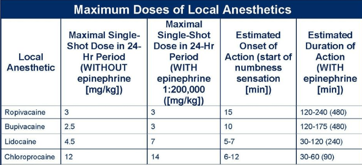
During a bedside laceration repair on a 70-kg adult, which of the following represents the maximum recommended dose of lidocaine when combined with epinephrine?
lidocaine with epinephrine at 7 mg/kg (not to exceed 500 mg total) and without epinephrine at 4.5 mg/kg (not to exceed 300 mg total).
300
A 48-year-old man presents to the ED with hematemesis. He has a history of heavy alcohol consumption but has been abstinent since a bout of severe pancreatitis 14 months ago. CT imaging at that time showed occlusion of the splenic vein with a patent portal vein. He stabilizes after resuscitation with crystalloid and blood transfusions. Upper endoscopy now shows slow bleeding from isolated gastric varices. What is the best definitive management of this patient?
Splenectomy
This patient’s history of pancreatic inflammation and isolated gastric varices is due to a splenic vein thrombus (ie, left-sided portal HTN). This most frequently occurs in patients with either pancreatic inflammation or neoplasm. Because the venous HTN is isolated to the gastrosplenic veins, only gastric varices are formed, because the splenic flow cannot drain via the occluded splenic vein’s being diverted to local collaterals. Definitive management of gastric varices due to splenic vein occlusion is splenectomy, as this eliminates the splenic inflow. A TIPS procedure is not indicated, as removing the spleen will decrease left-sided portal hypertension without the need for an additional procedure. A partial gastrectomy will not correct the occluded splenic venous drainage, and blood will still back up into the gastrosplenic veins, causing gastric varices. Ligation of gastric varices does not correct the underlying pathology causing the gastric varices, and a liver transplant also will not correct the underlying pathology if the spleen is unable to drain.
400
A 48-year-old female presents with a 6-month history of progressive right upper quadrant pain and early satiety. Vitals are BP 122/78 mmHg, HR 76 bpm, Temp 37.0°C. Physical exam reveals a palpable, non-tender mass in the right upper quadrant. A CT scan of the abdomen demonstrates a 12 cm well-defined, hypodense lesion in the right lobe of the liver without an enhancing rim, septations, or solid components. Hydatid serology is negative. What is the most appropriate definitive management for this patient?
Laparoscopic cyst fenestration
This patient has a large, symptomatic simple hepatic cyst (well-defined hypodense lesion without an enhancing rim). The definitive treatment for a symptomatic simple cyst is laparoscopic cyst fenestration. The excised capsule should be sent for pathology to rule out malignancy
BTK
400
A 58-year-old man underwent partial gastrectomy with Billroth II reconstruction for peptic ulcer disease 4 months ago. He now reports episodes of sweating, tremulousness, light-headedness, palpitations, and confusion that reliably begin about 2 hours after eating, particularly after carbohydrate-rich meals. He denies crampy abdominal pain, bloating, or diarrhea at the time of these episodes. Symptoms resolve promptly after he eats a snack.
Which of the following best explains the pathophysiology of this patient's symptoms?
A. Rapid passage of a hyperosmolar food bolus into the small intestine, causing a fluid shift into the lumen with resultant distention
B. Release of serotonin, bradykinin-like substances, neurotensin, and enteroglucagon shortly after the meal
C. Rapid delivery and absorption of carbohydrates causing hyperglycemia, excessive insulin release, and subsequent reactive hypoglycemia
D. Bacterial overgrowth in the afferent limb producing bile acid deconjugation
E. Bile reflux into the gastric remnant causing mucosal inflammation
Answer: C
Rationale:
Timing and symptom profile identify late dumping. Symptoms occurring 1 to 3 hours after a meal that are predominantly vasomotor/neuroglycopenic (diaphoresis, tremulousness, light-headedness, tachycardia, confusion) and triggered by carbohydrate-rich meals characterize late dumping syndrome. The relief with eating reflects correction of hypoglycemia.
Choice C is the mechanism of late dumping: rapid gastric emptying delivers carbohydrates quickly to the proximal small bowel, producing hyperglycemia, an exaggerated insulin surge, and subsequent reactive hypoglycemia that activates catecholamine release.
Choice A describes early dumping (within ~30 minutes): the hypertonic bolus draws extracellular fluid into the lumen, causing luminal distention and combined GI plus vasomotor symptoms.
Choice B describes the proposed humoral mediators of early dumping (serotonin, bradykinin-like substances, neurotensin, enteroglucagon).
Choices D and E describe afferent (blind) loop syndrome and alkaline (bile) reflux gastritis, respectively—other postgastrectomy complications, not dumping.
Key teaching distinction: both early and late dumping share rapid gastric emptying as the underlying defect, but early dumping is driven by an osmotic fluid shift within 30 minutes, whereas late dumping is driven by reactive hypoglycemia 1–3 hours postprandially. First-line management for both is dietary modification (small frequent meals low in simple sugars, rich in protein/fat/fiber, separating liquids from solids), with octreotide reserved for refractory cases.
400
A patient bleeding immediately after an IV bolus of 5,000 units of unfractionated heparin requires reversal. Approximately how much protamine sulfate should be administered?
One milligram of protamine sulfate neutralizes approximately 100 units of unfractionated heparin, so 5,000 units requires roughly 50 mg.
400
A 38-year-old male with a history of well-controlled ulcerative colitis and primary sclerosing cholangitis (PSC) undergoes a screening right upper quadrant ultrasound. He is currently asymptomatic, and his physical exam is unremarkable. Ultrasound reveals a 4 mm immobile, non-shadowing echogenic lesion attached to the gallbladder wall. No gallstones are seen, and the gallbladder wall thickness is 2 mm. What is the most appropriate management for this patient?
Laparoscopic cholecystectomy
The 2022 ESGAR/EAES/EFISDS/ESGE joint guidelines and the Society of Radiologists in Ultrasound (SRU) consensus recommend cholecystectomy for any gallbladder polyp in patients with Primary Sclerosing Cholangitis, regardless of size or symptoms. The risk of gallbladder carcinoma is significantly elevated in this population, and even polyps smaller than 5 mm have been found to harbor high-grade dysplasia or invasive adenocarcinoma at the time of resection
500
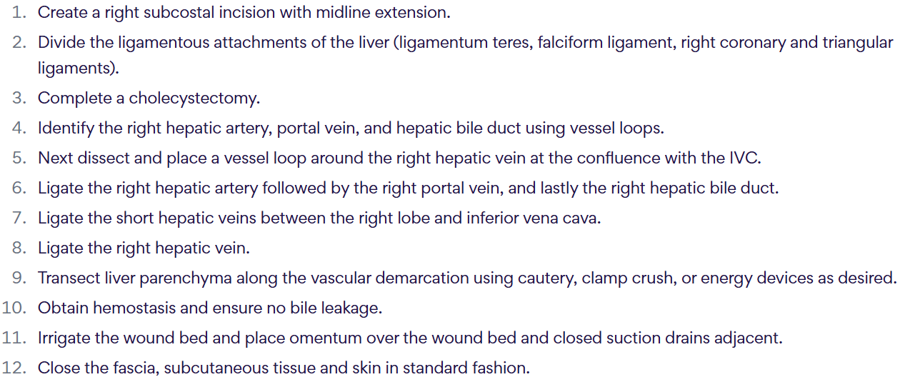
How do you complete an open right hepatectomy?
500
What are the steps of a transduodenal sphincteroplasty?
A transduodenal sphincteroplasty involves the following steps:
- Perform an extensive Kocher maneuver.
- Make a longitudinal duodenotomy on the lateral duodenal wall at the junction of the lower one-third and upper two-thirds of the duodenum.
- Identify the papilla.
- Cut the ampullary sphincter in the 11 o'clock position on the papilla using cautery or scissors.
- Extend this to include the entire common tract of the sphincter of Oddi.
- Extract all stones (if common bile duct stones are present).
- Suture the wall of the common bile duct to the duodenal mucosa using interrupted fine absorbable sutures.
- Close the lateral duodenotomy in a transverse fashion. Take care to protect the pancreatic sphincter to prevent postoperative complications.
As noted above, duodenotomy should be made longitudinally and closed transversely to avoid lumen narrowing. Division of the pancreatic sphincter would not improve the patient's problem; furthermore, the pancreatic sphincter is protected during a transduodenal sphincterotomy. The other answer options are not recognized treatments or components of the procedure.
500
A 16-year-old boy with hundreds of colorectal adenomas on colonoscopy is found to carry a germline pathogenic variant. Which gene and inheritance pattern are responsible, and what is the untreated lifetime colorectal cancer risk?
Classic FAP is caused by germline pathogenic variants in the APC tumor suppressor gene, is autosomal dominant, and carries a colorectal cancer risk approaching 100% if untreated
500
What is the treatment for each of the following choledochal cysts?
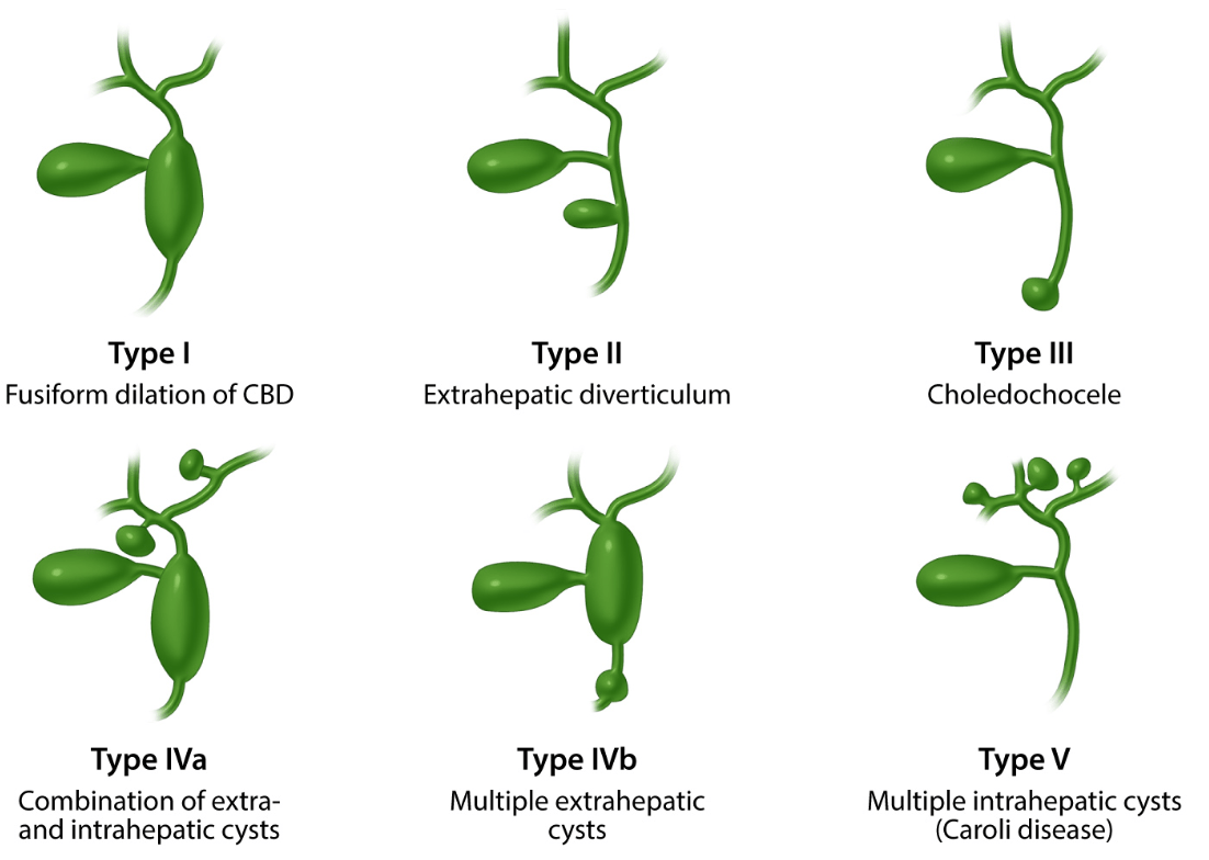
- Type I: cyst excision followed by Roux-en-Y hepaticojejunostomy or hepaticoduodenostomy
- Type II: resection of the diverticulum off the side of the common bile duct
- Type III: endoscopic sphincterotomy versus transduodenal excision and sphincteroplasty
- Type IV: cyst excision followed by Roux-en-Y hepaticojejunostomy or hepaticoduodenostomy. Hepatic resection should be considered if cysts are limited to one lobe.
- Type V: typically, liver transplant required to treat entire disease