Muscle Locations
Muscle Actions
Muscle Fibers
Muscle Contraction/Sliding filament theory
Muscle Contraction Types
Axial Skeleton
Appendicular Skeleton
Joints/Joint Movement
100
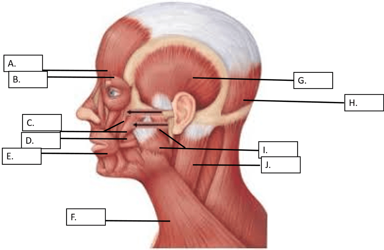
This muscle is indicated by the letter J in the diagram above
What is the Sternocleidomastoid
100
This muscle is responsible for raising the lateral corners of the mouth (smiling muscle)
What is the Zygomaticus
100
The tissue surrounding an individual muscle fiber
What is the endomysium
100
The region that is shown below: 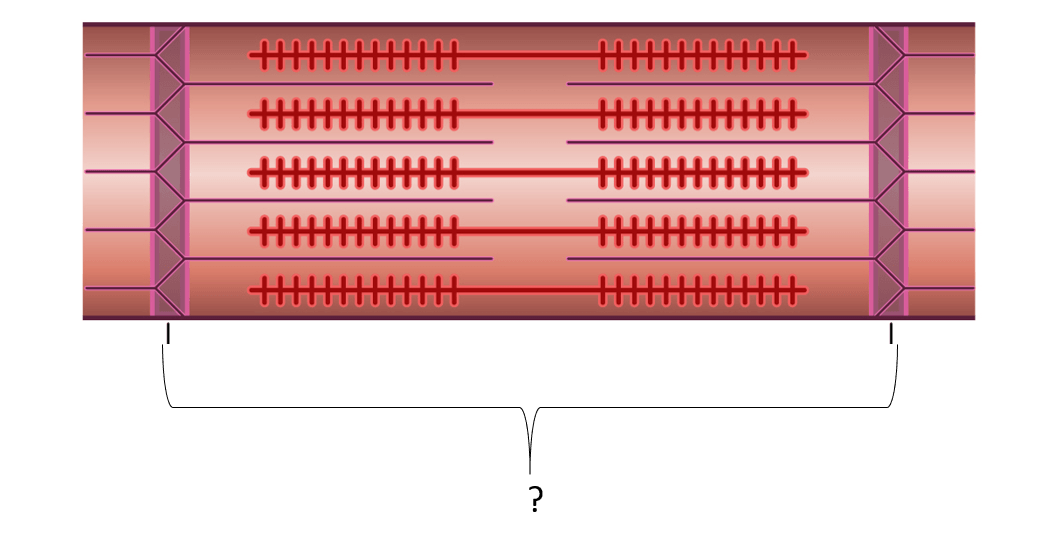
What is the sarcomere
100
An isotonic contraction where the muscle shortens.
What is a concentric contraction
100
The axial skeleton consists of the skull, hyoid bone ___________ ___________ and the thorax (ribs and sternum)
What is the Vertebral Column
100
These two bones make up the antecubital region of the arm
What are the radius and ulna
200
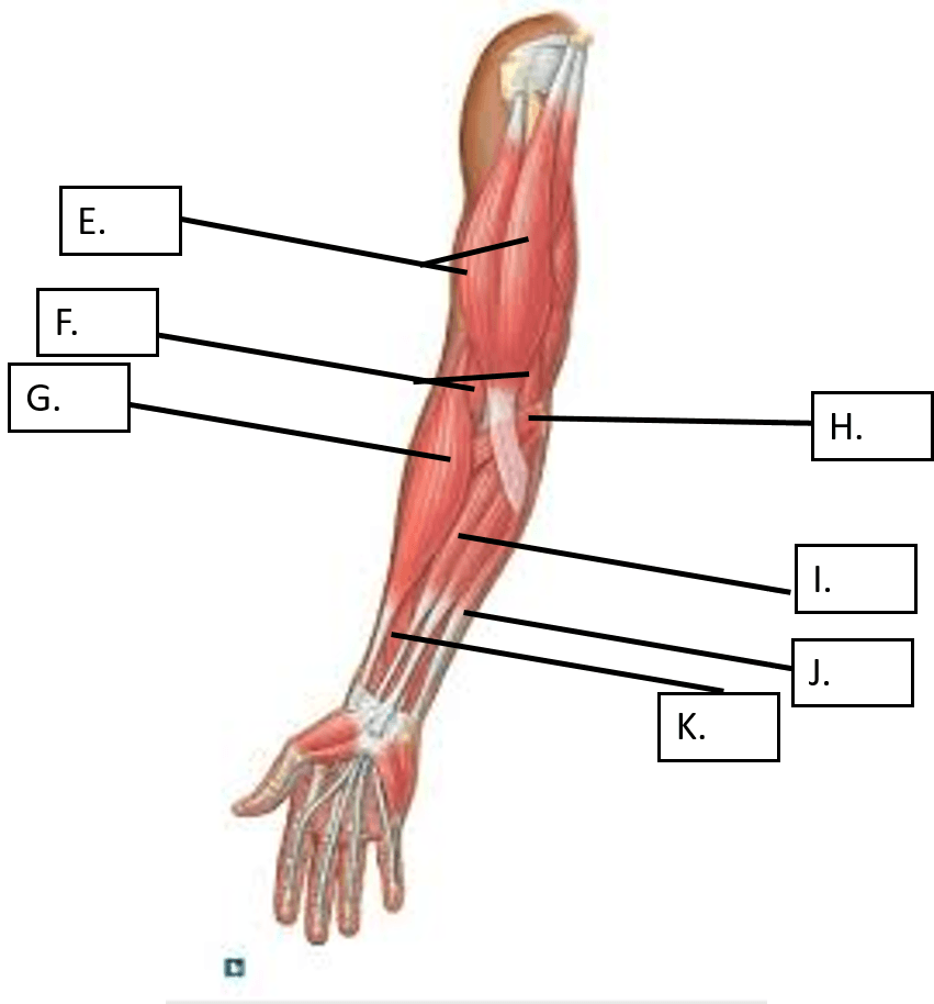
This is the muscle indicated by the letter G in the diagram above.
What is the Brachioradialis
200
This muscle's action is pronation of the forearm.
What is the Pronator Teres
200
The tissue surrounding a group of muscle fibers
What is the perimysium
200
These two myofilaments are the main protein filaments responsible for muscle contraction
What are myosin and actin
200
A neuron and all the muscle cells it stimulates make up this
What is a motor unit
200
The bone indicated by the I in the diagram.
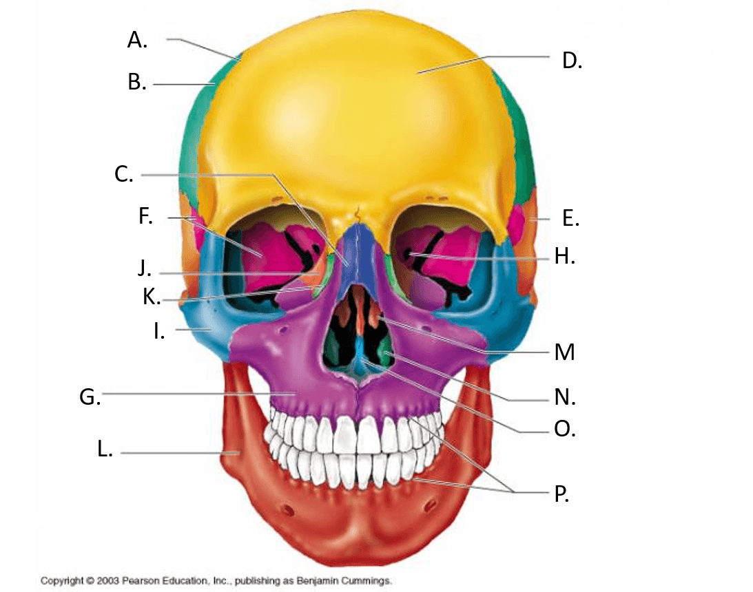
What is the zygomatic bone
200
This bone connects the appendicular skeleton to the axial skeleton in the diagram below. 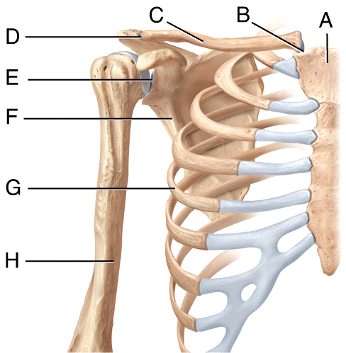
What is the clavicle
300
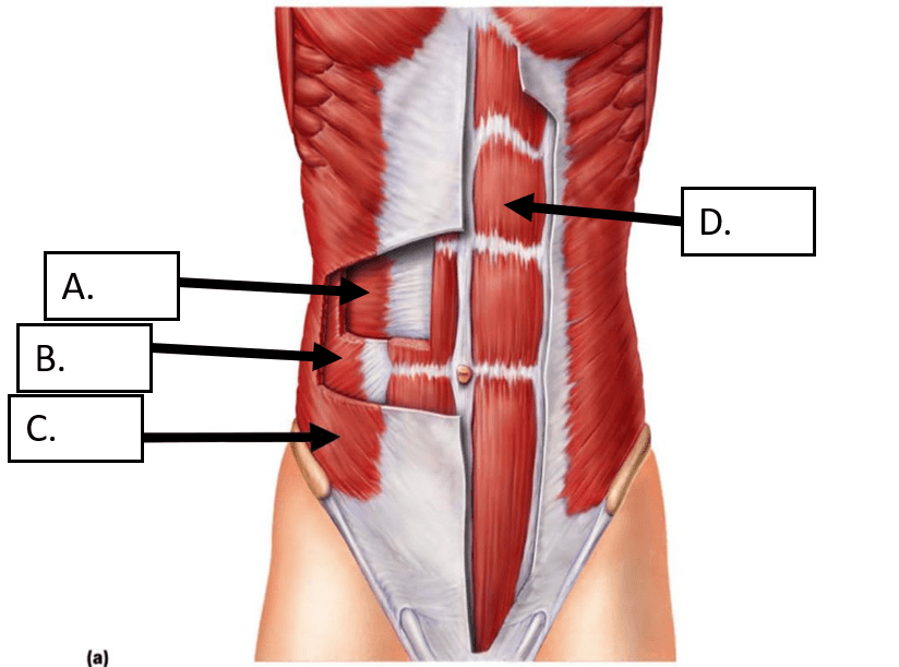
This is the muscle indicated by the letter D. in the image above
What is the rectus abdominis
300
This posterior muscle of the forearm extends and abducts the wrist
What is the Extensor Carpi Radialis Longus
300
The epimysium is a layer of connective tissue that surrounds the muscle and forms ___________ which connect the muscles to the bones.
What are tendons
300
This is the neurotransmitter that is released from the axon terminal into the synaptic cleft in a neuromuscular junction.
What is acetylcholine
300
single contractions of muscle fibers caused by single threshold stimulus; rare
What is twitch
300
The part of the sternum indicated by number 2 in the diagram. 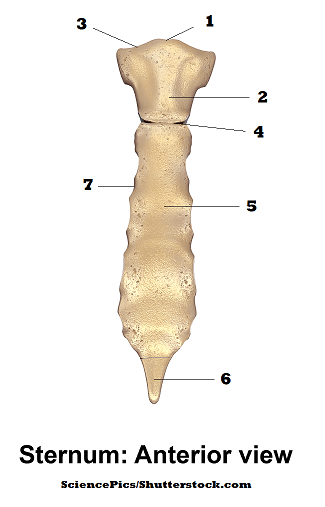
What is the manubrium
300
The names given to the three bones that make up the pelvic bone. 
What are the illium, ischium and the pubic bones
400
These THREE muscles are indicated by the letters B, C and D in the image above.
What are the Rectus Femoris, Vastus Lateralis and the Vastus Medialis
400
These two muscles work to elevate the mandible. (chewing)
What are the masseter and the temporalis muscles
400
Muscle fibers are made up of groups of these smaller pieces
What are myofibrils
400
In order for myosin to be able to reach out and attach to actin this molecule must be broken down into ADP + P. This energizes the myosin heads.
What is ATP
400
The minimal level of stimulation required to cause a muscle fiber to contract.
What is the threshold stimulus
400
Name all of the four sutures of the cranium
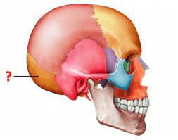
What are the lambdoidal suture, the saggital suture, coronal suture and the squamous suture
400
This joint is the area where the bones of the pelvis connect to the bones of the spine. 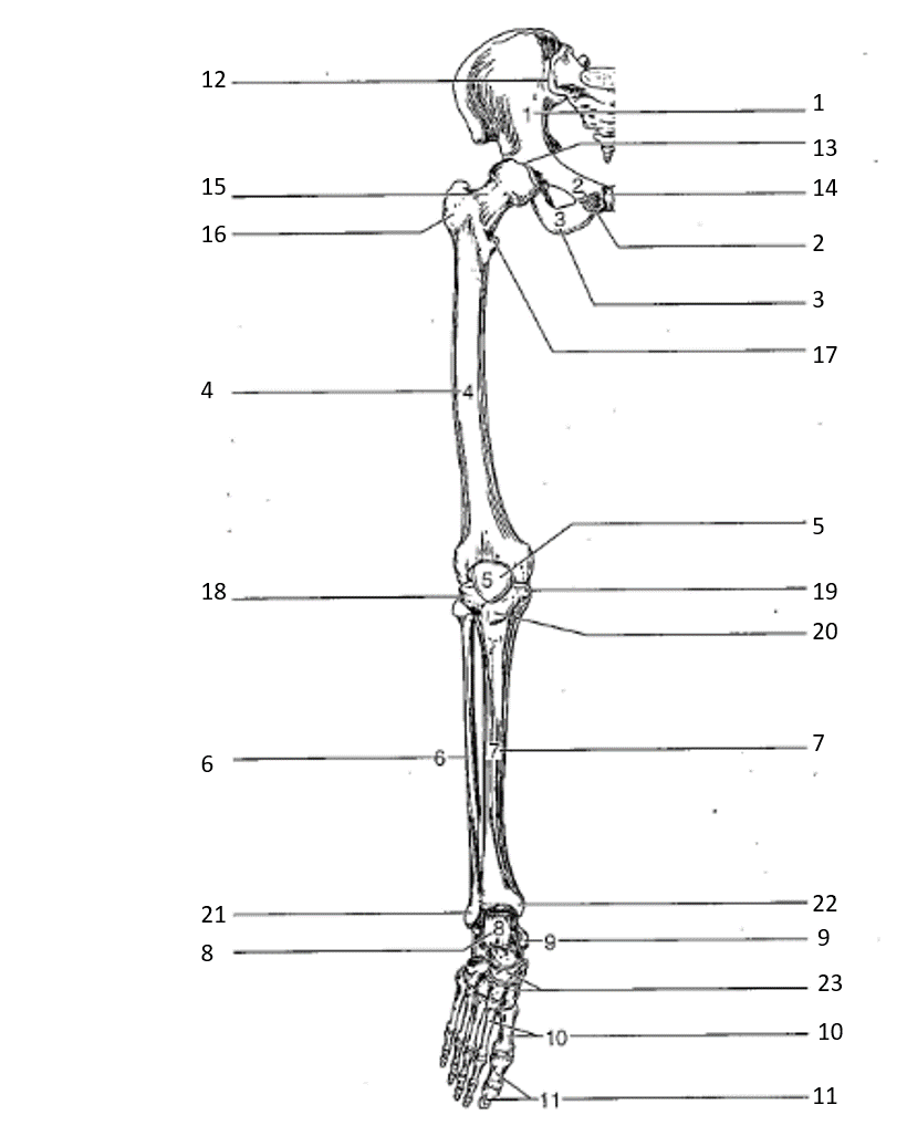
What is the sacroiliac joint
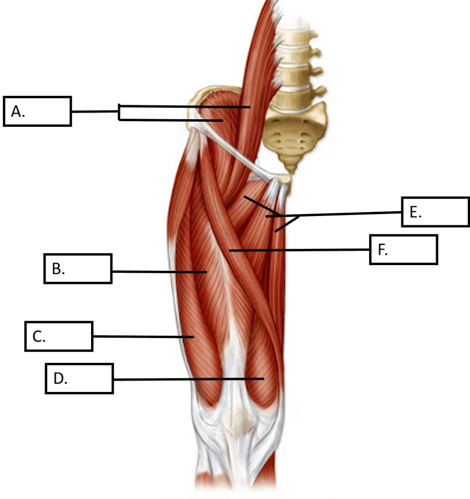
500
These two muscles are indicated by the letters F and G in the diagram.
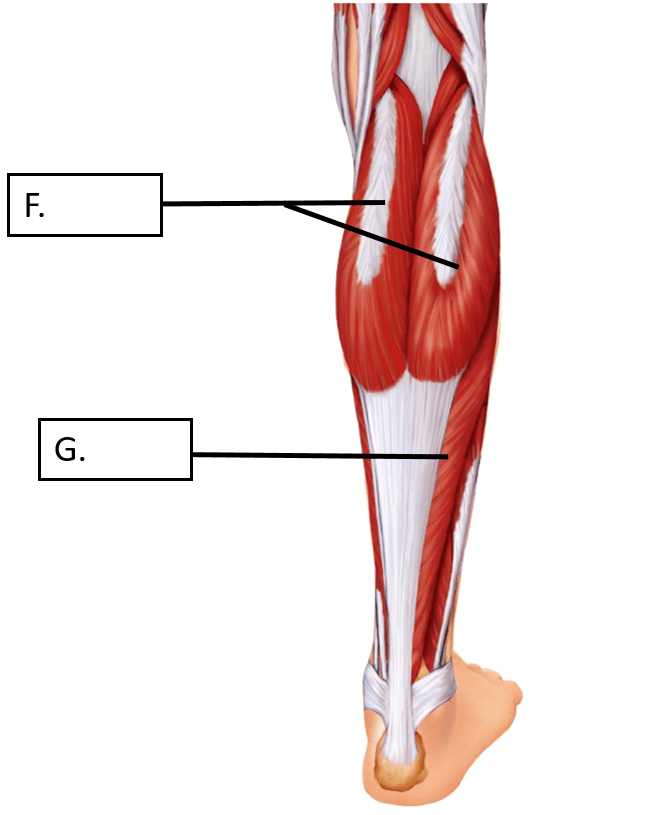
What are the gastrocnemius (F) and the soleus (G)
500
These THREE muscles of the thigh are responsible for adduction, medial rotation and flexion of the thigh.
What are the Adductor Longus, Magnus and Brevis
500
Groups of muscle fibers surrounded by the perimysium make up these
What is a fascicle
500
These ions are released from the sarcoplasmic reticulum and help to move tropomyosin and troponin off of the actin myofilament.
What is Calcium or Ca2+
500
sustained, steady muscular contractions caused by a series of stimuli bombarding a muscle in rapid succession (not necessarily a maximal contraction)
What is tetanus
500
These bones have foramen in their transverse processes.
What are cervical vertebrae
500
This is the structural name given to the joints of the spine
What is cartilaginous joints
600
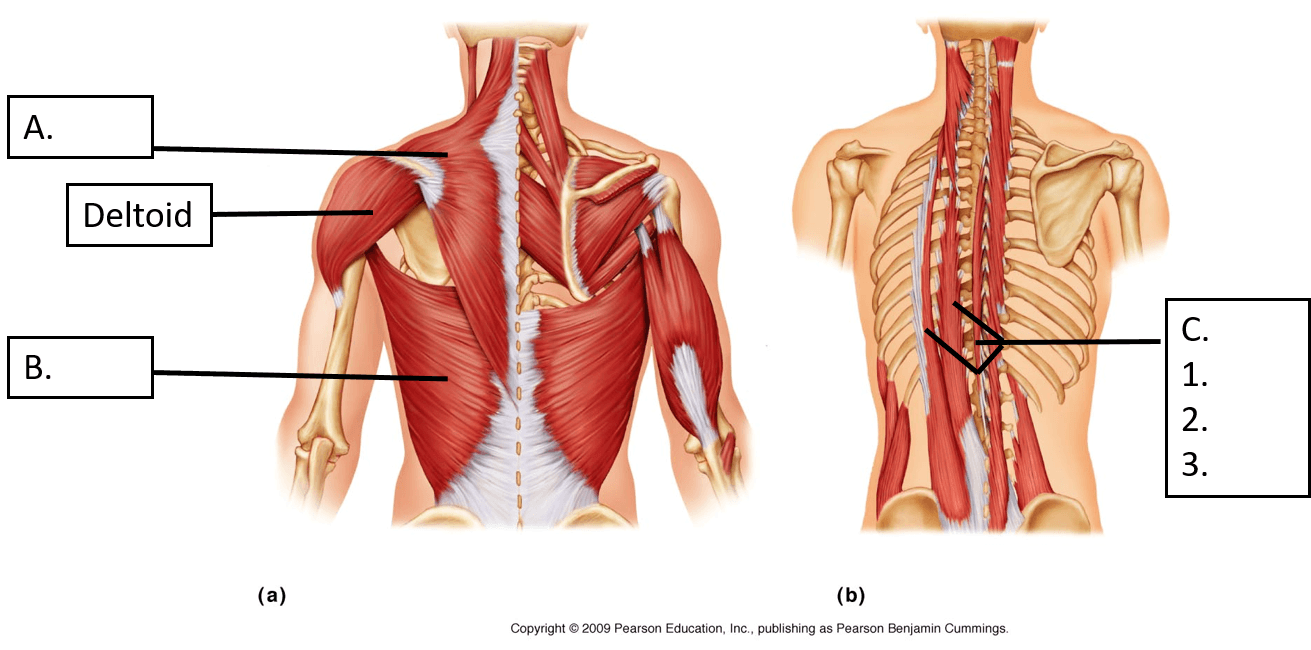
These three muscles make up the Erector spinae (1,2 and three in the image above)
What are the iliocostalis, longissimus, and the spinalis.
600
These three muscles work together to extend at the hip and flex at the knee.
What are the Biceps Femoris, Semitendinosus, and the Semimembranosus. (Hamstrings)
600
Muscle fibers have multiple ___________ and a plasma membrane called a _______________.
What are nuclei and sarcolemma.
600
Once the channel proteins on the sarcolemma are changed by the acetylcholine this allows for these two ions to rush in and out of the muscle cell causing depolarization of the motor end plate.
What are sodium (Na+) and potassium (K+)
600
Muscle contractions that do not produce movement.
What are isometric contractions
600
This bone articulates with ALL bones of the cranium
What is the sphenoid bone
600
Name the two processes and the fossa found on the scapula
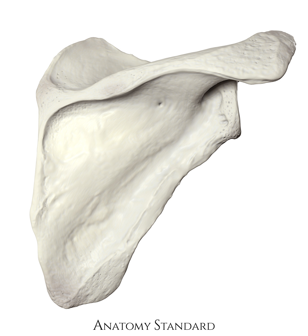
What are the corocoid process, acromion process and the glenoid fossa