Ventricles Structures
Ventricles Functions
Meninges and their Spaces
Dural Extensions
Structures in the Diencephalon
100
How many ventricles are in the brain?
4 ventricles
(Ch. 1&2 PowerPoint, slide 74)
100
True or False: Ventricles protect healthy cell function in both the brain and the spinal cord.
TRUE
(Ch. 1&2 PowerPoint, slide 75)
100
Which meninges layer is very thin and delicate?
Pia Mater
(Ch. 1&2 PowerPoint, slide 86)
100
What are three dural extensions?
The Falx Cerebri, Tentorium Cerebelli, and the Falx Cerebelli
(Bhatnagar page 67-68)
100
What are the two major structures in the diencephalon?
The Thalamus and the Hypothalamus
(Bhatnagar page 44)
200
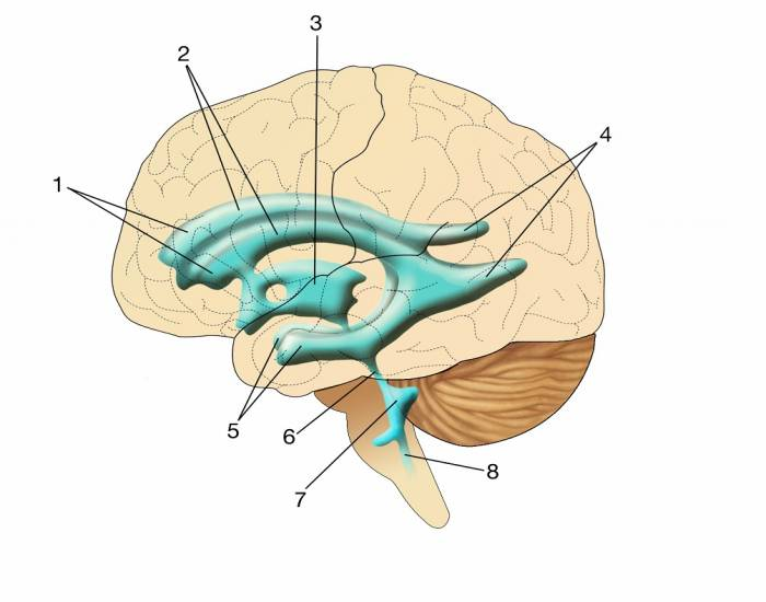 What is #7 pointing to?
The Fourth Ventricle
(Bhatnagar page 61)
200
What is the Primary Function of the Ventricles?
To store and circulate Cerebrospinal Fluid (CSF)
(Bhatnagar page 60)
200
What is the space called between the Dura Mater and the skull? (2 names)
Epidural Space (above) and Subdural Space (below)
(Ch. 1&2 PowerPoint, slide 84)
200
Which dural extension is the largest?
The Falx Cerebri
(Bhatnagar page 67)
200
True or false: A major function of the Hypothalamus is to relay sensorimotor information to the cortex.
FALSE (correct answer is the Thalamus)
(Bhatnagar page 45)
300
What are the names of Structures 1,2, and 4 and which ventricle are they connected with?
Lateral Ventricle
1= anterior horn
2= body of the lateral ventricle
4= posterior horn
(Bhatnagar page 61)
300
What does CSF stand for and what is its function?
CSF= Cerebrospinal Fluid
CSF is a clear fluid that forms a cushion in the subarachnoid space to protect the brain and CNS.
(Bhatnagar page 456)
300
What are the three layers of tissue in order of innermost to outermost?
Pia Mater, Arachnoid membrane, and Dura Mater
(Ch. 1&2 PowerPoint, slide 80)
300
 Which Dural Extension is letter D pointing to?
Falx Cerebelli
(Bhatnagar page 69)
300
The diencephalon in an inner part of the brain that lies between which two structures?
The cerebral hemispheres and the midbrain
(Bhatnagar page 459)
400
How are ventricles connected?
Ventricles are connected via Foramen/ aqueducts
(Ch. 1&2 PowerPoint, slide 74)
400
Where is CSF produced?
CSF is produced in the Choroid Plexus (which line the ventricles)
(Ch. 1&2 PowerPoint, slide 75)
400
What is the space called between the arachnoid and the pia mater?
The Subarachnoid space
(Bhatnagar page 69)
400
Which dural extension separates the cerebral hemispheres?
The Falx Cerebelli
(Bhatnagar page 68)
400
What connects the diencephalon to the spinal cord?
The Brainstem
(Bhatnagar page 46)
500
What is the roof of the Lateral Ventricles formed by?
The Lateral Ventricles are formed by the fibers of the corpus callosum.
(Bhatnagar page 61)
500
The walls of the ventricle cavities are lined with a layer of ependymal cells. What is the function of these cells?
The function of ependymal cells prevents diffusion of substances from the CSF into the brain.
(Bhatnagar page 61)
500
When the pia mater and the arachnoid membrane are together, they are called ____?
Leptomeninges
(Bhatnagar page 70)
500
What is the space called above the Tentorium Cerebelli and what does it house?
The area is called the Supratentorial Space and it houses the Cerebral Cortex
(Bhatnagar page 68)
500
What is the main function of the Hypothalamus?
The main function is to secrets hormones that regulates several things including feeding behaviors (water and food), reproduction, and regulating body temperature.
(Bhatnagar page 46)