Which neurotransmitter is decreased in early Parkinson's?
Dopamine
Sx's: shuffling gait, resting tremor, masked faces
Nexus Criteria for clinical clearance of c-spine injury include specific findings on history or exam to determine whether the c-spine requires imaging for clearance. Name these criteria.
Focal neurologic deficit present
Midline spinal tenderness present
Altered level of consciousness present
Intoxication present
Distracting injury present
A 45-year-old female presents to the emergency department with a 3-day history of severe headache, fever, and progressive swelling and redness around her right eye. She reports a history of a recent upper respiratory tract infection that was treated with antibiotics. On examination, she has proptosis, chemosis, and ophthalmoplegia of the right eye. Her right pupil is dilated and non-reactive to light. Fundoscopic examination reveals papilledema. Laboratory tests show elevated white blood cell count and inflammatory markers. What is the most likely diagnosis?
Cavernous sinus thrombosis
The patient's presentation is highly suggestive of sinus cavernous thrombosis (CST), a serious condition characterized by:
- Severe headache, fever, and periorbital symptoms: These are classic symptoms of CST.
- Progressive swelling and redness around the eye (periorbital edema): Indicates involvement of the cavernous sinus.
- Proptosis, chemosis, and ophthalmoplegia: These findings are due to cranial nerve involvement within the cavernous sinus (CN III, IV, V1, V2, and VI).
- Dilated, non-reactive pupil: Suggests involvement of cranial nerve III.
- Papilledema: Indicates increased intracranial pressure.
- Recent upper respiratory tract infection: Often a preceding factor, as infections can spread to the cavernous sinus.
Prior to immunization against H. influenzae type B, this was a more common cause of what neurological infectious disease?
Bacterial meningitis
A 7-year-old child presents with worsening headaches, morning vomiting, and ataxia. Magnetic resonance imaging (MRI) reveals a midline posterior fossa mass compressing the fourth ventricle. Histological examination of the tumor reveals small, round, blue cells with Homer-Wright rosettes. Which of the following is the most likely diagnosis?
A) Glioblastoma multiforme
B) Ependymoma
C) Medulloblastoma
D) Meningioma
E) Craniopharyngioma
Correct answer: C) Medulloblastoma
Explanation: Medulloblastoma is a malignant brain tumor commonly found in children, particularly in the posterior fossa. It typically presents with symptoms such as worsening headaches, morning vomiting (due to increased intracranial pressure), and ataxia. On MRI, a midline posterior fossa mass compressing the fourth ventricle is often observed. Histologically, medulloblastomas consist of small, round, blue cells with Homer-Wright rosettes.
Glioblastoma multiforme is a malignant glioma typically seen in adults and presents differently on histology.
Ependymomas are tumors arising from ependymal cells and typically occur in the ventricles or central canal of the spinal cord.
Meningiomas are usually benign tumors arising from the meninges.
Craniopharyngiomas arise from remnants of Rathke's pouch and often present with symptoms related to hypothalamic-pituitary dysfunction.
Cobalamin deficiency manifests as these symptoms
What is fatigue, pallor, glossitis, paresthesia, decreased vibratory sense
B12 deficiency
What is xanthochromia, and what condition is it associated with?
Yellow CSF discoloration of an LP associated with SAH.
A healthy 35-year-old presents to your clinic with gradually worsening severe headaches, decreased visual acuity, speaking difficulties, and severe imbalance which are now all very bothersome. A tangle of vascular channels is seen on head MRI, and enlarged draining veins are seen on non-contrast head CT. Pending further studies, your diagnosis is:
Name 3 different types of meningitis
Bacterial Meningitis: Caused by bacterial infections, with common pathogens including Streptococcus pneumoniae, Neisseria meningitidis, and Haemophilus influenzae.
Viral Meningitis: Often less severe and caused by viruses such as enteroviruses, herpes simplex virus, and West Nile virus.
Fungal Meningitis: Caused by fungal infections, commonly seen in immunocompromised individuals. Cryptococcus neoformans is a typical causative agent.
Tuberculous Meningitis: Caused by Mycobacterium tuberculosis. This form of meningitis is often chronic and can occur in individuals with or without active tuberculosis.
Parasitic Meningitis: Caused by parasites such as Naegleria fowleri (primary amoebic meningoencephalitis) or Angiostrongylus cantonensis (eosinophilic meningitis). This type is relatively rare compared to bacterial or viral meningitis.
Which disease process is associated with reduced dystrophin on muscle biopsy and presents as progressive muscle degeneration and weakness with difficulty standing, walking, getting up from sitting?
Duchenne Muscular Dystrophy
What are other common presentations/lab finding
- Calf hypertrophy (pseudohypertrophy due to fat and connective tissue replacing muscle)
- Elevated serum creatine kinase
This tremor does not appear at rest and often improves with etoh intake.
Essential tremor
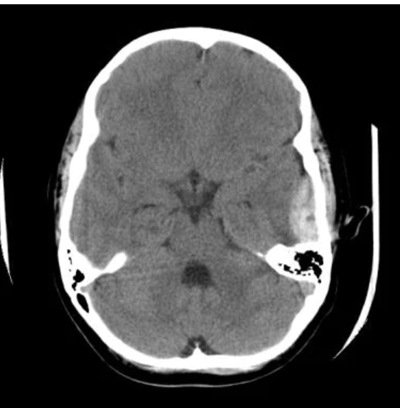
A 35-year-old male construction worker presents to the emergency department after a fall from a height at a construction site. He was wearing a hard hat but lost consciousness briefly after the fall and complained of severe headache on regaining consciousness. He denies any loss of memory regarding the event but reports feeling increasingly drowsy. On examination, his Glasgow Coma Scale (GCS) score is 13 (Eyes 3, Verbal 4, Motor 6). His pupils are equal and reactive to light. There are no signs of external trauma to the head apart from a scalp laceration over the right temporal area. 
Epidural Hematoma
A 7-year-old girl is brought to the pediatric clinic by her parents who are concerned about her frequent "daydreaming" episodes. The parents report that she often stares blankly for about 10 seconds and is unresponsive during these episodes, which occur multiple times a day. There is no associated postictal confusion or loss of muscle tone. Her teacher has also noticed these episodes in school, particularly during periods of inactivity. The girl has no history of head trauma, and her developmental milestones are normal. Which of the following is the most appropriate next step in management?
EEG
Bonus: What is the diagnosis
Absence seizure
A ventriculoperitoneal (VP) shunt is a surgical treatment used to relieve pressure on the brain caused by the accumulation of cerebrospinal fluid (CSF) in the ventricles, a condition known as this term. The shunt system diverts the excess CSF from the ventricles in the brain to the peritoneal cavity, where it can be absorbed.
What is hydrocephalus.
A 75 year old female is brought in by her daughter who has noticed she is walking as if her feet are glued to the floor, urinary incontinence, and confusion. Her urine dip is negative for blood, leuks, nitrites. This neurological disorder should be on your differential
What is NPH
Normal pressure hydrocephalus
A 42-year-old male is brought to the emergency department after a motor vehicle accident (MVA). He complains of weakness and loss of sensation below his chest. Examination reveals intact proprioception but loss of pain and temperature sensation bilaterally below the level of T6. Motor strength is preserved in the upper extremities but decreased in the lower extremities. Babinski reflex is present bilaterally.
Which of the following vascular structures is most likely affected in this patient?
Anterior artery
Dx: Anterior Cord Syndrome
Bonus: Describe a patient's symptoms with VBA dissection
What is one historical differentiates VBA from Cavernous Sinus Thrombosis?
Neck Pain: VBA dissection typically presents with acute or subacute neck pain, which is not a prominent feature in CST unless associated with underlying sinusitis or other infections.
Orbital and Ocular Symptoms: CST prominently features periorbital swelling, proptosis, and ophthalmoplegia, which are not typical findings in VBA dissection.
Systemic Signs: CST may present with fever and signs of infection, whereas VBA dissection does not typically present with systemic signs unless complicated by associated conditions like infection.
Neurological Symptoms: VBA dissection can lead to focal neurological deficits due to ischemia in the vertebrobasilar circulation, which may be absent or less pronounced in CST, where symptoms are primarily related to cranial nerve involvement.
A 35-year-old male presents to the neurology clinic with episodes of sudden, intense déjà vu and a rising epigastric sensation that lasts about 1-2 minutes. During these episodes, he remains fully conscious and aware of his surroundings, but cannot respond until the sensation passes. He reports that these episodes occur several times a month and started about six months ago. There is no history of head trauma or significant medical illness. His neurological examination is normal. What is the most appropriate diagnostic test to confirm the diagnosis?
A. Brain MRI
B. Electroencephalogram (EEG)
C. Serum glucose levels
D. Holter monitor
E. Lumbar puncture
Correct Answer: B. Electroencephalogram (EEG)
Explanation: The patient's description of sudden, intense déjà vu and a rising epigastric sensation with preserved consciousness is indicative of a focal aware seizure, likely originating from the temporal lobe. An EEG is the most appropriate diagnostic test to confirm the diagnosis as it can capture the characteristic electrical activity during a seizure.
Foil Explanations:
A. Brain MRI: While a brain MRI can help identify any structural abnormalities that might be causing the seizures, it does not confirm the diagnosis of a seizure disorder. It is often used in conjunction with an EEG.
C. Serum glucose levels: Checking glucose levels can help rule out hypoglycemia as a cause of altered mental status, but it is not specific for diagnosing seizures.
D. Holter monitor: A Holter monitor is used to record cardiac rhythms and would not be useful in diagnosing seizures.
E. Lumbar puncture: A lumbar puncture is used to diagnose infections or other conditions involving the central nervous system, but it is not specific for diagnosing seizures.
A 2-year-old child presents with developmental delay, spasticity, and poor coordination. The child's medical history is significant for a severe infection with a prolonged febrile illness and seizures at 8 months of age. Neuroimaging reveals periventricular leukomalacia. Based on this presentation, what is the most likely infectious agent that led to the new diagnosis of cerebral palsy?
A. Group B Streptococcus
B. Herpes Simplex Virus
C. Cytomegalovirus
D. Varicella-Zoster Virus
E. Neisseria meningitidis
Correct Answer: C. Cytomegalovirus
Explanation: Cerebral palsy (CP) can result from various prenatal, perinatal, and postnatal factors, including infections. Cytomegalovirus (CMV) is a common congenital infection associated with significant neurodevelopmental sequelae, including CP. CMV can cause periventricular leukomalacia, a type of white matter brain injury often associated with CP. The history of a severe infection with prolonged febrile illness and seizures aligns with the typical clinical course of a CMV infection leading to neurological damage.
TORCH Toxo Other Rubella CMV Herpes Simplex 1 & 2
These symptoms are likely describing a patient with this condition.
- Localized headache: Commonly on one side of the head, often near the temples.
- Scalp tenderness: Pain when touching the scalp.
- Jaw claudication: Pain in the jaw with chewing.
- Systemic symptoms: Such as fatigue and malaise.
- History of polymyalgia rheumatica: a condition characterized by muscle pain and stiffness.
What is Giant Cell Arteritis
A 45-year-old female presents with a three-month history of progressive weakness, particularly involving her ocular muscles. She reports experiencing double vision that worsens with prolonged use of her eyes and improves with rest. On examination, there is bilateral ptosis and ophthalmoplegia. The rest of the neurological examination is normal. Serologic testing reveals elevated levels of acetylcholine receptor antibodies confirming your suspected diagnosis.
What is myasthenia gravis
Serologic testing for acetylcholine receptor (AChR) antibodies: Elevated levels of AChR antibodies are found in the majority of patients with MG.
Name the Cranial Nerve associated with each of the following exam findings:
A. Mydriasis
B. Facial droop
C. Ipsilateral monocular vision loss
D. Loss of blink
E. Decrease in facial movement
A. Mydriasis: Cranial nerve III (the oculomotor nerve) is responsible for innervating the muscles that control most eye movements, the levator palpebrae superioris muscle (which raises the eyelid), and the sphincter pupillae muscle (which constricts the pupil). A dysfunction of cranial nerve III can result in mydriasis (dilated pupil) because the parasympathetic fibers that constrict the pupil are affected. This finding is particularly diagnostic as the other options are related to different cranial nerves:
B. Facial droop: This is typically associated with dysfunction of cranial nerve VII (the facial nerve).
C. Ipsilateral monocular vision loss: This finding is associated with lesions affecting the optic nerve (cranial nerve II) rather than the oculomotor nerve.
D. Loss of blink: The blink reflex involves both the trigeminal nerve (cranial nerve V) and the facial nerve (cranial nerve VII). The oculomotor nerve is not primarily responsible for the blink reflex.
E. Decrease in facial movement: This is associated with dysfunction of cranial nerve VII (the facial nerve), not cranial nerve III.
A 33 year old male presents with double vision. During extraocular movement testing, he is noted to have delayed adduction of the right eye. Visual acuity is 20/20 right, left, and bilateral. The rest of the examination is unremarkable for motor, sensory, reflex or gait deficits. He does not feel or appear ill. You order an LP and CSF analysis shows the presence of Oligoclonal Bands confirming your diagnosis of this disease.
What is MS?
Choose one of the following LP findings and name the most likely associated diagnosis.
A. Abnormally low CSF glucose level
B. Abnormally low CSF total protein level
C. Predominance of CSF mononuclear cells (lymphocytes)
D. Elevated CSF opening pressure
E. Presence of diplococci in pairs on gram stain
A. Abnormally low CSF glucose level: Bacterial Meningitis
- Low glucose levels in cerebrospinal fluid (CSF) are often indicative of bacterial meningitis because bacteria metabolize glucose.
B. Abnormally low CSF total protein level: Normal Pressure Hydrocephalus (NPH)
- Low CSF protein levels are less common, but in the context of NPH, the CSF protein level may be low due to increased CSF turnover.
C. Predominance of CSF mononuclear cells (lymphocytes): Viral Meningitis
- A predominance of lymphocytes in the CSF typically points to viral meningitis, also known as aseptic meningitis.
D. Elevated CSF opening pressure: Idiopathic Intracranial Hypertension (IIH)
- Elevated CSF opening pressure can be seen in idiopathic intracranial hypertension (IIH), also known as pseudotumor cerebri.
E. Presence of diplococci in pairs on gram stain: Pneumococcal Meningitis
- The presence of diplococci in pairs, particularly if they are gram-positive, is highly suggestive of infection with Streptococcus pneumoniae, leading to pneumococcal meningitis.
A 60-year-old male with a 15-year history of poorly controlled type 2 diabetes mellitus presents to the clinic with complaints of burning pain, tingling, and numbness in his feet, which has progressively worsened over the past year. He also reports a loss of sensation that has caused him to sustain several unnoticed injuries to his feet. On physical examination, he has diminished ankle reflexes and decreased sensation to light touch and vibration in a stocking-glove distribution. His recent HbA1c level was 9.2%. Which of the following is the most likely diagnosis?
A. Peripheral arterial disease B. Guillain-Barré syndrome C. Charcot-Marie-Tooth disease D. Diabetic neuropathy E. Spinal cord compression
Correct Answer: D. Diabetic neuropathy
Explanation: The patient's symptoms and history are characteristic of diabetic neuropathy, a common complication of long-standing and poorly controlled diabetes mellitus. Key features supporting this diagnosis include:
- Burning pain, tingling, and numbness: These are classic symptoms of neuropathy.
- Progressive worsening: Chronic condition, often worsening over time.
- Loss of sensation leading to unnoticed injuries: Indicative of sensory neuropathy.
- Diminished ankle reflexes and decreased sensation: Common findings in diabetic peripheral neuropathy.
- Stocking-glove distribution: Typical pattern of sensory loss in diabetic neuropathy.
- Poor glycemic control: As evidenced by a high HbA1c level, which is associated with an increased risk of neuropathy.
Foil Explanations:
A. Peripheral arterial disease: This condition can cause pain and numbness in the extremities, but it is typically associated with intermittent claudication, and physical examination would reveal diminished pulses rather than diminished reflexes and sensation.
B. Guillain-Barré syndrome: This acute condition presents with rapidly progressive weakness and areflexia, often starting in the legs and ascending, but it is not typically associated with a long history of diabetes or a stocking-glove distribution of sensory loss.
C. Charcot-Marie-Tooth disease: A hereditary neuropathy that presents with similar symptoms, but it usually starts in childhood or adolescence and has a family history. It is less likely given the patient's age and history of diabetes.
E. Spinal cord compression: This condition can cause sensory and motor deficits, but it would usually present with more localized symptoms depending on the level of compression, and it would not cause a stocking-glove distribution of sensory loss.
A 72-year-old male presents to the clinic with a history of progressive cognitive decline over the past year. His wife reports that he experiences vivid visual hallucinations, fluctuating levels of alertness and attention, and episodes of disorganized thinking. He also has developed parkinsonian motor symptoms such as bradykinesia, rigidity, and a shuffling gait. On examination, the patient is alert but has difficulty with visuospatial tasks and exhibits a resting tremor. What is the most appropriate diagnostic test to order?
Brain MRI
Explanation:
Brain MRI is the most appropriate diagnostic test to order for a patient with suspected Lewy body dementia (LBD) based on the clinical presentation. MRI can help to rule out other potential causes of dementia, such as vascular lesions, tumors, or normal pressure hydrocephalus, and can also provide supportive evidence for the diagnosis of LBD through imaging findings that are characteristic of the disease.
Lewy body dementia (LBD) is characterized by the following key features:
- Progressive cognitive decline: Similar to other dementias but with specific characteristics.
- Visual hallucinations: Vivid and detailed, occurring early in the disease.
- Fluctuating levels of alertness and attention: Periods of confusion that alternate with periods of clarity.
- Parkinsonian motor symptoms: Bradykinesia, rigidity, shuffling gait, and resting tremor, which are common in LBD.
- Difficulty with visuospatial tasks: Common cognitive symptom in LBD.