Fluids/Compartments
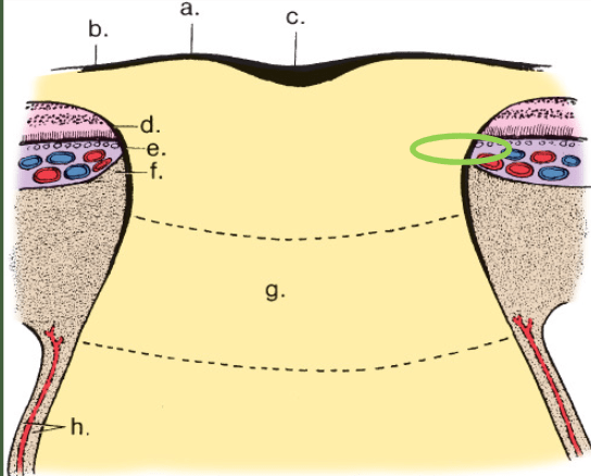
Conjunctiva
Epithelium and Bowman's Layer
Stroma, Descemet's Membrane, and Endothelium
Inflow & Outflow
Lens
Vitreous
ONH
Vascular Circulation
Blood & Ocular Barriers
100
Passive movement (non-energy requiring) involving movement of small molecules from an area of high concentration to an area of low concentration
- Spontaneous
-Automatic
Diffusion
100
Inflammation at level of superficial episcleral vessels
Episcleritis
100
These cells are only found in the corneal periphery and are responsible for antigen recognition and processing
Langerhan's cells
100
Hydrophilic components in the corneal stroma that create a strong osmotic draw into it
Dictates structural matrix of lamellar spacing
GAGs
100
For IOP to remain stable, aqueous inflow should equal
Aqueous outflow
100
Proteins that make up the zonules
Fibrilins
100
The major structural protein of the vitreous
Collagen
100
These cells make myelin in the CNS
Oligodendroglia
100
White infarcts are from this
Arterial occlusion
100
It is crucial to keep plasma protein levels low in the aqueous humor. Low protein content minimizes this issue
Light scattering
200
An integral membrane protein that functions in transport
- 3 Na+ out of the cell
- 2 K+ into the cell
Higher concentration of extracelluar Na+ provides electrochemical energy for many other nearby cellular transports to occur
Essential for aqueous production and maintaining corneal transparency
Na+/K+ ATPase Pump
200
Posterior conjunctival arteries
Anterior ciliary arteries
Vascular supply of the conjunctiva
200
The corneal epithelial pump is under this type of control
Sympathetic
200
What counteracts the osmotic draw created by the GAGS in the corneal stroma, preventing it from excessive swelling and loss of transparency
Deturgescence
200
Average episcleral venous pressure
~9 mmHg
200
Complete dislocation of the lens
Luxation
200
The major GAG in the vitreous
Hyaluronic Acid (HA)
200
This forms the internal limiting membrane of the optic nerve head
Astrocytes
200
Large vessels can be found within this layer of the retina
Nerve Fiber Layer (NFL)
200
These vessels are non-fenestrated and have tight junctions between interendothelial spaces in order to prevent leakage of plasma proteins into the iris stroma, which is filled with aqueous humor
Iris capillaries
300
1 of the 2 extracellular compartments of water (~6 L total volume)
Includes:
- Blood
- Lymphatic fluid volumes
-Aqueous humor
-Vitreous humor
Intravascular component
300
Produces the bulk of the mucus layer of the tear film
Goblet cells
300
The remarkable resilience of this corneal layer is why/how patients can tolerate the use of a fast-spinning Alger brush/corneal burr
Bowman's Layer
300
MMPs and TIMPs are produced by?
Keratocytes
300
The part of the outflow network that sees the most flow
Posterior meshwork
300
This feature in the lens epithelium make it possible for innermost layers of the lens to receive nutrients, materials, and to remove waste
Gap junctions
300
A centrally located "collagen free" region that contributes significantly to maintaining high transparency
Cloquet's canal
300
Here, there is no central supply from the central retinal artery so astrocytes reach out to peripheral blood vessels to bring nutrients
Laminar region
300
This area of the retina is at greater risk for a vascular occlusion because it relies on a vascular supply with less capacity for collateral flow
Peripapillary retina
300
Because the choroidal vessels are fenestrated, it is this which acts as the blood-retinal barrier.
Prevents non-specific diffusion of plasma constituents to reach sensory retina
RPE
400
Serves as the anchor negative ion inside the cell around which smaller ions must adjust
(Na+ does the same function for extracellular space)
Proteins
400
The most mitotically active location on the ocular surface
Limbus
400
Corneal layers with no innervation
Descemet's membrane
Endothelium
400
What is the only glucose transporter observed within corneal cells?
GLUT1
400
Goldmann Equation
F = Ctm(Pi - Pe)
F = Aqueous outflow (uL/min)
Ctm = Facility of trabecular outflow (uL/min/mmHg)
Pi = Intraocular pressure (mmHg)
Pe = Episcleral pressure (mmHg)
***Flow also equals change in pressure over resistance***
400
Aggregations of this within the crystalline lens fibers will result in high molecular weight aggregation and, eventually, cataracts
Disulfide linkages between neighboring crystalline protein thiol groups
400
The firmest attachment of the posterior vitreous face is here
Adjacent to optic nerve
400
Much research indicates that this feature is compromised in glaucoma patients. As IOP rises and falls throughout the day, the vessels are unable to do this to ensure continued and constant flow
Autoregulation
400
The capillary beds of the retinal microvasculature span from the ganglion cell later to the junction between these 2 layers
Inner Nuclear Layer
Outer Plexiform Layer
400
This region of the eye will always have plasma protein free aqueous humor
Posterior chamber
500
SGLT-1 in the intestinal epithelium is an example of this type of transport protein
Symporter
500
Enables surface tolerance, memory, and communication throughout the entire conjunctiva
- Exposure of 1 mucosa will initiate memory of that event to provide acquired immunity response at next encounter or reinforce tolerance at next counter (at all mucosal surfaces simultaneously - not just the surface that contacted the pathogen)
MALT (CALT) System
500
Controls stromal hydration under normal conditions
Corneal endothelium
500
O2 supply of both distal portion of stroma and epithelium
Tears
500
This class of pharmacological drug controls IOP by increasing uveoscleral outflow
Does so via the dissolution of connective tissue matrix in ciliary muscle, making it more permeable
Prostaglandin Analogues (e.g., Xalatan)
500
Essential antioxidant that limits H2O2 damage (resulting from ascorbate oxidation) in the lens
Reduced Glutathione (GSH)
500
Locations (2) of highest collagen content - where the vitreous remains a gel
Vitreous cortex
Vitreous base
500
What 2 proteins are uniquely found at the lamina cribrosa (and not the sclera)?
Laminin
Type IV collagen
500
The OPL in the FAZ is known as this
Nerve Fiber Layer of Henle
500
Where in the eye is the concentration of plasma protein 75% of that found in the plasma itself?
Ciliary body stroma
600
Formed by sphingolipids (membrane lipids with larger hydrophilic moieties)
- Involved in cell signal transduction
- Cell-cell communication
- Endocytosis and uptake of bacteriaLipid rafts
600
Provides antimicrobial efficacy by binding to free iron (reducing availability of iron, which is necessary for microbial growth)
- Also inhibits biofilm formation
- Plays role in protecting contact lens surfaces
Lactoferrin
600
Sympathetic pathway control of Cl- transport suggests that the corneal epithelial pump is particularly important in what times?
Times of corneal stress (e.g., erosion)
600
Corneal stroma water content percentage that results in optimal optical clarity
75-80%
600
Ouabain reduces aqueous secretion specifically by inhibiting this pump in the non-pigmented ciliary epithelium
Na+/K+ ATPase pump
600
Present in the aqueous humor, this component sacrifices itself to reduce UV damage in the eye, creating H2O2 in the process
Ascorbate
600
The vitreous can act as this, which slows the bulk movements of substances between the anterior and posterior segment of the eye.
**This is why the vitreous acts as a slow-release depot for drug delivery and this is also why topically administered eye drops generally have no efficacy on the retina and optic nerve**
Diffusion barrier
600
There is increased interest in changes that occur in glaucoma at this specific location of the optic nerve head
Border tissue of Jacoby
600
Result from microvascular occlusion and ischemia in the nerve fiber layer
Found predominately in peripapillary retina
Stains cell-like but does not stain blue like actual nuclei
Cotton Wool Spots
600
Tight junctions within this region of the eye prevent plasma proteins from entering the posterior chamber
Non-pigmented ciliary epithelium
700
Process that allows circulation of aqueous humor and drainage of metabolic waste within the anterior chamber
Faster circulation than simple diffusion
- Circulation of aqueous humor driven by temperature differences between the warmer iris and cooler cornea
Convective Flow
700
Secretory component that stabilizes IgA and masks proteolytic sites, making it less vulnerable to host and pathogen proteases
Neutralizes pathogens by preventing their attachment to host cells
Can also bind to adhesin molecules on pathogens, causing their aggregation and entrapment
Secretory IgA (sIgA)
700
Chronic RGP lenses wearers may stop producing these (on the corneal epithelial surface) as a response to constant injury
Mircoplicae
700
This ion (moving against its concentration gradient) is symported with Na+ (moving along its concentration gradient) into the aqueous humor space via a corneal endothelial cell membrane transporter
Bicarbonate (HCO3-)
700
This class of pharmaceutical drug reduces IOP by reducing aqueous humor production
Achieves this by inhibiting monoamine transporter (responsible for norepinephrine uptake)
reduced NE reuptake --> vasoconstriction --> decreased fluid available to produce AH
Alpha-Agonists
700
This type of cataract will cause a hyperopic shift in refractive error
Cortical cataract
700
Separation of the posterior vitreous face from the internal limiting membrane of the retina due to shrinking vitreous
Liquid vitreous partitions from gel
Posterior vitreous detachment (PVD)
700
The dominant astrocyte in the optic nerve head
Type 1B astrocytes
700
Region of the choroid where the medial and lateral posterior ciliary arteries meet
- Diminished perfusion in this area - area that will suffer the most if either the medial or lateral PCA was compromised
Watershed zones
700
Restricting entry of this material into the aqueous humor may also potentially prevent entry of antigenic agents that typically travel along with it
Albumin
800
Used to calculate equilibrium potential for a single ion
Allows us to compare the strength of an electrical concentration vs the force of its concentration gradient for an individual ion
Nernst Equation
E = -2.3 RT/zFlog10(Ci/Ce)
E = Equilibrium potential
2.3 RT/F = Constant (60 mV at 37 deg C)
z = Charge of ion
Ci = Intracellular concentration of the ion (mmol/L)
Ce = Extracellular concentration of the ion (mmol/L)
800
Condition that is a result of drugs with cationic, amphiphilic properties penetrating lysosomes and binding with cellular lipids
Vorticeal Keratopathy
800
Aging, extreme cold, UV light exposure, oxygen reduction due to contact lens wear, and/or neurotropic viral epithelial disease are all factors contributing to this
Decreasing corneal sensitivity
800
Frequent anaerobic respiration of glycogen in the corneal endothelium will create hypoxic conditions resulting in corneal edema due to buildup of this molecule
Lactate (lactic acid)
800
Clinical flare can result from drugs that suppress production of aqueous humor because of the disrupted balance of concentrations between aqueous humor and ________
Plasma protein (created separately from AH - continues to rise while AH decreases)
800
This important product of the Pentose Phosphate Shunt that is necessary to keep glutathione in a reduced state
NADPH
800
Gradual shift from gel vitreous to liquid vitreous
Vitreous syneresis
800
Patients with progressive glaucoma were found to have this symptom which is not observed in patients with stable glaucoma
Lower systemic blood pressure (especially at night)
800
Trypsin digests show loss of this type of cell from the walls of the microvasculature in the case of diabetic microaneurysms.
Pericytes
800
Clinical test that assesses integrity of blood-retinal barrier
Fluorescein Angiogram
900
Equilibrium of electrical gradient and concentration gradient driving individual ions that can move
- At equilibrium, the concentration and electrical gradients are equal and opposite
Equilbrium Potential = Charge difference membrane at which this ^ occurs
Gibbs-Donnan Equilibrium
900
Parasympathetic neurotransmitters that stimulate mucin secretion (Goblet cells; conjunctival epithelium)
Acetylcholine
Vasoactive intestinal peptide (VIP)
900
The osmotic difference created by high concentration of this ion on the tear side of the corneal epithelium draws water out of the cornea
Cl-
900
Critical Oxygen Tension (COT) (AKA, the tension below which corneal dysfunction occurs) in air?
10% O2
900
Carbonic anhydrase inhibitors used in the treatment of glaucoma inhibit these components of the posterior ciliary epithelium
NHE-1 and AE2 antiports
900
Type of cataract due to abnormal migration of lens epithelial cells to posterior pole when they should have converted into lens fibers at the lens equator
Causes disproportionately more impaired vision at near compared to distance
Posterior subcapsular cataract
900
The ratio of gel to liquid in the vitreous will be, on average, 50/50 at approximately this age
80
900
Research indicates focal ischemia of the optic nerve head may be a causative factor for glaucoma in some cases. What class of drug will counteract these effects by generating vasodilators within the ONH?
Nitric oxide donors (NOD drugs)
Example = Vyzulta
900
What part of the eye is particularly vulnerable to temporal arteritis (AKA, Giant Cell Arteritis)?
Muscular arteries
900
Plasma derived proteins are only added to aqueous by diffusion from this region of the eye
The ciliary body stroma
1000
How would you treat a patient with an IOP > 40 mmHg?
- Both standard way and way for diabetic patients
Standard way - Oral glycerin. Draws in water - dramatically increases osmotic pressure in bloodstream (drawing water out of vitreous - decreasing IOP)
For diabetic patients - Administer IV mannitol. Similar function/process to oral glycerin
1000
Despite having ACE2 receptors in the mucosal lipid raft of the conjunctiva, the COVID-19 virus cannot enter through this layer. This is because this component (which is responsible for priming the spike protein in order for the viral membrane to be able to attach to the host cell membrane) is not present in the conjunctival mucosa
TMPRSS2
1000
Autoimmune response to components of hemidesmosomes of corneal and conjunctival epithelium
Ocular cicatricial pemphigoid
1000
Average adult corneal endothelial density
2500 cells/mm2
1000
Glaucoma patients have a higher than normal concentration of this in their aqueous humor
TGF-b
1000
In diabetic patients, there is an excess amount of glucose in the blood. Excess glucose goes through alternative pathways (known as the Polyol pathway) when there is too much for only hexokinase to convert it into G6P.
Accumulation of this product in the lens (from the Polyol pathway) also results in water being drawn into the lens (osmostic force is created since it is too big to pass through the lens membrane). This results in swelling and formation of a cataract
^What is the major product in the Polyol pathway that causes this to happen?
Sorbitol
1000
A vitrectomy increases the risk of this
Iris neovascularization
1000
Formula for Mean Ocular Perfusion Pressure (MOPP)
MOPP = (2/3)[DBP + (1/3)(SBP - DBP)] - IOP
MOPP = Mean ocular perfusion pressure
DBP = Diastolic blood pressure
SBP = Systolic blood pressure
IOP = Intraocular pressure1000
Clinical consequence of lack of autoregulation in the choroid
Choroidal effusion/expulsive choroidal hemorrhage
1000
The blood aqueous barrier is complete from the back of the eye all the way up to this landmark
(Beyond this landmark environment is more permissive)
Pupil margin