Views From the 6
(not so) Happily Ever After
Approach With Caution
Lemme see that pelvis
I'm In Danger
100
The posterior column can be best visualized by this radiographic view
Obturator oblique
100
In addition to age >40 and associated fracture patterns, this is a risk factor for DJD
Concomitant femoral head injury
100
The Kocher-Langenbeck approach allows you to access these parts of the pelvis/acetabulum (2)
Posterior column, posterior wall
100
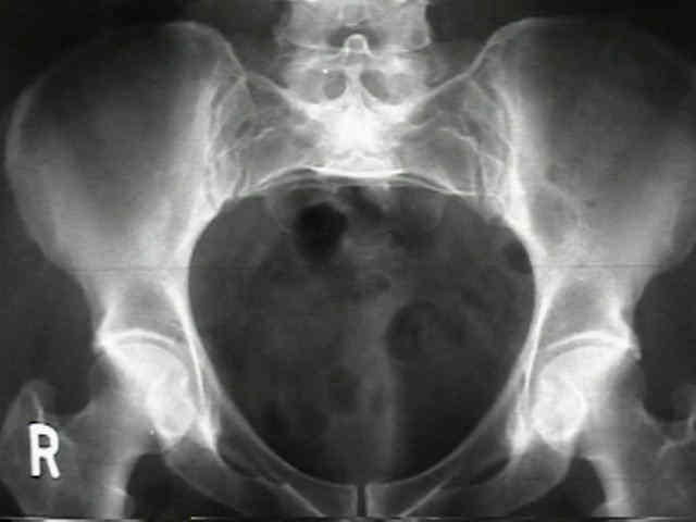
These images are a representation of this pelvic ring fracture pattern
LC 2 (rami fx, IL posterior ilium Fx-Dlx/crescent)
100
The corona mortise is the anastomosis of these two arteries
Internal (obturator) and external iliac
200
This radiographic view is used to assess anterior posterior location when placing an SI screw
Inlet
200
This pelvic ring fracture pattern has the highest risk for sexual dysfunction
APC/saddle horn injuries
200
The only single incision approach that allows for direct visualization of both columns
Extended iliofemoral
200
This radiographic findings has a name and is indicative of what?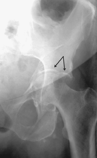
Gull sign - posterior wall fracture
200
This acetabular fracture pattern has the highest association with neurovascular injury
Transverse + posterior wall
300
The obturator outlet view, commonly used for supra-acetabular ExFix placement is obtained using this these angles
~45deg roll over towards injured side plus ~30deg of caudual tilt
300
The weight bearing recommendation after ORIF of an acetabular fracture
TTWB/FFWB (joint reactive forces)
300
This approach is associated with the lowest rate of HO
Ilioinguinal
300
The image demonstrates this type of acetabular fracture![]()
Transverse
300
The LFCN is at greatest which with this surgical approach
Ilioinguinal
400
This feature suggests a quality inlet radiograph
The S1 body overlies the S2 body to make a crisp line
400
The #1 cause of mortality in these injuries is hemorrhage and closed head injuries, respectively
APC (hemorrhage), LC (closed head injury)
400
The two prophylactic treatments for HO
Indomethacin, single dose of radition
400
This acetabular fracture pattern is associated with disruption of both the iliopectineal and ilioischial lines, a fractured obturator ring, and an intact iliac wing

T-type
400
This nerve structure is at risk with abberantly placed SI screws
L5 nerve root
500
This radiographic view is most helpful for assessing supra-acetabular pin placement in the cranial caudal plane
Hint: it helps you verify you are not in the sciatic notch
Iliac oblique
500
After a pelvic ring injury, DVT rates are as high as this percentage
60%
If VTE PPX COI due to head injury, get them a vena cava filter
500
This approach is ideal for an associated acetabular fracture pattern >3 weeks old
Extended iliofemoral
500
This image demonstrates this particular acetabular fracture![]()
Anterior column + Posterior hemi-transverse
Lines disrupted?: 2
Obturator ring?: fractured
Iliac wing?: fracture
Must be ACPHT or ABC
500
With this approach, the corona mortis must be exposed and ligated
Modified Stoppa