Tracts of my tears
Rad
You CN do this
Stroke of Misfortune
Neck deep in anatomy
100
This ascending spinal cord pathway carries vibration, fine touch, and proprioception information to the brain.
What is the dorsal column-medial lemniscus pathway?
100
On a brain MRI, these paired cerebrospinal fluid-filled structures are enlarged in a patient with hydrocephalus and are located deep within the cerebral hemispheres.
What are the lateral ventricles?
100
This cranial nerve is responsible for motor innervation of the tongue.
What is the hypoglossal nerve (CN XII)?
100
This artery is most commonly involved in strokes producing aphasia in the dominant hemisphere.
What is the middle cerebral artery?
100
This large superficial neck muscle divides the anterior and posterior cervical triangles.
What is the sternocleidomastoid muscle?
200
Damage to this visual pathway structure causes loss of the left visual field in both eyes.
What is the right optic tract?
200
These endothelial-lined venous channels drain blood and cerebrospinal fluid from the brain into the internal jugular veins.
What are the dural venous sinuses?
200
Damage to this cranial nerve causes inability to abduct the eye laterally.
What is the abducens nerve (CN VI)?
200
This type of aphasia is characterized by fluent but nonsensical speech with impaired comprehension.
What is Wernicke aphasia?
200
This cranial nerve innervates the trapezius muscle and may be injured during surgery in the posterior triangle of the neck.
What is the accessory nerve (CN XI)?
300
This descending pathway controls voluntary skeletal muscle movement and decussates in the medulla.
What is the corticospinal tract?
300
This posterior brain structure, shown degenerating in the image below, coordinates balance, fine motor movement, and motor learning and is visible on this MRI.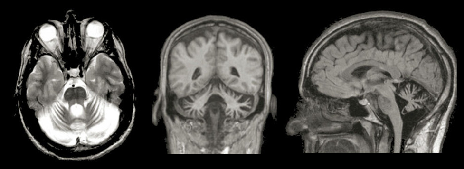
What is the cerebellum?
300
This cranial nerve is commonly affected in Ramsay Hunt syndrome and causes ipsilateral facial paralysis.
What is the facial nerve (CN VII)?
300
This hemorrhage classically appears lens-shaped on CT imaging.
What is an epidural hematoma?
300
This major artery ascends through the neck and bifurcates into internal and external branches near the level of C4.
What is the common carotid artery?
400
This ascending pathway carries pain and temperature sensation and typically crosses within 1–2 spinal cord levels after entering the cord.
What is the spinothalamic tract?
400
This paranasal sinus appears opacified on the right side of the MRI in a patient with facial pain, nasal congestion, and tenderness over the cheek.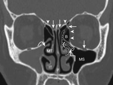
What is the right maxillary sinus?
400
A patient with anosmia (no sense of smell) after facial trauma most likely damaged this bony structure.
What is the cribriform plate?
400
This artery is most commonly torn in an epidural hematoma following temporal bone trauma.
What is the middle meningeal artery?
400
This autoimmune disease causes hypothyroidism and is associated with a firm, diffusely enlarged thyroid gland.
What is Hashimoto thyroiditis?
500
A patient with Brown-Séquard syndrome from a left spinal cord hemisection at T10 would develop ipsilateral loss of vibration and motor function with contralateral loss of this sensory modality below the lesion.
What is pain and temperature sensation?
500
This condition, caused by a fluid-filled cavity within the spinal cord, classically produces bilateral loss of pain and temperature sensation over the shoulders and upper limbs.
What is syringomyelia?
500
A lesion of the right hypoglossal nerve would cause the tongue to deviate in this direction when protruded.
What is toward the right side?
500
A stroke affecting this artery can produce prosopagnosia, making patients unable to recognize familiar faces.
What is the posterior cerebral artery?
500
Injury to this branch of the vagus nerve during thyroid surgery may cause hoarseness due to paralysis of most intrinsic laryngeal muscles
What is the recurrent laryngeal nerve?