Radiographic Basics
Obtaining and Interpreting Radiographs
Technique
Recognition of Normalities
and Abnormalities
and Abnormalities
Principles of Radiophysics and Radiobiology
100
Three purposes of dental radiographs in dental hygiene
What is to detect dental caries, periodontal disease, and other oral conditions?
100
These are two common techniques used for obtaining bitewing radiographs.
What are parallelling and bisecting angle techniques?
100
This technique error will result with overlapped contacts which makes the radiographic image nondiagnostic.
What is improper horizontal angulation?
100
This radiopaque normal landmark is often seen in the maxillary central incisor image.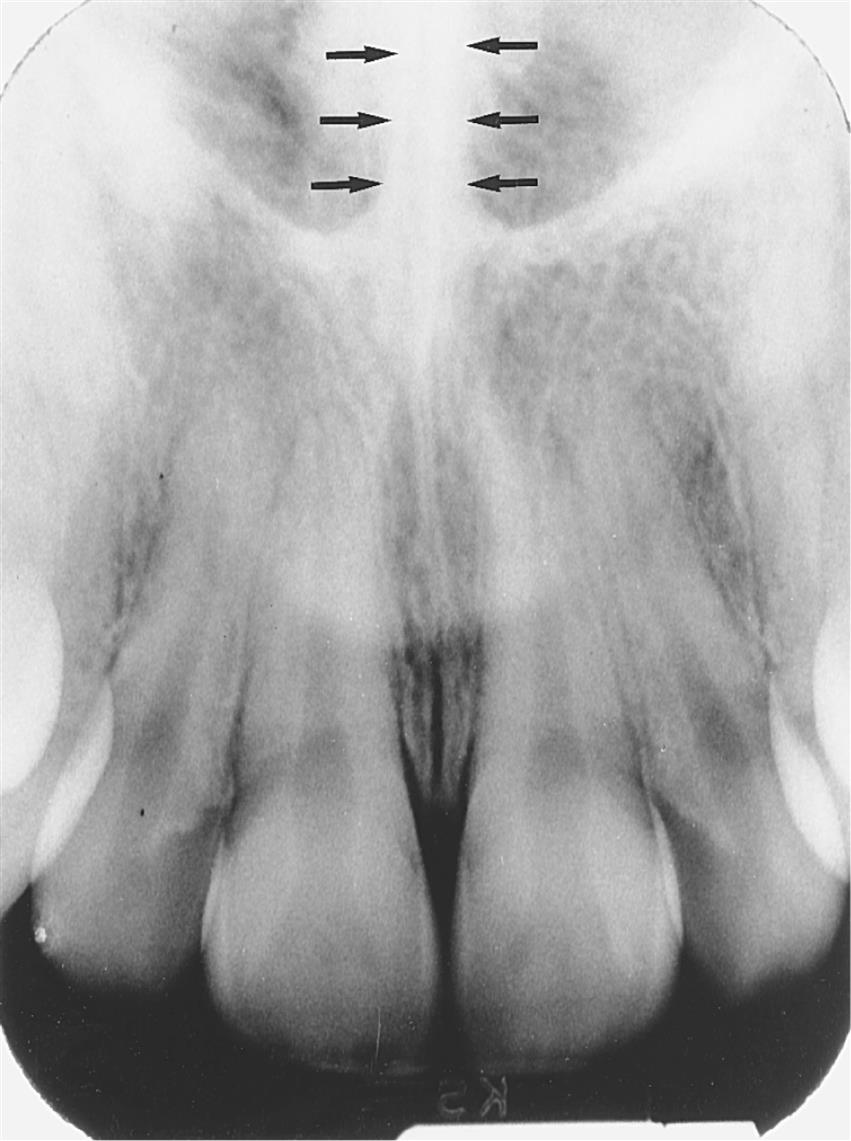
What is the nasal septum?
100
Oral structures that are _________ will appear radiopaque on the image because they absorb more x-rays.
What is more dense?
200
These words make up the ALARA concept, and it refers to minimizing the patient's radiation exposure.
As Low As Reasonably Achievable
200
This helps to stabilize the sensor in the correct position, reduces the risk of distortion, and ensures consistent image quality.
What is and XCP instrument or sensor holding device?
200
These are the 3 exposure factors which influence the quality of dental images.
What is time (impulses), milliamperage (mA), and kilovolts (kVp)?
200
A normal radiolucent landmark with a radiopaque border in the mandible.
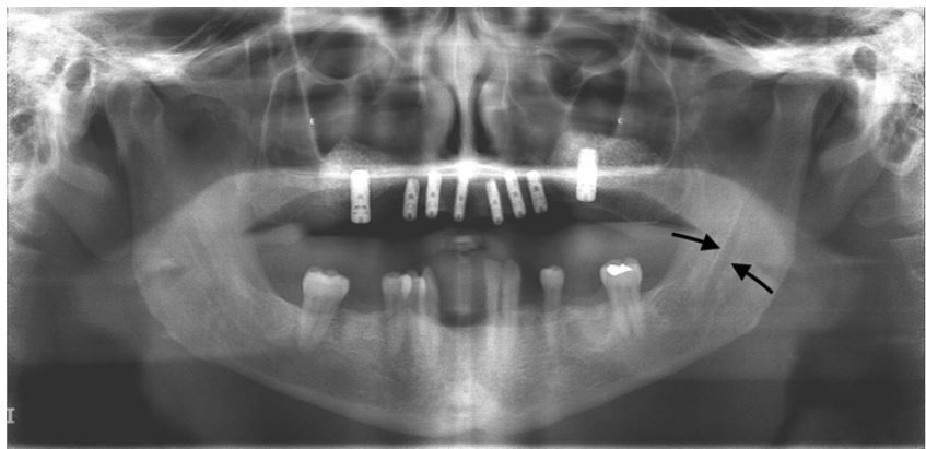
What is the mandibular canal?
200
This traditional unit of exposure for x-rays has limitations because it is unable to measure radiation absorbed by the body.
What is roentgen (R)?
300
This refers to the degree of blackness or opacity on a radiograph, and depends on the amount of x-ray absorption by the tissues.
What is radiographic density?
300
This type of radiograph is for visualizing the entire tooth, including the root and surrounding bone.
What is a periapical radiograph?
300
Your radiographic image may be too light due to this error.
What is inadequate or too little exposure time? (not enough radiation)
300
The inverted Y seen on this image is made up of these two normal landmarks.
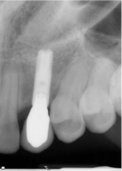
What is the floor of the nasal cavity and the maxillary sinus?
300
These effects are the result of a radiation injury that do not involve the reproductive organs and therefore is only seen in the person being irradiated.
What are somatic effects?
400
According to the principles of Shadow Casting, this type of distortion happens to an image when the target (focal spot) and receptor distance is too short.
What is magnification?
400
This type of collimator benefits the patient's safety and the image quality.
What is a rectangular collimator?
400
This error occurs when the radiation doesn't expose the entire sensor.
What is a cone cut?
400
The extracted tooth in this image.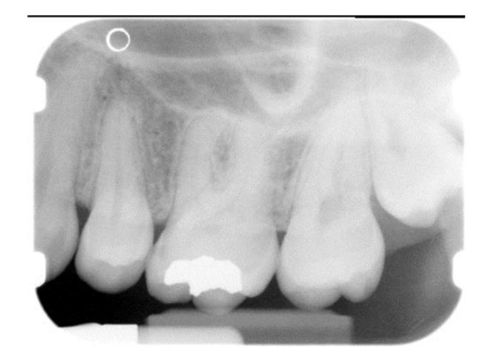
What is tooth #12?
400
Ionizing radiation has this primary biological effect on living tissues.
What is damage to cellular DNA which leads to mutations, cell death, or cancer?
500
The use of this device helps to demonstrate the densities and contrast scales and monitor the qualities of the film, the film processing, and the digital sensor, as well as calibration of the x-ray machine.
What is a stepwedge?
500
This term refers to the portion of a processed dental image that is dark or black.
What is Radiolucent?
500
When using the bisecting angle technique on a patient with a shallow palate, we must adjust this angulation to compensate and achieve proper image alignment and minimize distortion.
What is the vertical angulation?
500
This round radiolucency is seen in the apical area of the mandibular premolar area.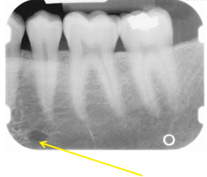
What is the mental foramen?
500
This form of secondary radiation and is the result of an x-ray that has been deflected from its path by the interaction with matter. It is detrimental to both the patient and the radiographer.
What is scatter radiation?