HL
Weird S***
See Eye
Anatomy
Hard
100
Name an autosomal dominant cause of hearing loss.
WANT CBS
- W — Waardenburg syndrome
- A — Apert syndrome
- N — Neurofibromatosis (Type 1 or 2)
- T — Treacher Collins syndrome
- C — Crouzon syndrome
- B — Branchio-oto-renal (BOR) syndrome
- S — Stickler syndrome
100
A surfer from Santa Cruz California (notorious for cold ocean water presents to clinic), name the pathology affecting the EAC.
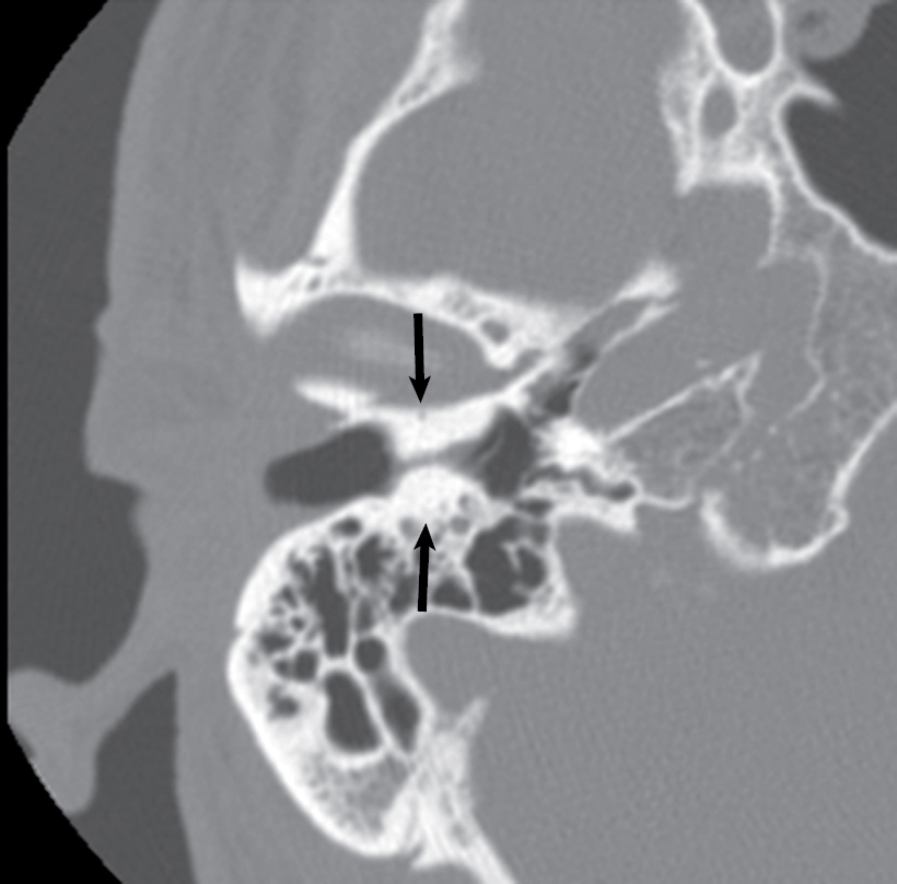
What is surfer's ear or exostosis?
100
This condition causes profound deafness or total hearing loss from birth, severe balance and vestibular problems, and night blindness that typically appears by age 10, eventually leading to tunnel vision or severe sight loss.
Must name condition and type for points.
What is Usher syndrome type 1
100
Name the three vestibular organs (must name all three for credit)
What are the utricle, saccule, and semicircular canals?
100
Broadmann's area 41 represents this auditory portion of the cerebral cortex which resides in the temporal lobe, close to the sylvian fissure.
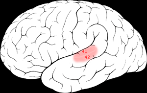
What is the primary auditory cortex?
200
MELAS syndrome is inherited with this pattern:
Mitochondrial inheritance (maternally inherited)
(Mitochondrial Encephalomyopathy, Lactic Acidosis, and Stroke-like Episodes): Linked commonly to the m.3243A>G mutation; features stroke-like events, muscle weakness, seizures, lactic acidosis, and progressive sensorineural hearing loss (SNHL).
200
Genetic mutations affecting prestin production (SLC26A4/PDS gene) lead to deficient mechanoelectrical transduction by outer hair cells manifesting as sensorineural hearing loss, a feature of this autosomal recessive syndrome.
What is Pendred syndrome?
Causes euthyroid goiter and early HL.
200
The head impulse test is used to test this reflex:
What is the vestibulo-ocular reflex?
200
The mandibular condyle articulates with this structure of the temporal bone.
What is the glenoid or mandibular fossa of the temporal bone?
200
Speed of sound in dry room temperature air.
Both teams text Julliette closest wins.
340 m/s
300
Because the left and right ears are located at the opposite sides of the head, the amount of time it takes for a sound stimulus to arrive at each ear is governed by the distance from the sound source to that particular ear. This can be used as a cue for sound localization. This is important for low-frequency sound localization.
What is the Interaural time difference (ITD)?
300
This is the most common type of cochlear dysplasia with involvement of the pars inferior (saccule and cochlear duct). The membranous cochlea is malformed, which results in high frequency hearing loss mostly and less frequently low frequency hearing loss.
What is Scheibe’s aplasia?
300
The number of turns in the cochlea
The cochlea is a snail-shaped structure that has a wide diameter at the base and narrows over 2.25 turns until it reaches its apex.
Will also accept 2.75 turns as this was quoted in the module.
300
This named area is the apex of the cochlea at which the Scala vestibuli and Scala tympani communicate.
What is the helicotrema?
At the apex of the cochlea is the helicotrema, where the Scalae vestibuli and tympani communicate.
300
The medial geniculate body is the auditory relay center that receives auditory information from the inferior colliculus and is housed within this portion of the brain. It has three divisions: ventral, dorsal, and medial.
The ventral division of the medial geniculate body projects to the primary auditory cortex, whereas the dorsal division projects to the auditory association cortex.
What is the thalamus?
400
These are low-intensity sounds generated by the cochlea either spontaneously or in response to external sound stimulation. They are believed to be related to the process of cochlear amplification. Contraction of the outer hair cells generates displacement waves in the basilar membrane that travel back through the middle ear and emerge in the ear canal, where they can be detected by a sensitive microphone. They are valuable clinical indicators of cochlear health.
What are Otoacoustic emissions (OAEs)?
400
This syndrome is the least severe form of inner ear dysplasia, affecting the cochlear duct and the basilar membrane and causing high frequency SNHL
What is Alexander syndrome?
400
The average length of the cochlear duct in mm, according to Cummings.
Both teams text Julliette guesses, closest wins.
You will have 2 minutes to text an answer, you will not lose points for guessing.
The cochlear duct length ranges from approximately 28.0 to 40.1 mm, with an average length between 33.13 and 34.0 mm. Cochlear duct length variability is relevant for cochlear implant electrode insertion depth and coverage of stimulable elements within the organ in rehabilitation of sensorineural hearing loss.
400
This is highly porous bone at the central core of the cochlea which allows the passage of auditory nerve fibers from the IAC to the hair cell synapses.
What is the modiolus?
The central core of the cochlea is the modiolus, a highly porous bone that allows the passage of auditory nerve fibers from the internal auditory meatus to the hair cell synapses.
400
Perilymph is found in the Scala vestibuli, Scala tympani, and internal spaces of the organ of Corti. It is high in sodium, low in potassium and communicates with CSF via which structure.
Communicates with cerebrospinal fluid via cochlear aqueduct (perilymphatic duct).
500
CDH23 and PCDH15 are calcium-sensitive cadherin proteins that connect tips of adjacent sterocilia and are essential for mechanoelectrical transduction.
Mutations in these genes are known cases of this genetic syndrome (of which there are four types: Ia, Ib, II, and III)
Cadherin 23 (CDH23) and protocadherin 15 (PCDH15), both members of the calcium-sensitive cadherin family of proteins, are key components of the hair bundle tip links in hair cells. These tip links, oriented obliquely, connect the tips of adjacent stereocilia and are essential for mechanoelectrical transduction. Several studies have identified mutations in the genes encoding CDH23 156 , 157 and PCDH15 158 , 159 as causes of Usher syndrome
500
This area housed within the petrous portion of the temporal bone is the hardest bone in the human body, with hardness equivalent to ivory.
What is the bony labyrinth consisting of the cochlea, semicircular canals, and vestibule?
500
Cochlear implants electrically stimulate these neurons of the cochlear nerve, bypassing the cochlea (and thus the most common location of SNHL).
A cochlear implant delivers sound information by electrically stimulating the primary afferent (spiral ganglion) neurons of the cochlear nerve, bypassing the cochlea where most sensorineural hearing loss (SNHL) occurs because of auditory hair cell loss or dysfunction. It is paired with an external sound processor, microphone, and power supply.
500
Name the membranes that border the Scala media superiorly and inferiorly, must name both for points.
The scala media is bordered:
Superiorly by the Reissner membrane
Inferiorly by the basilar membrane
Laterally by a portion of the outer cochlear wall
500
With regard to T1 and T2-weighting, contrast, and diffusion restriction, describe cholesteatoma enhancement patterns on MRI.
Both teams have 2 minutes to come up with a response and text to Julliette. Must answer all 4 for points. Will NOT lose points for incorrect answer.
Cholesteatoma has distinct imaging characteristics on magnetic resonance imaging (MRI). The lesion is isointense to hypointense on T1-weighted imaging, is intermediate on T2-weighted imaging, and does not enhance with addition of contrast medium. Most importantly, the lesion will restrict on diffusion-weighted imaging. MRI techniques for detection of residual cholesteatoma in lieu of staged surgery are evolving and promising.