Anatomy
Identification
Wild Card
100
What is the periodontal structure that normally appears as a thin, radiolucent line of uniform thickness around the root?
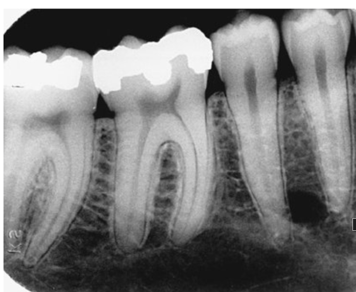
Periodontal ligament space
100
Horizontal bone loss is identified when bone loss occurs where?
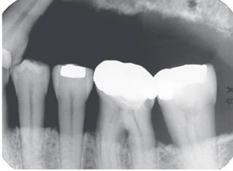
Parallel to adjacent CEJs
100
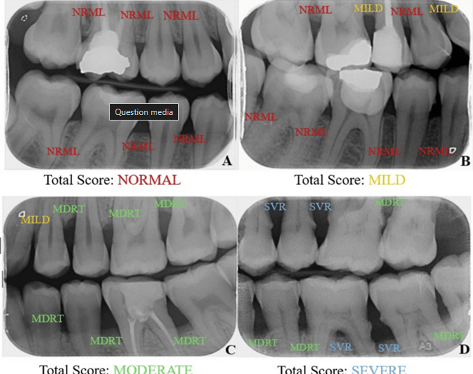
Of the 4 images below, which is the IMAGE OF CHOICE for evaluating Periodontal Disease?
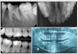
Periapical Image
200
What is the primary feature used to identify periodontal disease?
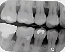
Changes in the alveolar crest
200
TRUE OR FALSE: Vertical bone loss appears in an uneven direction
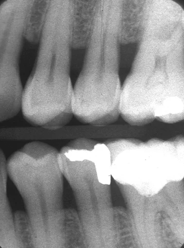
TRUE
200
TRUE OR FALSE: Dental images provide information about supporting bones that cannot be obtained clinically
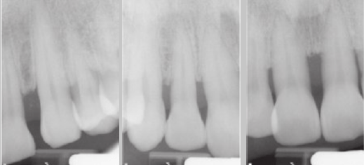
TRUE
300
Where is the alveolar crest is normally located?
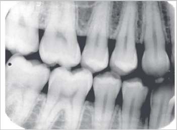
1.5 to 2 mm apical to the CEJ
300
hat type of bone loss is used to describe bone loss that is distributed evenly throughout dental arches with more than 30% of the sites involved?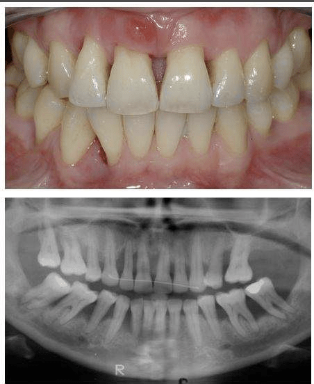
Generalized bone loss
300
A Flat, smooth, and parallel line joining adjacent CEJs indicates…
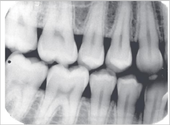
A healthy posterior alveolar crest
400
What are 3 features best describe the alveolar crest in the anterior region?
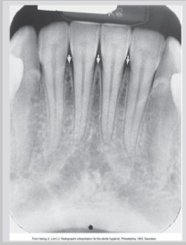
Pointed, sharp, very radiopaque
400
Bone loss extending to the mid third of a tooth with furcation involvement is what stage of periodontitis?
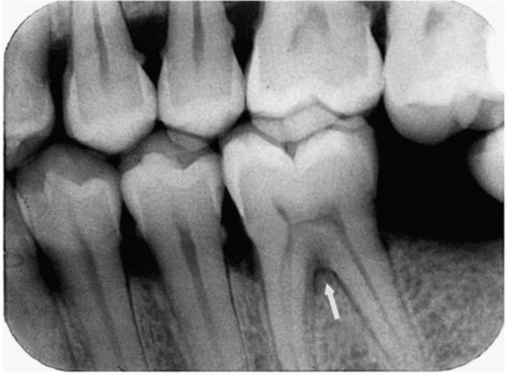
Stage III
400
What radiographic finding is MOST suggestive of furcation involvement?
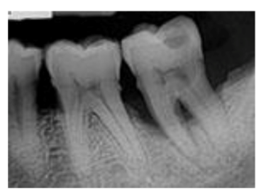
Radiolucency in the furcation area
500
Early stages of periodontal disease typically manifest at the alveolar crest as…
Fuzziness and loss of sharpness of the lamina dura
500
Which ADA Case Type is associated with no radiographic bone loss?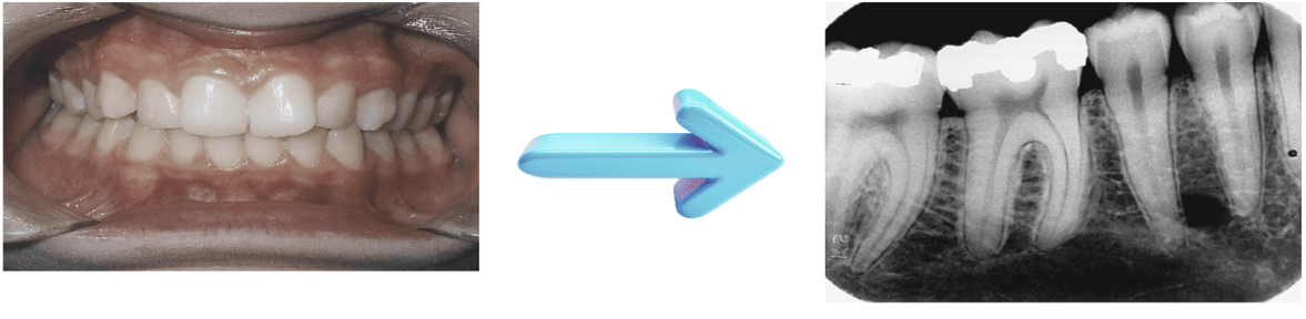
Type I
500
Defective restorations can act as local contributing factors for periodontal disease because they are
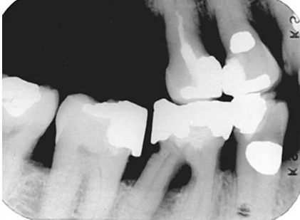
Severe plaque traps (which cause localized bone loss)