Who? what? Where?
What the Fundus?
Artifact Attack!!
Protocol Schmotocol
What did you call me??
OCT the good the bad and the ugly
100
A retinal detachment patient comes in what imaging would be taken?
Oct ou and Fundus photos ou
100
What is the MOST important part of taking Fundus photos on a retinal tear?
S-W-E-E-P-S
100
What is the top 3 artifacts seen in optos fundus photos? Bonus points on how to fix them!
Dust-Clean the mirror!!!!!!
Lashes- hold lids
Faceplate- Turn the patient further to each side
100
A patient comes in for HVF 30-2 what is the correct timing of dilation?
what is After the visual field is complete
100
What is the correct meaning of the acronym AF?
Autofluorescence Fundus photo
100
For an OCT Cube what should be the center of the scan?
what is the Macula/fovea
200
You pick up a chart with nothing circled the note reads "NP ref to Dr.Schneider for AMD ou" What would be the appropriate imaging? Bonus points for being extra specific!!
OCT ou OCTA ou (ORCC pdf) FP optos
200
What is the first thing you should check for when pulling a patient for Clarus fundus photos?
what is Dilation
200
New patient for Dr.Busbee was referred for DME you have completed the scans and Clarus fundus photos there is edema and dot blot hemes what is your next step?
What is alert scribes to review the imaging and ask about a FA
200
What are the 3 types of RVO
CRVO BRVO HRVO
200
What does OCT-A stand for?
Optical Coherence Tomography Angiography
300
if a brand new patient comes in for Dr. Dib for flashes of light what testing would be ordered?
OCT ou 21 line raster and 5 line raster OU Optos FP ou with SWEEPS
300
What is the difference between an RGB optos and the silverstone optos?
BONUS POINTS! Does any Dr. have a preference between them?
what is the RGB produces a true color photo
Dr. Thomas
300
What are the top diagnosis that would qualify for an OCTA?
AMD, CNV, RVO, Diabetic, CSR
300
What does RNFL and Optic disc cube 200x200 have in common?
what is it is the same scan over the nerve
400
" (Uveitis)3 mth rtn w/ Dr.Thomas exam ou dilate ou oct ou octA ou Fp Optos ou..." What testing should you do for this patient?
OCT ou OCTA ou Optos FP Optos AF ou
400
Can you came what can be improved in this photo?
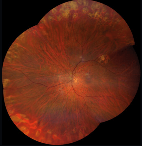
Not far enough out. Glare in each photo of the montage. not consistent coloring due to alignment or dialtion.
400
What camera can you do an HD radial? What diagnosis is it mostly used for?
what is cirrus OCT and Mac hole
500
Lattice vs schesis- what is it and what test would you do?
lattice-thinning of the prefereal retina tissue
schesis- Layers of the retina that have separated
Optos sweeps woul be appropriate for both!