Lung
Cardio 1
VExUS
Name that vessel
Signs
100
Name the findings shown in Blue:
A- Lines
100
The structure shown here in yellow:

Mitral valve
100
What vessel is being imaged here and is the doppler tracing normal or abnormal?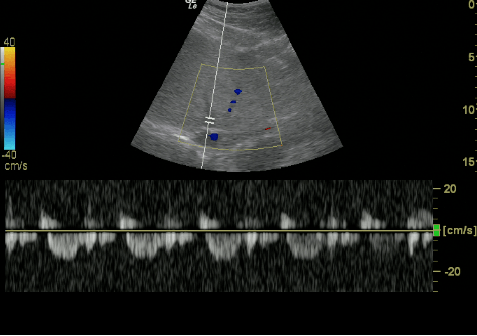
Hepatic Vein, Normal
100
Name the vessel with the color doppler in this Right LE vascular scan: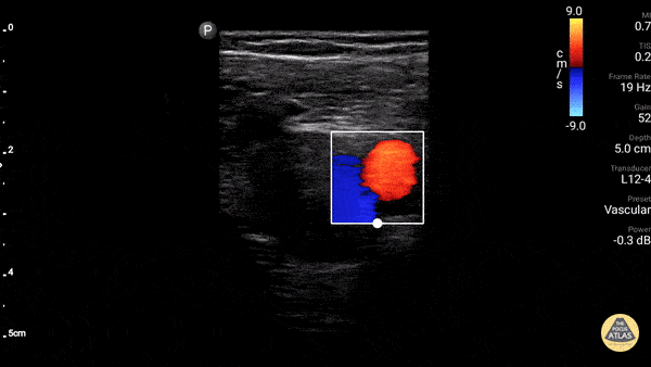
GSV
100
Seashore sign
200
Name the structure shown in Red:
Diaphragm
200
Chamber shown here in Green:

Right Atrium
200
Which Vessel and is the trace normal or abnormal?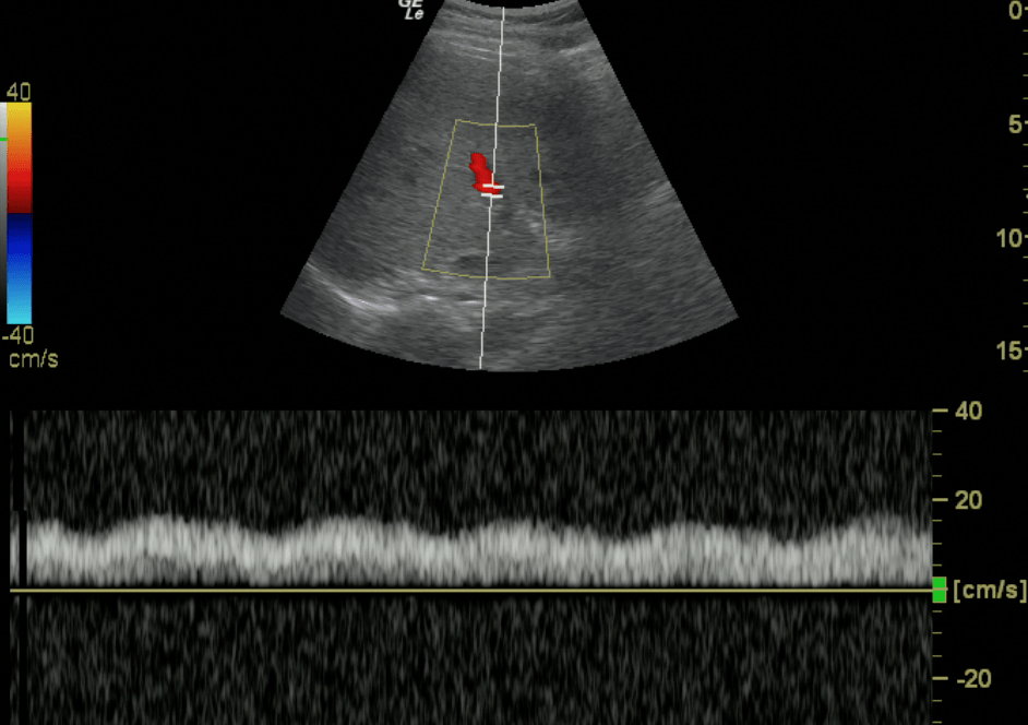
Portal vein, normal trace
200
Name the vessel with the Red color in this scna of the left side of the neck:
Carotid
200
What are those "dynamic" hyperechoic spots called:
Dynamic air bronchograms
300
Prominent finding(s) seen here:
Large pleural effusion, atelectatic lung
300
Name the view and levels shown here: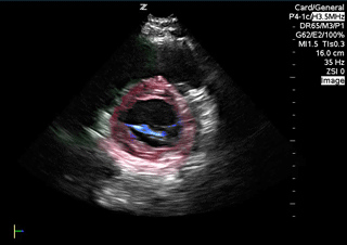
PSSX Mitral valve level and mid-ventricular or papillary muscle level
300
What vessel and what is your fluid assessment based on this solitary scan: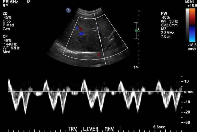
Hepatic vein, moderate congestion
300
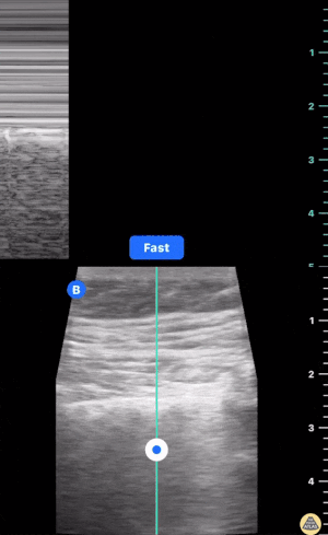
name the vessel being compressed in this scan of the Left leg: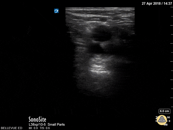
Popliteal vein
300
Sign shown in the RUQ scan:
Spine sign
400
Prominent finding seen here:
Absence of lung sliding
400
Name this view:
Apical 5 chamber
400
Which vessel, and volume assessment?
Portal vein and moderate to severe congestion
400
What vessel is the DVT in:
CFV
400
Lung related sign seen adjacent to the heart:
Jelly fish sign
500
76 y/o M with DOE, no hx of CHF, no signs of infection, smoking hx. What is the finding AND Likely diagnosis after seeing this scan of the Left lung base:
Septated pleural effusion likely exudative, Suspected malignancy
500
Name the view and the reason for the unusual finding seen here:
Subcostal 4 chamber, Agitated saline flush/air bubbles
500
Next best step in a patient with AKI and this renal doppler finding?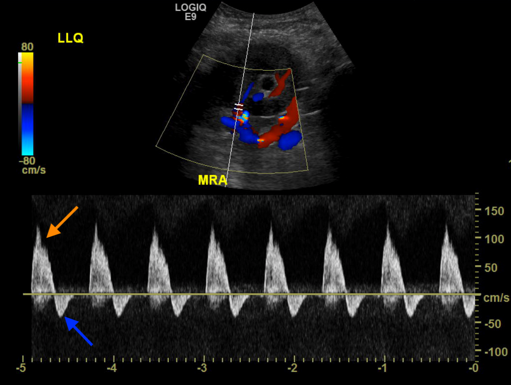
Diuresis
500
25yo had left molar extracted 10 days prior, presents with four days of subjective fever, malaise, and increasing pain to L neck.
POCUS scans through the jugular vein are shown below, what is this disease called?
Lemierre syndrome
500
What is this sign for PNA called:
Shred sign