Shoulder Complex
Elbow/Forearm
Wrist/Hand
Face/Neck
Nerves
Misc.
100
A patient demonstrates inability to initiate shoulder movement for the first 15 to 30 degrees while doing jumping jacks.
1. What motion is occurring at the shoulder, and in what plane?
2. What muscle is most involved?
3. Where is it's insertion?
1. Abduction, in the Coronal/Frontal Plane
2. The Supraspinatus Muscle
3. Greater Tubercle of the Humerus
100
Doing a push up, causes what movements at the elbow?
What plane do these osteokinematics occur in?
Elbow Flexion/Extension
They occur in the Sagittal Plane
100
1. The Hook of this carpal and the most "protruding" carpal bone on the palmar side are connected by a ligament that houses this nerve
2. What muscles are innervated by this Nerve?
1. The Ulnar Nerve
2. Flexor Carpi Ulnaris (FCU): Flexes and adducts the wrist.
- Flexor Digitorum Profundus (FDP - Medial/Ulnar Half): Flexes the distal interphalangeal joints of the ring and little fingers. [1, 2, 3]
Hand Muscles Supplied (Intrinsic)
Hypothenar Muscles (via deep branch):
- Abductor digiti minimi
- Flexor digiti minimi brevis
- Opponens digiti minimi
- Interossei Muscles (Deep branch):
- Four Dorsal Interossei: Abduct the fingers (DAB).
- Three Palmar Interossei: Adduct the fingers (PAD).
100
Pain and clicking during jaw opening suggests dysfunction of this TMJ structure
The articular disc
100
A fracture at the surgical neck of the humerus leads to impaired shoulder abduction and loss of sensation over the lateral shoulder.
1. Which Nerve is involved?
2. Which muscles are supplied by this nerve?
1. The Axillary Nerve
2. Deltoid and Teres minor
100
1. This plane divides the body into Right and left halves
2. This plane divides the body into a top and bottom halves
3. This plane divides the body into front and back halves
1. Sagital Plane
2. Transverse Plane
3. Frontal/Coronal Plane
200
Injury to this nerve will limited Scapular Protraction, and cause Scapular winging observed during a wall push-up.
Bonus: Where is the insertion of the muscle that is affected?
Long Thoracic Nerve
- Innervates the Serratus Anterior
Bonus:
Medial Border of the scapula (anterior surface)
Slide: Serratus Anterior
200
A patient cannot stabilize the radial head during Forearm Pronation following injury. Which Ligament is most likely involved?
The Annular Ligament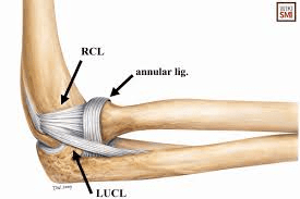
200
A patient presents with an injury to this tendon
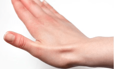 1. What is it?
2. What "region" of the body does this muscle create the border of ?
3. Which Carpal Bone can you palpate in this region?
1. Extensor Pollicis Longus
2. Anatomical Snuff Box
3. The Scaphoid
200
Deviation of the jaw to one side during opening indicates weakness of this muscle on the opposite side
What is the lateral pterygoid
So if the jaw deviates to the right, which side is affected?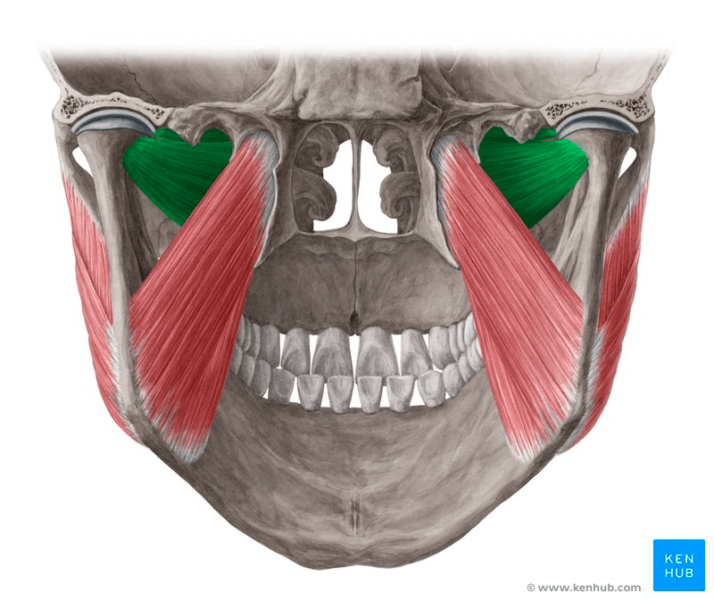
200
Compression beneath the Flexor Retinaculum leads to sensory deficits in the Volar Side of the thumb, index, middle, and lateral half of the ring finger.
1. Which Nerve is Involved?
2. What is the area beneath the flexor retinaculum called? (The area that houses all the flexor tendons, nerves)
1. The Median Nerve
2. The Carpal Tunnel
200
What is the difference between Type 1 and Type 2 Collagen?
Type I is densely packed for strength in skin, bones, and tendons, comprising 90% of the body's collagen, while Type II consists of looser, thinner fibers designed to provide flexible, shock absorbing cushion in cartilage and joints (Elastic cartilage, particularly in knees, shoulders, and other joints, as well as the trachea and ribcage.)
300
A patient is recovering from shoulder surgery and is having difficulty with bringing their arm to the side. Every time that they do, the PTA can tell that the tendon of this muscle is hitting the acromion process and causing pain.
1. Which muscle is involved?
2. Which joint(s) is not moving properly?
The supraspinatus muscle. The tendon is getting impinged under the acromion because the Scapulothoracic joint is not _______ rotating. 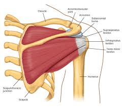
300
1. Which muscles are involved in supination?
2. Which of those is the most powerful when the elbow is flexed to 90 deg?
1. Supinator, Biceps Brachii, (and the brachioradialis assists)
2. The Biceps Brachii
300
1. This muscle inserts to the primarily into the pisiform bone, the hook of the hamate bone, and the base of the fifth metacarpal bone
2. What is the primary action of this muscle
1. The Flexor Carpi Ulnaris
2. Wrist Ulnar Deviation > Wrist Flexion
300
A patient was punched in the face, sustaining a blow to the mandible. The patient lost sensation of the face, as well as the ability to chew food. What structure (nerve) is affected?
The Trigeminal Nerve ( Cranial Nerve #5)
300
Weak wrist extension with wrist drop indicates injury to this nerve.
Bonus: Injury to which part of the humerus could cause this?
The Radial Nerve
Bonus: Radial Groove of the Humerus
300
A patient has sustained an injury to Frontalis Muscle, resulting in paralysis. What motion will they not be able to complete?
Elevating the eyebrows
400
A patient cannot extend the shoulder from a flexed position and has difficulty with powerful pulling movements.
1. Which Muscle is involved?
2. Which Nerve Innervates this muscle?
3. Which other muscles do Shoulder Extension?
1. Latissimus dorsi
2. Thoracodorsal Nerve
3.
Deltoid (posterior fibers)
Teres major
Triceps brachii (long head)
* Pectoralis major (lower fibers)
400
1. What is the anatomical region of the elbow crease called?
2. What can you palpate superior to this region that you will need for vitals?
1. Antecubital fossa
2. Brachial artery/Brachial Pulse
400
1. Which muscle is labeled letter A?
2. Give me another muscle that shares the same origin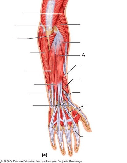
1. Extensor Digitorum
2. ECU, ECR (B), Extensor Digiti minimi
(Brachioradialis and ECRL originate right above these ones
Supinator also orginates from the lateral epicondyle, but does not share the common tendon)
400
1. What do the anterior scalenes do bilaterally?
2. What do all the scalenes ( anterior, middle, and posterior) do on one side (unilaterally)?
1. Cervical Flexion
2. Contralateral (opposite side) Cervical rotation and Ipsilateral (same side) lateral Cervical Flexion
400
Name the (4) major Spinal plexuses of the body
Cervical, Brachial, Lumbar and Sacral (the last two do have overlap and you will also see Lumbosacral Plexus)
400
The popliteal fossa is _______ to the Inguinal region (Provide anatomical descriptors)
Appropriate choices:
1. Inferior
2. Posterior
3. Caudal
500
1. This is the type of joint of the Sternoclavicular Joint:
2. This is the type of joint of the Glenohumeral Joint:
3. This is the type of joint of the Acromioclavicular Joint:
1. Saddle Joint
2. Ball-and-socket joint
3. Synovial plane (or planar) joint
500
A patient falls and fractures their olecranon process. Which Action will be most affected and why?
Elbow extension; the Triceps insertion is on the olecranon
500
Name the muscle. How do you know which muscle this is?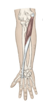
This is the Flexor Carpi Radialis
- It is a flexor as it is originating on the medial epicondlye
-It only crosses the wrist joint ( not a finger muscle mover)
-Its inserting on the radial side
500
Forward/Flexed cervical posture places increased strain on these cervical muscles
Upper Trapezius, Levator Scapulae
500
This is the name of the nerve that innervates the diaphragm ( Hint: It comes off of the cervical plexus, and the saying is " __, ___, ___ keeps the diaphragm alive"
What is the Phrenic Nerve: it innervates our pimary inspiratory muscle; the diaphragm. The saying is about its roots:" C3, C4, C5: keeps the diaphragm alive"
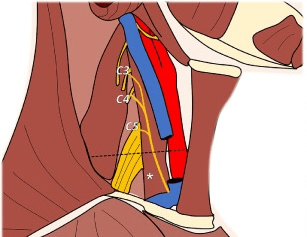
500
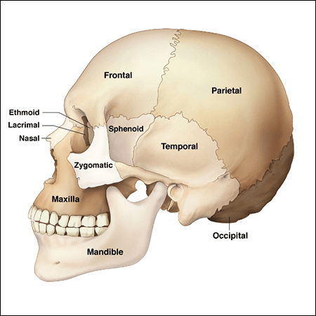
The mastoid process, the styloid process, the zygomatic arch, and mandibular fossa; are all parts of which bone?