Pathophysiology
Clinical Presentation
Diagnostics/Labs
Treatment
Double Trouble
100
Which of the following is the highest-risk period for developing a pulmonary embolism (PE) in pregnancy?
A) First trimester
B) Second trimester
C) Third trimester
D) Postpartum period
D) Postpartum period
100
What are some contents that a pulmonary embolism can be composed of? What is the most common?
Fat
Air
Thrombus (most common)
Bacteria
Amniotic Fluid
Tumor
Mnemonic: FATBAT
Bonus: In which clinical context are air emboli most commonly encountered?
100
What is the primary diagnostic imaging test for detecting a pulmonary embolism?
A) Chest X-ray
B) Ultrasound
C) CT pulmonary angiography (CTPA)
D) MRI
C) CT pulmonary angiography (CTPA)
100
A 29-year-old woman with a history of provoked DVT in her prior pregnancy is now 10 weeks pregnant. She has no known thrombophilia.
Which of the following is the best management approach for thromboprophylaxis during her current pregnancy?
A. No anticoagulation is needed since the prior event was provoked
B. Initiate prophylactic-dose LMWH antepartum and continue for 6 weeks postpartum
C. Initiate full therapeutic-dose LMWH and continue for 6 months postpartum
D. Use aspirin alone for thromboprophylaxis unless symptoms develop
Correct Answer: B. Initiate prophylactic-dose LMWH antepartum and continue for 6 weeks postpartum
Women with a prior provoked DVT (e.g., pregnancy-related) are at increased risk for recurrence in subsequent pregnancies. Prophylactic-dose LMWH antepartum and postpartum for 6 weeks is the recommended approach.
100
Which of the following mechanisms best explains how obesity contributes to an increased risk of venous thromboembolism (VTE)?
A. Enhanced fibrinolysis secondary to adipocyte-derived tissue plasminogen activator
B. Increased venous compliance leading to faster venous flow
C. Chronic low-grade inflammation and venous stasis due to physical compression and decreased venous return
D. Excess adipokines that reduce platelet activity and lower coagulation potential
E. Autoimmune-mediated destruction of prothrombotic factors
C) Obesity increases VTE risk through chronic low-grade inflammation and venous stasis (partial compression of veins and reduced venous return). This milieu leads to a hypercoagulable state, fulfilling key components of Virchow’s triad and predisposing to thrombosis.
200
Which of the following sets of risk factors most closely aligns with the development of our patient’s pulmonary embolism?
A. Atrial fibrillation, pneumonia, recent smoking cessation
B. Allergic reaction, peripheral arterial disease, essential hypertension
C. Prolonged immobility, hypercoagulable state, endothelial damage
D. Eczema, type 2 diabetes mellitus, vitamin D deficiency
C. Prolonged immobility, hypercoagulable state, endothelial damage
200
How would you compare the symptoms of a low-risk PE with symptoms of a massive PE?
Answer: Low-risk = asymptomatic/potential borderline hypotension
Massive = acute onset of chest pain, dyspnea, severe hypotension/shock
Discussion/bonus question: What type of PE did you think our patient had, and why?
200
A 45-year-old man with no significant medical history presents with sudden-onset dyspnea and pleuritic chest pain after a 12-hour flight. His vitals show a heart rate of 125 bpm, blood pressure of 110/75 mmHg, and oxygen saturation of 91% on room air. His ECG shows sinus tachycardia, and a D-dimer test is elevated. What is the most likely finding on CT pulmonary angiography?
A) A wedge-shaped infarction distal to an occluded pulmonary artery
B) Ground-glass opacities
C) Honeycombing pattern in both lungs
D) Bilateral pleural effusions
A) A wedge-shaped infarction distal to an occluded pulmonary artery
200
A 40-year-old man with a history of obesity and smoking is brought to the emergency department with severe shortness of breath and altered mental status. His blood pressure is 85/55 mmHg despite fluid resuscitation, heart rate is 130 beats/min, and oxygen saturation is 88% on 10 L of oxygen by face mask. A CT pulmonary angiogram shows a large, bilateral pulmonary embolus involving >50% of the pulmonary vasculature. Echocardiography reveals significant RV dilatation and dysfunction.
Question:
What is the most appropriate initial management step for this patient?
A. Begin low molecular weight heparin (LMWH) subcutaneously
B. Immediate surgical embolectomy
C. Thrombolytic therapy (e.g., alteplase)
D. Place an inferior vena cava (IVC) filter
E. Oral anticoagulation with a direct oral anticoagulant (DOAC)
Correct Answer: C. Thrombolytic therapy (e.g., alteplase)
In massive PE with hemodynamic instability (persistent hypotension, shock), systemic thrombolysis is recommended to rapidly dissolve the clot and improve hemodynamics, unless contraindications to thrombolytics exist.
200
Which of the following best explains how advanced maternal age can increase the risk of PE in pregnancy?
A. Decreased venous compliance promoting stasis and clot formation
B. Heightened fibrinolytic activity that rapidly clears forming clots
C. Reduced hypercoagulability due to lower hormonal levels
D. Improved arterial elasticity preventing venous pressure changes
E. Decreased likelihood of comorbidities (e.g., hypertension, obesity)
A)In the setting of advanced maternal age, multiple factors—including vascular changes, comorbid conditions, and baseline pregnancy hypercoagulability—all converge to heighten the risk of a venous thromboembolism (VTE), including PE.
300
A 40-year-old woman with systemic lupus erythematosus (SLE) presents with acute onset pleuritic chest pain and shortness of breath. She has been on chronic corticosteroids and has known antiphospholipid antibodies. Her ECG shows S1Q3T3 pattern, and a CT pulmonary angiogram confirms bilateral pulmonary emboli.
Which factor in Virchow’s triad is particularly relevant given her antiphospholipid antibody status?
A. Endothelial damage
B. Stasis of arterial blood
C. Turbulent blood flow
D. Hypercoagulability
D. Hypercoagulability
300
A 55-year-old male presents with sudden-onset dyspnea and syncope. He is found to be in cardiogenic shock with BP 75/50 mmHg. Which of the following is the most likely location of this patient’s PE?
A) Lobar PE
B) Segmental PE
C) Saddle PE
D) Subsegmental PE
C) Saddle PE
300
Why does a pulmonary embolism cause pleuritic chest pain?
A) It compresses the coronary arteries
B) It causes inflammation of the pleura due to lung tissue ischemia
C) It increases oxygen delivery to the lungs
D) It affects the diaphragm, leading to muscle spasm
B) It causes inflammation of the pleura due to lung tissue ischemia
300
A 60-year-old man with a history of hypertension and atrial fibrillation presents with acute dyspnea and hypotension. A CT pulmonary angiogram confirms a massive pulmonary embolism. The patient’s BP is 78/50 mmHg. Given his hemodynamic instability, thrombolytic therapy is considered.
Which of the following is an absolute contraindication to tPA administration in this patient?
A) History of ischemic stroke 5 years ago
B) Active gastrointestinal bleeding
C) Atrial fibrillation with left atrial thrombus
D) Uncontrolled hypertension with BP 150/90 mmHg
Correct Answer: B) Active gastrointestinal bleeding
Thrombolytic therapy (tPA, alteplase) is indicated for massive pulmonary embolism (PE) with hemodynamic instability (e.g., hypotension, shock). However, there are absolute contraindications to thrombolysis due to the high risk of life-threatening bleeding.
Active gastrointestinal bleeding is an absolute contraindication because tPA breaks down fibrin clots, increasing the risk of severe or fatal hemorrhage. A patient with ongoing GI bleeding is at extreme risk of massive hemorrhage if given thrombolytics.
300
A 52-year-old man with a history of recent lower limb orthopedic surgery presents to the emergency department with sudden onset shortness of breath, pleuritic chest pain, and lightheadedness. An electrocardiogram (EKG) is shown below.

Which of the following best explains these finding in the context of his clinical presentation?
A) left ventricular strain
B) right ventricular strain
C) Aortic dissection
D) Pericarditis
The S1Q3T3 pattern on an EKG—characterized by an S wave in lead I, a Q wave in lead III, and an inverted T wave in lead III—is a classic, albeit non-specific, sign of acute right ventricular strain. In this patient, the clinical context (postoperative status with risk factors for venous thromboembolism) along with the sudden onset of respiratory symptoms strongly suggests an acute pulmonary embolism. This embolic event can abruptly increase pulmonary vascular resistance, leading to right ventricular dilation and dysfunction, a condition known as acute cor pulmonale.
400
A 70-year-old man hospitalized for hip replacement surgery develops sudden shortness of breath and cyanosis on postoperative day two. Physical examination reveals a loud P2 (pulmonic component of the second heart sound) and jugular venous distension. Bedside echocardiography suggests right ventricular dilation.
What best explains the rapid development of right ventricular dilation in acute pulmonary embolism?
A. Rapid rise in pulmonary vascular resistance
B. Increased left ventricular preload
C. Massive fluid overload from IV fluids
D. Coronary artery spasm
A. Rapid rise in pulmonary vascular resistance
400
Identify the pathology in the CT angiogram below, and determine the most likely severity. Did our patient experience this exact pathology?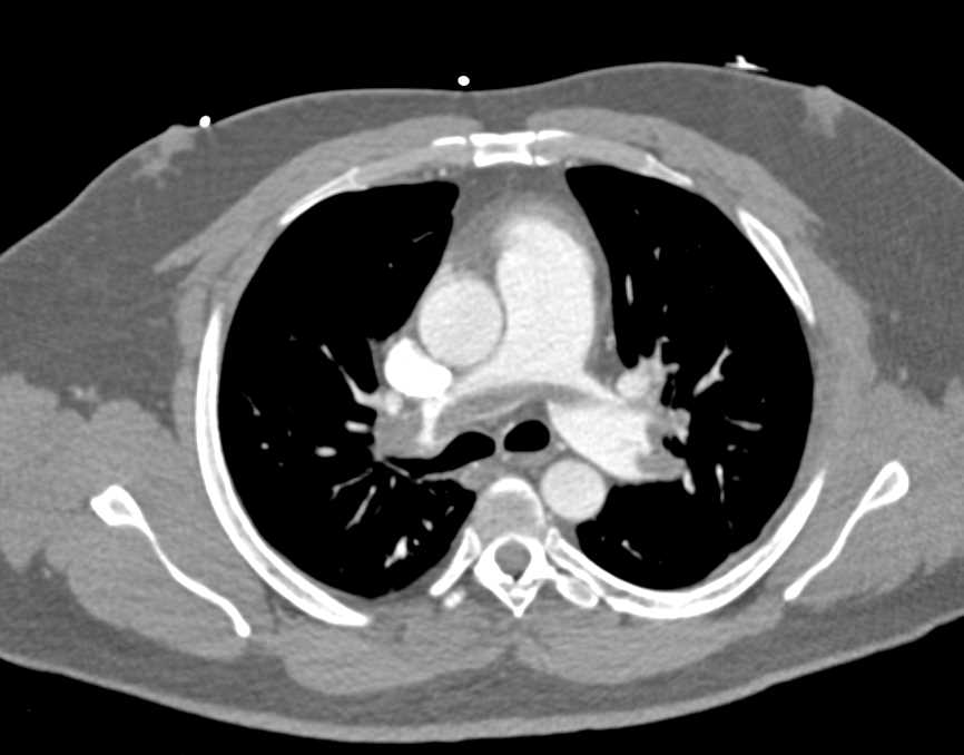
Saddle PE; Massive (High-risk) PE
Most likely present with acute onset hypotension/shock, sharp stabbing chest pain, (pleuritic), tachypnea (high RR)
No! Our patient's symptoms were gradual and not as sudden, alluding to a lobar, segmental, or subsegmental PE.
400
A 45-year-old obese woman presents to the emergency department with sudden-onset shortness of breath, pleuritic chest pain, and syncope. Her blood pressure is 85/60 mmHg, and her heart rate is 120 bpm. An EKG shows sinus tachycardia with a new right bundle branch block (RBBB). Which of the following best explains the development of RBBB in this patient?
A) Ischemia of the left ventricle due to coronary artery occlusion
B) Direct embolic occlusion of the bundle branches
C) Hypoxia-induced myocardial stunning affecting the left bundle branch
D) Increased right ventricular pressure impairing septal conduction
Increased right ventricular pressure impairing septal conduction
400
A 72-year-old man with metastatic colon cancer and recurrent venous thromboembolism (VTE) presents with acute right lower extremity swelling and pain. A duplex ultrasound confirms an extensive femoropopliteal DVT. His medical history includes a recent intracranial hemorrhage (ICH) two weeks ago, which has not fully resolved on repeat imaging. Despite best supportive measures and intermittent pneumatic compression devices, his leg swelling has worsened over the past 48 hours. He currently has stable hemodynamics, but you are concerned about the risk of a large pulmonary embolism (PE).
Which of the following is the most appropriate next step in management?
A. Initiate full-dose low-molecular-weight heparin (LMWH)
B. Insert a retrievable inferior vena cava (IVC) filter
C. Place a permanent superior vena cava (SVC) filter
D. Perform surgical thrombectomy of the right femoral vein
E. Resume warfarin at a reduced dose
Correct Answer: B. Insert a retrievable inferior vena cava (IVC) filter, In a patient with recent, unresolved intracranial hemorrhage and a new DVT in the setting of high PE risk, a retrievable IVC filter is the safest and most appropriate intervention to prevent life-threatening embolic events until anticoagulation can be safely restarted. SVC could be used for upper extremity risks
400
A 34-year-old woman in labor suddenly develops respiratory distress, hypotension, and signs of disseminated intravascular coagulation (DIC). What best explains her condition?
A. Septic shock from intrauterine infection results in DIC
B. A deep vein thrombosis (DVT) embolizes to the lungs, causing right heart failure and DIC.
C. An anaphylactic reaction to oxytocin leads to shock and DIC.
D. Fat embolism from pelvic trauma triggers DIC.
E. Amniotic fluid enters the maternal circulation, triggering an inflammatory and coagulopathic response.
E, Amniotic fluid embolism is a rare but catastrophic event that presents with sudden respiratory distress, hypotension, and DIC during labor. The pathophysiology involves amniotic fluid entering the maternal circulation and triggering an intense inflammatory response, making Option A the best explanation for the scenario.
500
A 45-year-old woman presents with sudden shortness of breath, tachypnea, and mild hemoptysis. She has a recent history of a hysterectomy for fibroids three weeks ago. On auscultation, breath sounds are diminished in the right lower lung zone. Further investigation reveals a wedge-shaped opacity on chest imaging in the right lower lobe. Which mechanism most accurately explains why pulmonary infarction may occur in this patient?
A. Complete interruption of bronchial artery blood supply
B. Blockage of distal pulmonary arteries leading to ischemia and necrosis
C. Direct invasion of the lung parenchyma by bacteria
D. Autoimmune inflammation triggered by surgery
B. Blockage of distal pulmonary arteries leading to ischemia and necrosis
500
A 64-year-old woman presents to the emergency department with sudden-onset dyspnea, pleuritic chest pain, and syncope. Her vital signs are BP 78/50 mmHg, HR 135 bpm, RR 30 breaths/min, and SpO₂ 89% on room air. Bedside echocardiography shows right ventricular dilation and septal bowing. Diagnosis of patients is High risk (massive) PE.
Which characteristic in this case classifies the patient’s pulmonary embolism as high-risk?
A) Hypotension
B) Tachycardia
C) Right ventricular dilation
D) Septal bowing
A) Hypotension
500
Explain or outline on the markerboard how a massive pulmonary embolism causes hypotension.
A massive PE obstructs pulmonary circulation -> leads to right ventricular strain and failure -> this reduces blood returning to the left heart -> lowering cardiac output (recall: CO=SV*HR) -> causing hypotension (recall: BP = COxSVR) -> can progress to shock if untreated
500
A 65-year-old woman with a history of recent major abdominal surgery (2 days ago) presents with acute onset of severe dyspnea, hypotension (blood pressure 85/50 mmHg), and profound tachycardia (heart rate 140 bpm). She appears diaphoretic and confused. Bedside transthoracic echocardiography reveals a severely dilated right ventricle (RV) with significant hypokinesis and a small, underfilled left ventricle. A CT pulmonary angiogram confirms a large saddle embolus straddling the main pulmonary artery bifurcation. Given her hypotension and end-organ hypoperfusion, you suspect massive PE. Laboratory workup shows a normal hemoglobin, but platelet count is slightly low at 110,000/µL. She has no active bleeding but is deemed a very high bleeding risk due to her recent surgery.
Which of the following is the most appropriate next step in management?
A. Intravenous thrombolytic therapy (e.g., alteplase)
B. Catheter-directed thrombolysis
C. Intravenous unfractionated heparin infusion only
D. Emergent surgical embolectomy
E. Placement of an inferior vena cava (IVC) filter
D) Emergent surgical embolectomy is indicated in massive, life-threatening PE when thrombolysis is contraindicated or has failed, and the patient is in shock or near shock. In this case, the recent major surgery (high bleeding risk) and profound hemodynamic instability make surgical embolectomy the priority intervention.
500
A 27-year-old male is brought to the emergency department 36 hours after a high-speed motor vehicle collision. He has multiple fractures, including bilateral femoral shaft fractures and a right humeral shaft fracture. Over the past 12 hours, he has developed the following:
Neurologic: Confusion and intermittent disorientation
Respiratory: Increasing shortness of breath, tachypnea (RR 30/min), and oxygen saturation of 91% on room air
Dermatologic: A petechial rash scattered over his neck, axillae, and upper arms
On auscultation, you note diffuse crackles bilaterally. A chest radiograph reveals bilateral fluffy infiltrates most prominent in the perihilar regions. Laboratory tests show an elevated serum lactate dehydrogenase (LDH).
Which of the following best explains the pathophysiology behind his pulmonary and neurological manifestations?
A. Micro emboli of fat globules from the marrow enter the venous circulation, lodge in the pulmonary capillary beds, and travel to cause neurologic findings.
B. Septic thrombi originating from open fractures that seed the lungs and central nervous system (CNS).
C. Direct alveolar hemorrhage secondary to the mechanical laceration of pulmonary vessels by bone fragments.
D. Amniotic fluid emboli that trigger disseminated intravascular coagulation and alveolar damage.
E. Cholesterol crystal emboli from atherosclerotic plaques that preferentially occlude small cerebral and pulmonary arterioles.
A) In the setting of multiple long-bone fractures, a petechial rash, acute respiratory distress, and neurological changes are highly suggestive of fat embolism syndrome. The pathophysiology involves fat globules from the marrow traveling to the pulmonary circulation (and possibly through a right-to-left shunt), causing widespread inflammatory and embolic phenomena.