Asthma & COPD
Pulmonary Embolism
Ventilation and Oxygenation
Ultrasound
Pulmonary Potpourri
100
What is the first-line treatment for Asthma? (Name four things)
Oxygen, inhaled bronchodilators, ipratropium, and systemic steroids.
100
What biochemical markers should be ordered in a patient being evaluated for PE?
Troponin & BNP
100
Adjusting this ventilator setting directly controls PaCO₂.
Minute ventilation (respitory rate x tidal volume)
100
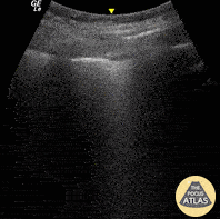
What is seen in this ultrasound?
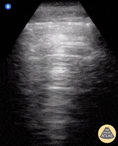
Normal lung sliding and A-lines.
A-lines are defined as horizontal, hyperechoic reverberation artifacts that appear at regular intervals below the pleural line, representing normal air-filled lung parenchyma. The presence of both lung sliding and A lines is considered the normal sonographic pattern of a well-aerated lung
100
What is the treatment for bronchiolitis?
Nasal suctioning and supplemental oxygen as needed
200
What is the only therapy proven to improve mortality in stable COPD?
Supplemental oxygen
200
What sign is seen in this US?

D sign
Flattening of the interventricular septum, giving the left ventricle a D-shaped appearance in the parasternal short-axis view
200
Plateau pressure should be kept below this to avoid barotrauma.
30cm H2O
200
What is seen in this US?
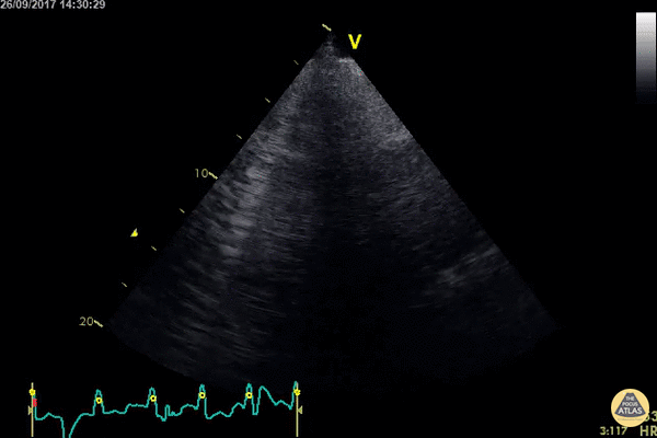
B-lines
B-lines on thoracic ultrasound should be described as vertical, hyperechoic artifacts that arise from the pleural line, extend to the bottom of the screen without fading, and move synchronously with lung sliding. The presence of multiple B-lines (≥3 per intercostal space) is indicative of alveolar-interstitial syndrome
200
What score can help to identify patients with pneumonia at high risk for morbidity and mortality?
CURB-65 Score
The Pneumonia Severity Index is best at identifying patients at low risk for mortality who are safe for outpatient treatment. The Infectious Diseases Society of America-American Thoracic Society recommends using this score over CURB-65; however, it requires an arterial blood gas (ABG).
300
What is an appropriate I:E ratio for an intubated asthmatic?
For patients who are mechanically ventilated for an acute asthma exacerbation, it should be set to 1:3 or longer.
The inspiratory flow rate should be set to 60-80 L/minute with a decelerating waveform.
300
Why do you want to avoid or delay intubation in a critical patient with a PE when possible?
Positive pressure ventilation and sedation may threaten hemodynamics (worsen RV preload and cardiac output), potentially leading to cardiovascular collapse.
300
What are the typical ventilator setting for a patient that was intubated for airway protection? The patient is 70 kg.
AC/VC
RR: 12-16
TV: 6-8 cc/kg IBW
PEEP: 5
FiO2: 60%
300
What is seen in this ultrasound?
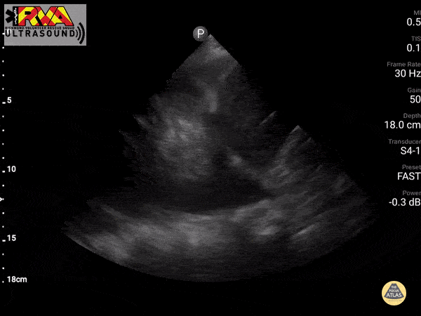
Pleural effusion.
On ultrasound, a pleural effusion is described as an anechoic (black) or hypoechoic space above the diaphragm, often triangular in shape. Key sonographic features include the visualization of the diaphragm, adjacent organs (liver or spleen), and the "spine sign"—where vertebral bodies are seen extending above the diaphragm due to the presence of fluid.
300
What is seen in this US?
PLAPS pneumonia
400
In a patient on a ventilator, this change decreases auto-PEEP in obstructive lung disease
Prolonging the expiratory time
400
What sign is seen on this ultrasound?

McConnell Sign
Hypokinesis or akinesis of the mid and basal segments of the RV free wall with preserved or hyperkinetic motion of the RV apex, typically visualized in the apical four-chamber view. This regional wall motion abnormality is classically associated with acute PE, reflecting acute RV pressure overload.
400
When a patient on the ventilator suddenly desaturates, this mnemonic can help guide troubleshooting_____. What do the letters stand for?
D.O.P.E.S.
Displacement of tube, Obstruction of tube, Pneumothorax, Equipment failure (or Extubation), and Stacked breaths (auto-PEEP)
400
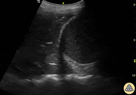
What finding is seen here?

Lung point
This represents the boundary where the collapsed lung intermittently contacts the parietal pleura during respiration, and is pathognomonic for pneumothorax.
400
What are indications for a chest tube (3) vs a percutaneous pigtail catheter (4)?
Chest tube:
Hemothorax
Traumatic pneumothorax with a large parenchymal or bronchial lung injury (bronchopleural or alveolar-pleural fistula)
Tension pneumothorax after a finger thoracostomy
Percutaneous pigtail catheter:
Most pneumothoraces
Small hemothorax
Pleural effusion
Empyema
500
_________ are a surrogate for lung volume at end-inspiration, which directly correlates to the risk of barotrauma.
Plateau pressures
500
What can be given to a patient in cardiac arrest where a PE is suspected?
Thrombolytics
500
A 55-year-old asthmatic on the vent suddenly becomes hypotensive with high peak pressures. What DOPE(S) complication is most likely?
Auto-PEEP
500
What sign is this?
Shred sign
Is a static sonographic sign of lung consolidation. Consolidated lung tissue appears as a subpleural hypoechoic region that has an irregular (shredded) deep border (fractal line) abutting normally aerated lung, which has echogenic artifacts.
500
What structures make up the “triangle of safety” when placing a chest tube?
Bordered anteriorly by the lateral edge of the pectoralis major, laterally by the lateral edge of the latissimus dorsi, inferiorly by the line of the fifth intercostal space, and superiorly by the base of the axilla.